New Yeast Species,
Malassezia dermatis
, Isolated from
Patients with Atopic Dermatitis
Takashi Sugita,
1* Masako Takashima,
2Takako Shinoda,
1Hajime Suto,
3Tetsushi Unno,
3Ryoji Tsuboi,
3Hideoki Ogawa,
3and Akemi Nishikawa
4Department of Microbiology1and Department of Immunobiology,4Meiji Pharmaceutical University,
Kiyose, and Department of Dermatology, School of Medicine, Juntendo University, Bunkyo-ku,3Tokyo, and Japan Collection of Microorganisms, RIKEN (The
Institute of Physical and Chemical Research), Wako, Saitama,2Japan
Received 4 September 2001/Returned for modification 31 December 2001/Accepted 2 February 2002
Malasseziaspecies are considered to be one of the exacerbating factors in atopic dermatitis (AD). During examination of the cutaneous colonization ofMalasseziaspecies in AD patients, we found a new species on the surface of the patients’ skin. Analysis of ribosomal DNA sequences suggested that the isolates belonged to the genusMalassezia. They did not grow in Sabouraud dextrose agar but utilized specific concentrations of Tween 20, 40, 60, and 80 as a lipid source. Thus, we concluded that our isolates were new members of the genus
Malasseziaand propose the nameMalassezia dermatissp. nov. for these isolates.
Malasseziaspecies are known causative factors in pityriasis versicolor, seborrheic dermatitis (SD), and atopic dermatitis (AD) (3). In the last decade, research has focused primarily on
isolatingMalasseziastrains and detecting specific
immunoglob-ulin E (IgE) antibodies from patients (9, 13, 14, 26). A
com-parison of the isolation rates ofMalassezia species from the
skin of AD patients and healthy control subjects detected a significantly higher rate for patients than for healthy subjects
(8). AD patients had specific IgE antibodies against
Malasse-zia, while healthy subjects did not. In recent years, studies have
increasingly been directed towards analyzing how the cutane-ous microflora at the species level are related to each disease type (pityriasis versicolor, SD, and AD) (1, 6, 7, 12, 16). We
previously compared the distribution ofMalasseziaspecies in
skin lesions of AD patients and in healthy subjects using a nonculture method (nested PCR) that is not affected by the isolating medium (21). Of the seven members of the genus
Malassezia,Malassezia globosaandM. restrictawere the species
most commonly associated with AD, whileM. obtusaand M.
pachydermatiswere not detected in AD. In our survey of
cu-taneousMalasseziamicroflora, we isolated newMalassezia
spe-cies from several patients with AD. In this paper, we propose
a new species,M. dermatis, for these isolates.
MATERIALS AND METHODS
Malasseziaisolates.Nineteen AD outpatients at Juntendo University Hospital
were included in the study. To obtain samples, OpSite transparent dressings (3 by 7 cm; Smith and Nephew Medical Ltd., Hull, United Kingdom) were applied to skin lesions (erosive, erythematous, and lichenoid) on the scalp, back, and nape of the neck of AD patients. Samples were then transferred onto Leeming and Notman agar (LNA) (10.0 g of polypeptone, 5.0 g of glucose, 0.1 g of yeast extract, 8.0 g of ox gall, 1.0 mg of glycerol, 0.5 g of glycerol stearate, 0.5 mg of Tween 60, 10 ml of cow’s milk [whole fat], and 12.0 g of agar per liter) plates
containing 50l of chloramphenicol (Sankyo, Tokyo, Japan) and incubated at 32°C for 2 weeks.
Direct DNA sequencing of rRNA genes of the isolates.Yeast isolates recovered from LNA medium were identified by analysis of rRNA gene sequences. Nuclear DNA of the isolates was extracted by the method of Makimura et al. (15). The D1 and D2 regions of 26S ribosomal DNA (rDNA) and internal transcribed spacer (ITS) regions in the rRNA gene were sequenced directly from PCR products using the primer pairs NL-1 (forward; GCATATCAATAAGCGGAG GAAAAG) and NL-4 (reverse; GGTCCGTGTTTCAAGACGG) (11) and pITS-F (forward; GTCGTAACAAGGTTAACCTGCGG) and pITS-R (reverse; TCCTCCGCTTATTGATATGC) (22), respectively. The PCR products were sequenced using an ABI 310 DNA sequencer and an ABI PRISM BigDye Terminator Cycle Sequencing kit (Perkin-Elmer Applied Biosystems, Foster, Calif.) according to the manufacturer’s instructions. Strains with 99% or more similarity of the D1 and D2 regions of 26S rDNA and the overall ITS sequences were defined as conspecific (18, 22). The sequence data were searched using the BLAST system (http://www.ncbi.nlm.nih.gov/BLAST/) at the National Center for Biotechnology Information, Bethesda, Md.
Molecular phylogenetic analysis.The sequences were aligned using ClustalW software (25). For neighbor-joining analysis (20), the distances between se-quences were calculated using Kimura’s two-parameter model (10). A bootstrap analysis was conducted with 100 replications (2).
Morphological, physiological, and chemotaxonomic characteristics. Morphol-ogy was examined on LNA after incubation at 32°C for 7 days. Tween 20, 40, 60, and 80 utilization, catalase reactions, and diazonium blue B reactions were performed as described by Guého et al. (4). Identification of ubiquinone was carried out using methods described by Nakase and Suzuki (17). The nuclear DNA base composition (moles percent G⫹C) was determined by high-pressure liquid chromatography after enzymatic digestion of DNA into deoxyribonucleo-sides (23, 24). The DNA-GC kit (Yamasa Shouyu, Chiba, Japan) was used according to the manufacturer’s instructions.
Nucleotide sequence accession numbers. The nucleotide sequences deter-mined in this study have been deposited with DDBJ (DNA Data Bank of Japan) under the accession numbers shown in Fig. 1.
RESULTS
Molecular phylogenetic analysis. All of the yeast isolates obtained from the 19 AD patients were identified by analysis of rRNA gene sequences (D1 and D2 regions of 26S rDNA and ITS). Of these, the DNA sequences of five isolates did not match sequences in the DNA sequence database. These iso-lates were designated M 9927, M 9928, M 9929, M 9930, and M
* Corresponding author. Mailing address: Department of Microbi-ology, Meiji Pharmaceutical University, 2-522-1 Noshio, Kiyose, Tokyo 204-8588, Japan. Phone: 81-424-95-8762. Fax: 81-424-95-8762. E-mail: sugita@my-pharm.ac.jp.
1363
on May 15, 2020 by guest
http://jcm.asm.org/
9931. The first three strains were isolated from a single patient, while the last two strains were isolated from one patient each. Sequence analysis of their 26S rDNA indicated that these
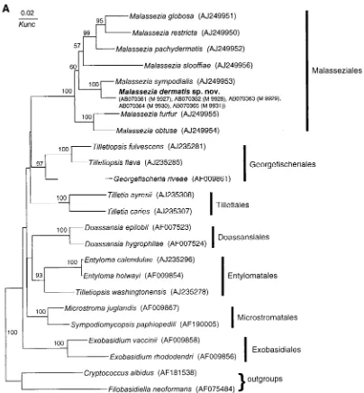
isolates belonged to the genusMalassezia(Fig. 1A). The
se-quences of five isolates were completely identical in both the
26S rDNA and ITS regions and clustered withM. sympodialis
with high bootstrap values (100%) (Fig. 1). Dissimilarities
be-tween these isolates and the M. sympodialis strain in their
D1 and D2 regions of 26S, ITS1, and ITS2 were 1.2% (7 of 578), 10.5% (17 of 162), and 10.3% (24 of 233), respec-tively. The overall dissimilarity of ITS regions was 10.4% (41 of 395).
Taxonomic characteristics.The characteristics that
differen-tiate the new species,M. dermatis, from the other seven known
Malasseziaspecies are summarized in Table 1. The
physiolog-ical characteristics ofM. dermatisare identical to those ofM.
furfur. However, the moles percent G⫹C ofM. dermatis
nu-clear DNA is 60.4% while that ofM. furfuris 66.0 to 66.7%. A
difference of more than 1 mol% G⫹C between two strains is
considered taxonomically significant (19, 24).
Latin description ofMalassezia dermatisSugita, Takashima, Nishikawa et Shinoda sp. nov.In LNA, post dies 7 ad 32°C, cellulae vegetativae sphaericae, ovoideae vel ellipsoideae (2–8)
⫻ (2–10) m. Cultura xanthoalba, semi-nitida aut hebetata,
butyracea et margo glabra aut lobulata. In agaro glucoso-pep-tonico Tween 20, 40, 60, 80 (0.1–1%) addito crecit. H2O2 hydrolysatur. Commutatio colori per diazonium caeruleum B
positiva. Proportio molaris guanini⫹cytosini in acido
deoxyri-FIG. 1. Molecular phylogenetic trees constructed using the sequences of D1 and D2 26S rDNA ofM. dermatissp. nov. and related
Ustilagi-nomycetesspecies (A) and the ITS1 region ofM. dermatissp. nov. and other member of the genusMalassezia(B). DDBJ/GenBank accession
numbers are indicated in parentheses. The numerals represent the confidence levels from 100 replicate bootstrap samplings (frequencies of less than 50% are not indicated). T, type strain.Knuc, Kimura’s parameter (10).
1364 SUGITA ET AL. J. CLIN. MICROBIOL.
on May 15, 2020 by guest
http://jcm.asm.org/
[image:2.587.89.493.72.508.2]bonucleico: 60.4 mol%. Ubiquinonum majus: Q-9.
Telemor-phis ignota. Typus: JCM 11348T, ex cute morbosa, Tokyo,
Japonia, 30.9.1999, T. Sugita (originaliter ut M 9927), conser-vatur in collectionibus culturarum quas ‘Japan Collection of Microorganisms,’ Saitama, Japan sustentat.
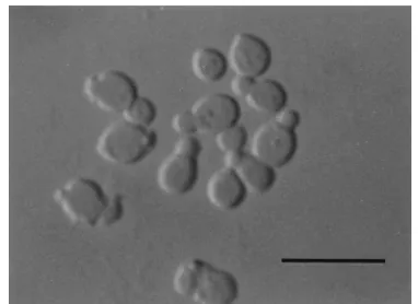
Description ofM. dermatisSugita, Takashima, Nishikawa et Shinoda sp. nov.After 7 days on LNA at 32°C, the vegetative
cells were spherical, oval, or ellipsoidal (2 to 8 by 2 to 10m),
and they reproduced by budding (Fig. 2). The colony was yellowish white, semishiny to dull, convex, and butyrous and had an entire or lobed margin. Filaments sometimes formed at the area of the origin of the bud. Growth was seen on glucose-peptone agar with either 0.1, 0.5, 1.0, 5.0, or 10% Tween 20, 40, 60, or 80 as sole source of lipid. Catalase reaction was positive.
The diazonium blue B reaction was positive. The G⫹C content
of nuclear DNA was 60.4 mol%, and the major ubiquinone was
Q-9. Teleomorph is unknown. JCM 11348T(originally M 9927)
isolated from skin lesions of an AD patient in Tokyo, Japan, by T. Sugita on 30 September 1999 is maintained in the Japan Collection of Microorganisms (JCM), Saitama, Japan. The other strains, M 9929 and M 9931 have also been deposited in the JCM, as JCM 11469 and JCM 11470, respectively. Etymol-ogy is from the Latin name for skin, from which this strain of the species was obtained.
DISCUSSION
The genus Malassezia is phylogenetically monophyletic
with a high bootstrap value (100%) and is positioned in the
classUstilaginomycetes(Fig. 1A). Our isolates clustered with
M. sympodialison the tree. We did not perform a nuclear
DNA-DNA hybridization study betweenM. sympodialisand
our isolates, but sequence analysis of rDNA (26S and ITS) strongly suggests that our five isolates represent a new
spe-cies in the genus Malassezia. At present, D1 and D2 26S
rDNA sequences are used from almost all yeasts for species identification or phylogenetic analysis. Peterson and Kurtz-man (18) correlated the biological species concept with the phylogenetic species concept by the extent of nucleotide substitutions in 26S rDNA sequences. Their study demon-strated that strains of a single biological species show less than 1% substitution in this region. Subsequently, Sugita et al. (22) also found that conspecific strains have less than 1% dissimilarity in the ITS1 and -2 regions after comparing the nuclear DNA-DNA hybridization values and similarity in the sequences of the entire ITS1 and -2 regions. According to the species concept used in the two reports cited above,
the divergence between M. sympodialis and our isolates is
sufficient to resolve them as individual species. In addition,
the approximately 1 mol% difference in the G⫹C content of
nuclear DNA that was found between these two species also has taxonomic significance (19, 24). Guillot et al. (5) de-scribed a practical approach (combination of Tween utiliza-tion and catalase tests) for easy and simple identificautiliza-tion of
Malassezia species. Unfortunately, M. dermatis cannot be identified by this system, despite having similar
characteris-tics to M. furfur. At present, sequence analysis of the D1
and D2 regions of the 26S rDNA or the ITS regions is the
most reliable and simplest method forM. dermatis
identifi-cation.
We previously demonstrated cutaneous Malassezia
[image:3.587.97.489.75.344.2]colo-nization in 32 AD patients by a nonculture method (nested
FIG. 1—Continued.
on May 15, 2020 by guest
http://jcm.asm.org/
PCR) and compared anti-Malassezia antibody levels with
those in healthy subjects. The detection ofM. globosaand
M. restrictain more than 90% of AD patients suggested that these two species play an important role in AD. Since we
were not aware of the existence ofM. dermatisat that time,
we did not detect the DNA of this species in our previous study. Although the present study examined a limited
num-ber of patients,M. dermatiswas found in 3 of 19 patients.
However, the degree ofM. dermatiscolonization of the skin
surface in the AD patients was not as great as that ofM.
globosaandM. restricta.
In conclusion, we described a novel species,M. dermatis,
isolated from skin lesions of AD patients. Further studies should examine whether this microorganism is responsible for AD or other skin diseases and whether it is specific to Japan.
ACKNOWLEDGMENT
This study was supported in part by a Grant for the Promotion of the Advancement of Education and Research in Graduate Schools from the Ministry of Education, Culture, Sports, Science, and Technology of Japan.
REFERENCES
1.Aspiroz, C., L. A. Moreno, A. Rezusta, and C. Rubio.1999. Differentiation of three biotypes ofMalasseziaspecies on human normal skin. Correspondence withM. globosa,M. sympodialisandM. restricta. Mycopathologia145:69–74. 2.Felsenstein, J.1985. Confidence limits on phylogenies: an approach using
the bootstrap. Evolution39:783–791.
3.Guého, E., T. Boekhout, H. R. Ashbee, J. Guillot, A. van Belkum, and J. Faergemann.1998. The role ofMalasseziaspecies in the ecology of human skin and as pathogens. Med. Mycol.36(Suppl. 1):220–229.
4.Guého, E., G. Midgley, and J. Guillot.1996. The genus Malasseziawith description of four new species. Antonie van Leeuwenhoek69:337–355. 5.Guillot, J., E. Guého, M. Lesourd, G. Midgley, G. Chevrier, and B. Dupont.
1996. Identification ofMalasseziaspecies. J. Mycol. Med.6:103–110. 6.Gupta, A. K., Y. Kohli, J. Faergemann, and R. C. Summerbell.2001.
Epi-demiology ofMalasseziayeasts associated with pityriasis versicolor in On-tario, Canada. Med. Mycol.39:199–206.
7.Gupta, A. K., Y. Kohli, R. C. Summerbell, and J. Faergemann.2001. Quan-titative culture ofMalasseziaspecies from different body sites of individuals with or without dermatoses. Med. Mycol.39:243–251.
8.Hiruma, M., D. J. Maeng, M. Kobayashi, H. Suto, and H. Ogawa.1999. Fungi and atopic dermatitis. Jpn. J. Med. Mycol.40:79–83.
9.Kim, T. Y., I. G. Jang, Y. M. Park, H. O. Kim, and C. W. Kim.1999. Head and neck dermatitis: the role ofMalassezia furfur, topical steroid use and environmental factors in its causation. Clin. Exp. Dermatol.24:226–231. 10.Kimura, M.1980. A simple method for estimation evolutionary rate of base
[image:4.587.325.516.71.210.2]substitutions through comparative studies of nucleotide sequences. J. Mol. E 16:111–120.
FIG. 2. Vegetative cells ofM. dermatisM 9927 (JCM 11348) grown in LNA for 7 days at 32°C. Bar, 10m.
TABLE 1. Taxonomic characteristics of M. dermatis and other Malassezia species Characteristic M. dermatis sp. nov. M. furfur a M. pachydermatis a M. sympodialis a M. globosa a M. obtusa a M. restricta a M. sloof fiae a Morphological characteristics Colony morphology
Convex, butyrous, entire
or lobed magin Mat, dull, smooth, umbonate or slightly folded with convex elevation Mat, convex, umbonate (sometimes) Glistening, smooth, flat or with a slight central elevation Raised, folded, rough Smooth, flat Dull, smooth to rough at the edges Rough but usually with fine grooves Cell shape (size [ m]) Spherical, oval, ellipsoidal (2.0 –8.0 by 2.0 –10.0) Oval, cylindrical (1.5 –3.0 by 2.5 – 8.0), spherical (2.5 –5.0) Oval (2.0 –2.5 by 4.0 –5.0) Oval, globosal (1.5 –2.5 by 2.5 –6.0) Spherical (2.5 –8.0) Cylindrical (1.5 –2.0 by 4.0 –6.0) Spherical, oval (1.5 –2.0 by 2.5 –4.0) Short cylindrical (1.0 –2.0 by 1.5 –4.0) Physiological characteristics b Growth on Sa at 32 °C ⫺ ⫺ ⫹ ⫺ ⫺ ⫺⫺⫺ Growth on mDixon 32 °C ⫹ ⫹ ⫹ ⫹ ⫹ ⫹⫹⫹ 37 °C ⫹⫹ ⫹ ⫹ ⫾ or ⫺⫾ or ⫹⫹ ⫹ 40 °C ⫹ ⫹ ⫹ ⫹ ⫺ ⫺⫺⫹ Catalase reaction ⫹⫹ ⫾ or ⫹ ⫹ ⫹ ⫹⫺⫹ Utilization of: Tween 20 ⫹ ⫹ ⫺ ⫺ ⫺ ⫺⫺⫾ or ⫹ Tween 40 ⫹ ⫹ ⫹ ⫹ ⫺ ⫺⫺⫹ Tween 60 ⫹ ⫹ ⫹ ⫹ ⫺ ⫺⫺⫹ Tween 80 ⫹ ⫹ ⫹ ⫹ ⫺ ⫺⫺⫺ Mol% G ⫹ C 60.4 66.0 –66.7 55.5 –56.0 61.9 –62.5 53.5 –53.7 60.5 –60.7 59.8 –60.0 68.5 –69.0 aData are from the work of G úeho et al. (4). bSa, Sabouraud dextrose agar; mDixon, modi fied Dixon agar; ⫹ ,positive; ⫺ ,negative; ⫾ ,weakly positive.
1366 SUGITA ET AL. J. CLIN. MICROBIOL.
on May 15, 2020 by guest
http://jcm.asm.org/
11.Kurtzman, C. P., and C. J. Robnett.1997. Identification of clinically impor-tant ascomycetous yeasts based on nucleotide divergence in the 5⬘end of the large-subunit (26S) ribosomal DNA gene. J. Clin. Microbiol.35:1216–1223. 12.Leeming, J. P., J. E. Sansom, and J. L. Burton. 1997. Susceptibility of Malassezia furfursubgroups to terbinafine. Br. J. Dermatol.137:764–767. 13.Leung, D. Y.1995. Atopic dermatitis: the skin as a window into the
patho-genesis of chronic allergic diseases. J. Allergy Clin. Immunol.96:302–318. 14.Lintu, P., J. Savolainen, and K. Kalimo.1997. IgE antibodies to protein and
mannan antigens ofPityrosporum ovalein atopic dermatitis patients. Clin. Exp. Allergy27:87–95.
15.Makimura, K., Y. S. Murayama, and H. Yamaguchi.1994. Detection of a wide range of medically important fungal species by polymerase chain reac-tion (PCR). J. Med. Microbiol.40:358–364.
16.Nakabayashi, A., Y. Sei, and J. Guillot.2000. Identification ofMalassezia species isolated from patients with seborrhoeic dermatitis, atopic dermatitis, pityriasis versicolor and normal subjects. Med. Mycol.38:337–341. 17.Nakase, T., and M. Suzuki.1986.Bullera megalospora, a new species of yeast
forming large ballistospores isolated from dead leaves ofOryza sativa, Mis-canthus sinensis, andSasasp. in Japan. J. Gen. Appl. Microbiol.32:225–240. 18.Peterson, S. W., and C. P. Kurtzman.1991. Ribosomal RNA sequence divergence among sibling species of yeasts. Syst. Appl. Microbiol.14:124– 129.
19.Price, C. W., G. B. Fuson, and H. J. Phaff.1978. Genome comparison in yeast
systematics: delimitation of species within the generaSchwanniomyces, Sac-charomyces,Debaryomyces, andPichia. Microbiol. Rev.42:161–193. 20.Saitou, N., and M. Nei.1987. The neighbor-joining method: a new method
for reconstructing phylogenetic trees. Mol. Biol. E4:406–425.
21.Sugita, T., H. Suto, T. Unno, R. Tsuboi, H. Ogawa, T. Shinoda, and A. Nishikawa.2001. Molecular analysis ofMalasseziamicroflora on the skin of atopic dermatitis patients and healthy subjects. J. Clin. Microbiol.39:3486– 3490.
22.Sugita, T., A. Nishikawa, R. Ikeda, and T. Shinoda.1999. Identification of medically relevantTrichosporonspecies based on sequences of internal tran-scribed spacer regions and construction of a database forTrichosporon iden-tification. J. Clin. Microbiol.37:1985–1993.
23.Takashima, M., and T. Nakase. 2000. Four new species of the genus Sporobolomycesisolated from leaves in Thailand. Mycoscience41:65–77. 24.Tamaoka, J., and K. Komagata.1984. Determination of DNA base
compo-sition by reversed-phase high-performance liquid chromatography. FEMS Microbiol. Lett.25:125–128.
25.Thompson, J. D., D. G. Higgins, and T. J. Gibson.1994. CLUSTAL W: improving the sensitivity of progressive multiple sequence alignment through sequence weighting, position-specific gap penalties and weight matrix choice. Nucleic Acids Res.22:4673–4680.
26.Wessels, M. W., G. Doekes, A. G. Van Ieperen-Van Kijk, W. J. Koers, and E. Young.1991. IgE antibodies toPityrosporum ovalein atopic dermatitis. Br. J. Dermatol.125:227–232.