Copyright © 1999, American Society for Microbiology. All Rights Reserved.
Detection of Viable
Mycobacterium tuberculosis
by Reverse
Transcriptase-Strand Displacement
Amplification of mRNA
T. J. HELLYER,
1†L. E. D
ESJARDIN,
1‡L. TEIXEIRA,
2M. D. PERKINS,
2M. D. CAVE,
3ANDK. D. EISENACH
1,4*
Departments of Pathology,
1Microbiology/Immunology,
4and Anatomy,
3University of Arkansas for Medical
Sciences, Little Rock, Arkansas, and Duke University Medical Center/Universidade
Federal do Espirito Santo, Vito´ria, Brazil
2Received 27 August 1998/Returned for modification 22 November 1998/Accepted 7 December 1998
Numerous assays have been described for the detection of DNA and rRNA sequences that are specific for the
Mycobacterium tuberculosis
complex. Although beneficial to initial diagnosis, such assays have proven
unsuit-able for monitoring therapeutic efficacy owing to the persistence of these nucleic acid targets long after
conversion of smears and cultures to negative. However, prokaryotic mRNA has a typical half-life of only a few
minutes and we have previously shown that the presence of mRNA is a good indicator of bacterial viability. The
purpose of the present study was to develop a novel reverse transcriptase-strand displacement amplification
system for the detection of
M. tuberculosis
a
-antigen (85B protein) mRNA and to demonstrate the use of this
assay in assessing chemotherapeutic efficacy in patients with pulmonary tuberculosis. The assay was applied
to sequential, noninduced sputum specimens collected from four patients: 10 of 11 samples (91%) collected
prior to the start of therapy were positive for alpha-antigen mRNA, compared with 1 of 8 (13%), 2 of 8 (25%),
2 of 8 (25%), and 0 of 8 collected on days 2, 4, 7, and 14 of treatment, respectively. In contrast, 39 of 44 samples
(89%) collected on or before day 14 were positive for
a
-antigen DNA. The loss of detectable mRNA
corre-sponded to a rapid drop over the first 4 days of treatment in the number of viable organisms present in each
sputum sample, equivalent to a mean fall of 0.43 log
10CFU/ml/day. Analysis of mRNA is a potentially useful
method for monitoring therapeutic efficacy and for rapid in vitro determination of drug susceptibility.
The continued worldwide dominance of tuberculosis as a
cause of morbidity and mortality (22) has fueled extensive
research into more rapid and reliable means of diagnosis.
Nu-merous systems for the amplification DNA or rRNA target
sequences that are specific for members of the
Mycobacterium
tuberculosis
complex have been described (9, 14, 16, 23, 25, 27).
However, while useful in reducing the amount of time required
for definitive diagnosis, these techniques have not been applied
successfully in the role of monitoring therapeutic efficacy.
Typically, successful treatment for pulmonary tuberculosis
results in conversion of smears and cultures to negative within
2 to 3 months (5). Recently, we have demonstrated that
DNA-based amplification assays are an inappropriate substitute for
conventional microbiological methods of patient follow-up,
since amplifiable nucleic acid may persist for long periods
beyond the point of smear and culture conversion (6, 12, 13).
Similarly, in patients receiving antituberculosis therapy, a poor
correlation has been observed between the results of
micros-copy or growth-based detection of
M. tuberculosis
and those of
assays for mycobacterial 16S rRNA (10, 19).
However, in contrast to DNA and rRNA, bacterial mRNA is
typically short-lived, with a half-life of only a few minutes (2,
26). Consequently, an mRNA-based assay is likely to detect
only living organisms and thus be a good indicator of
thera-peutic efficacy and/or susceptibility to antibacterial agents. We
have previously described a method for the efficient recovery
of mycobacterial RNA from clinical specimens, as well as the
use of reverse transcriptase (RT)-PCR assays to detect specific
RNA targets (7). The objectives of the present study were to
develop a novel RT-strand displacement amplification (SDA)
assay for the mRNA that encodes the
M. tuberculosis
complex
alpha-antigen (
a
-antigen), also known as 85B protein, and to
determine whether such an assay could provide a useful
alter-native to conventional means of patient follow-up in the
treat-ment of tuberculosis. The
a
-antigen target was selected
be-cause this protein is known to be produced in abundance by
M.
tuberculosis
both in broth cultures and in human mononuclear
phagocytes (11, 18, 28). The
a
-antigen may comprise up to
41% of the protein in culture supernatants, and it is reasonable
to predict that viable cells will possess a corresponding
abun-dance of the encoding mRNA. Here we describe the
applica-tion of the RT-SDA assay, together with a thermophilic SDA
(tSDA) assay for
a
-antigen DNA, to sequential sputum
spec-imens from four patients receiving treatment for pulmonary
tuberculosis.
MATERIALS AND METHODS
tSDA fora-antigen DNA.tSDA was performed in 50-ml volumes as follows. Target DNA was added to buffer containing (final concentrations) 52.5 mM K1PO4(pH 7.6); 12% dimethyl sulfoxide (DMSO); 7.7% glycerol; 500 nM SDA
primers S1 and S2; 50 nM bumper primers B1 and B2 (Table 1); 0.2 mM dATP, dGTP, and dUTP; 0.8 mM 29-deoxycytosine-59-O-(1-thiotriphosphate); 500 ng of human placental DNA; and 5mg of acetylated bovine serum albumin. Tubes were heated at 95°C for 2.5 min to denature the target and cooled to 45°C, and 1 U of uracil DNA glycosylase (UDG) was added to facilitate removal of contaminating amplicons. Incubation was continued at 45°C for 10 min before transfer to a heat block at 52.5°C and addition of 40 U ofBsoBI, 15 U of exonuclease-deficientBstpolymerase (both from New England Biolabs), 4 U of
* Corresponding author. Mailing address: John L. McClellan
Me-morial Veterans’ Hospital, Slot-151, 4300 West 7th St., Little Rock,
AR 72205. Phone: (501) 257-4827. Fax: (501) 664-6748. E-mail:
[email protected].
† Present address: Becton Dickinson Microbiology Systems, Sparks,
Md.
‡ Present address: University of Iowa, Iowa City, Iowa.
518
on May 15, 2020 by guest
http://jcm.asm.org/
UDG inhibitor, and 7 mM (final concentration) magnesium acetate. Reaction mixtures were incubated for 45 min, and reactions were terminated by heating at 95°C for 3 min.
RT-SDA fora-antigen mRNA.Reverse transcription was performed in 20-ml volumes as follows. Target RNA was added to buffer containing (final concen-trations) 30 mM K1PO4(pH 7.6); 12% DMSO; 12.5% glycerol 1,250 nM SDA
primer S2; 125 nM bumper primer B2; 12.5 nM B3; 1.25 nM B4 (Table 1); 300 ng of human placental DNA; 5mg of acetylated bovine serum albumin; 0.2 mM dATP, dGTP, and dUTP; 0.8 mM 29-deoxycytosine-59-O-(1-thiotriphosphate); 1 U of PRIME RNase inhibitor (5 Prime-3 Prime, Inc.); and 1 U of UDG. Use of three primers in the reverse transcription reaction mixture was designed to take advantage of the strand displacement activity of avian myeloblastosis virus RT (4) and facilitate the synthesis of multiple copies of cDNA from a single mRNA target molecule. Reaction mixtures were incubated at 45°C for 15 min to facil-itate removal of contaminating amplicons by the UDG enzyme before addition of 4 U of UDG inhibitor and 2.5 U of avian myeloblastosis virus RT (Boehringer Mannheim). Tubes were incubated for a further 15 min at 45°C before being equilibrated at 52.5°C for 3 min. SDA was then initiated at the same temperature in a final volume of 50ml through addition of K1PO4, DMSO, primers S1 and B1,
nucleotides, human placental DNA, magnesium acetate,BsoBI, andBst poly-merase to the final concentrations given above for the tSDA DNA assay. Am-plification was carried out for 45 min, and reactions were stopped by heating at 95°C for 3 min. Parallel reactions were performed with all clinical samples without addition of RT in order to monitor for DNA contamination of RNA extracts.
Detection of tSDA and RT-SDA products.The products of amplification were detected by primer extension or chemiluminescence assay. Primer extension analysis was performed with a32P-labeled D1 probe (Table 1) and
exonuclease-deficient Klenow polymerase. Specific oligonucleotide products of 43 and 62 bases were generated which were visualized by autoradiography following elec-trophoretic separation through 8% denaturing polyacrylamide gels. Chemilumi-nescent detection of tSDA and RT-SDA products was done with streptavidin-coated microtiter plates as previously described (24), by using a biotinylated capture probe and an alkaline phosphatase-conjugated detector probe (C1 and AP1, respectively; Table 1) which hybridize to the SDA amplicon in the inter-vening region between primers S1 and S2. SDA samples were diluted 1:10 in 50 mM K1PO4(pH 7.6) prior to mixing with the capture and detector probes.
Readings were taken by using a Labsystems Luminoskan luminometer, and all wells that gave values fivefold higher than the background were considered positive. This cutoff value was determined empirically by comparison of chemi-luminescence data with the results of primer extension analysis. Background readings were determined for each assay run by using dummy reaction mixtures containing all of the components of a tSDA reaction exceptM. tuberculosistarget DNA.
Amplification controls.Positive controls for tSDA ofa-antigen DNA com-prised reaction mixtures containing known amounts of purified DNA fromM. tuberculosisH37Rv. Initial experiments with the RT-SDA system employed pos-itive controls containing in vitro mRNA transcripts generated from a partial clone of theM. tuberculosisa-antigen gene. This clone comprised the central 584-bpEcoRV-SacII fragment of thea-antigen gene ligated into pBlueScript KS1(Stratagene). In vitro transcripts were generated from the T3 RNA poly-merase promoter by using an Ambion MEGAscript T3 Kit in accordance with the manufacturer’s instructions. Later experiments were performed by using transcripts derived from a full-length copy of theM. tuberculosisa-antigen gene
(11) cloned into theEcoRV site of the pBlueScript vector. No difference in analytical sensitivity was observed when transcripts derived from either the full-length or partiala-antigen clones were used. Negative controls contained all of the components necessary for DNA or RNA amplification with the addition of either water or target diluent (10-ng/ml yeast carrier RNA [Ambion] or human placental DNA).
Primer specificity.tSDA reactions were performed as described above, on purified DNA that was isolated from a representative panel of 31 organisms belonging to theM. tuberculosiscomplex. The S1, S2, B1, and B2 primer set was also tested for cross-reactivity with DNAs from 10 other species of mycobacteria and five closely related genera.
Analytical sensitivity.To determine the analytical sensitivities of the tSDA and RT-SDA assays, dilutions of purified M. tuberculosis H37Rv DNA and full-length in vitro transcripts of thea-antigen gene were amplified and detected by chemiluminescence assay. Results obtained by chemiluminescence assay were confirmed by primer extension analysis with a32P-labeled D1 probe.
Control cultures for nucleic acid extraction.M. tuberculosisH37Rv was ob-tained from the American Type Culture Collection (strain 27294). All cultures were incubated at 37°C in an atmosphere of 5% CO2. Liquid cultures were grown
in Dubos broth containing 10% Dubos medium albumin (both from Difco) and 0.1% Tween 80. Viable counts were performed by preparing serial 10-fold dilu-tions of cells in fresh broth and plating them on Dubos agar containing 10% Dubos oleic albumin complex (both from Difco) and 0.1% Tween 80. As a control for the efficiency of nucleic acid extraction, an aliquot of cultured cells was processed with each batch of clinical samples.
Nucleic acid extraction.Bacteria were lysed by using a FastPrep FP120 cell disrupter with Blue FastRNA Tubes (both from Bio 101), and RNA was isolated by using a modified guanidine-acid phenol procedure (8). DNA was recovered from the same samples by back extraction of the interface and organic layers with a basic salt solution (100 mM Tris-HCl [pH 8.0], 1 mM EDTA, 50 mM NaCl, 1% sodium dodecyl sulfate) and then precipitated with isopropanol (7). Extracted RNA and DNA were resuspended in 100ml of sterile distilled water.
In order to demonstrate the efficiency of nucleic acid recovery by using the guanidine-phenol extraction procedure, DNA and RNA were isolated from a logarithmically growing culture ofM. tuberculosisH37Rv. Five-hundred-micro-liter aliquots of a culture containing 1.233106CFU/ml were processed as
described above, and the relative yields of RNA and DNA were estimated by amplifying serial fourfold dilutions of the recovered nucleic acid.
Patients and specimens.Sequential noninduced sputum samples were ob-tained from four patients with newly diagnosed, smear-positive pulmonary tu-berculosis who attended the Hospital Universitario Cassiano Antonio de Mo-rase, Universidade Federal do Espı´rito Santo, Vito´ria, Brazil. Three specimens were collected on the day treatment was initiated (day 0) with a standard four-drug antituberculosis regimen comprising isoniazid, rifampin, ethambutol, and pyrazinamide. Subsequently, duplicate specimens were collected on days 2, 4, 7, 14, and 30. Samples were homogenized with 2.5%N-acetyl-L-cysteine and
glass beads as previously described (7) and 0.5-ml aliquots were frozen at270°C for nucleic acid extraction. Additional aliquots were decontaminated with NaOH-sodium citrate and plated on selective solid medium for quantitative culture (6). DNA and RNA were prepared from thawed specimens as described above.
RNA extracts of clinical samples frequently contained low levels ofM. tuber-culosisDNA. To prevent subsequent amplification, the extendable 39ends of contaminating DNA molecules were blocked by incubation with
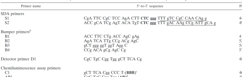
dideoxynucle-TABLE 1. Primer sequences for tSDA and RT-SDA of
M. tuberculosis
complex
a-antigen DNA and mRNA
Primer name 59-to-39sequence Positiona
SDA primers
S1
CgA TTC CgC TCC AgA CTT
CTC ggg
TTT gTC CgC CAA CAg g
445–459
S2
ACC gCA TCg AgT ACA TgT
CTC ggg
TTT gAC AAg CCg ATT gCA g
497–482
Bumper primers
bB1
ACC TTC CTg ACC AgC gAg
415–432
B2
AgA TCA TTg CCg ACg AgC
523–506
B3
gCT ggg ggT ggT Agg C
544–529
B4
CCg ACA gCg AgC Cg
571–558
Detector primer D1
CgC TgC Cgg Tgg gCT TCA Cg
481–462
Chemiluminescence assay primers
C1
gCT TCA Cgg CCC T-(
BBB
)
c469–457
AP1
CgC TgC Cgg Tgg-(
AP
)
d481–470
aPosition withina-antigen sequence ofM. tuberculosisstrain Erdman (8).
bBsoBI recognition sequences are in boldface. SDA primer target binding regions are underlined. cB, biotin.
dAP, alkaline phosphatase.
on May 15, 2020 by guest
http://jcm.asm.org/
[image:2.612.54.552.82.242.2]otide triphosphates and DNA polymerase. In brief, RNA extracts from clinical samples were diluted 1:10 in 10-ng/ml yeast RNA, and 5ml was then added to a mixture containing (final concentrations) 50 mM Tris-HCl (pH 8.0), 10 mM magnesium chloride, 25 mM each dideoxynucleotide triphosphate (ddATP, ddCTP, ddGTP, and ddTTP), 5 U of exonuclease-deficient Klenow DNA poly-merase, and 1 U of PRIME RNase inhibitor. Mixtures were incubated at 37°C for 30 min and diluted either 1:4 or 1:40 in 10-ng/ml yeast carrier RNA or human placental DNA prior to reverse transcription. Dilution of the target RNA was necessary in order to obtain a titration of signal over the course of therapy. To permit comparison, DNA extracts were diluted in a similar fashion prior to amplification. Separate experiments demonstrated that treatment of RNA ex-tracts with dideoxynucleotides under the above-described conditions did not adversely affect the analytical sensitivity of the RT-SDA assay (data not shown).
RESULTS
Primer specificity.
DNAs from 24 isolates of
M. tuberculosis
from North and South America, Asia, Africa, and Europe were
tested in tSDA reactions using primers S1, S2, B1, and B2. All
yielded specific products by primer extension analysis with the
32
P-labeled D1 probe, as did four North American isolates of
M. bovis
(including two of human origin),
M. bovis
bacille
Calmette-Gue´rin (Glaxo strain), and one strain each of
M.
africanum
and
M. microti
(data not shown).
No significant cross-reaction was observed among 36 strains
of nontuberculous mycobacteria comprising 10 isolates of
M.
avium-M. intracellulare, 7 of
M. fortuitum
5 of
M. xenopi, 4 of
M.
malmoense, 3 of
M. kansasii, 3 of
M. chelonei, and 1 each of
M.
scrofulaceum,
M. gordonae,
M. abscessus, and
M. celatum.
Sim-ilarly, no cross-reaction was detected with the phylogenetically
related organisms
Actinomyces israeli,
Corynebacterium
diph-theriae,
Nocardia braziliensis,
Rhodococcus rhodochrous, and
Streptomyces albus.
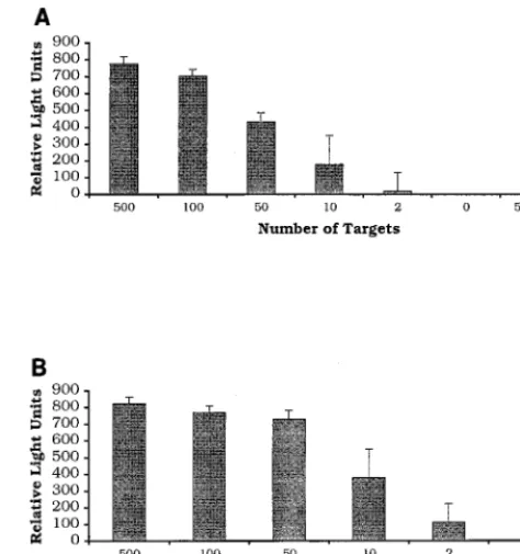
Analytical sensitivity.
The analytical sensitivities of the
tSDA and RT-SDA assays were determined by amplifying
di-lutions of purified
M. tuberculosis
H37Rv DNA and full-length
in vitro transcripts of the
a
-antigen gene, respectively (Fig. 1).
Results obtained by chemiluminescence assay were confirmed
by primer extension analysis with the
32P-labeled D1 probe
(data not shown). The analytical sensitivities of the two assays
were similar, on the order of 10 copies of target nucleic acid.
For the tSDA assay, two of three reaction mixtures containing
two input targets were also positive, as was one of three
RT-SDA reaction mixtures. No signal was detected from RT-RT-SDA
reaction mixtures prepared without addition of RT.
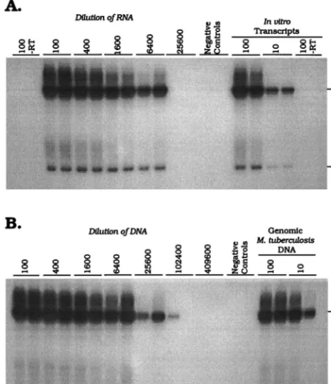
Nucleic acid extraction.
In order to demonstrate the
effi-ciency of nucleic acid recovery by the guanidine-phenol
extrac-tion procedure, DNA and RNA were isolated from a
logarith-mically growing culture of
M. tuberculosis
H37Rv. In Fig. 2A, a
signal was detected at a 1/6,400 dilution of the recovered RNA.
No signal was obtained from reaction mixtures prepared with
the 1/100 dilution of RNA in the absence of RT, indicating that
the extracted RNA was free of contamination with
M.
tuber-culosis
DNA. Figure 2B shows the results obtained by
ampli-fying dilutions of DNA isolated from the same culture sample.
Both of the tSDA reaction mixtures prepared with DNA at a
dilution of 1/25,600 were positive, as was one reaction mixture
prepared with a dilution of 1/102,400. Given that the tSDA and
RT-SDA assays have similar analytical sensitivities (above),
these data indicate an approximately fourfold greater efficiency
of recovery of DNA than RNA. This observation was
consis-tent between replicate extractions of cultured
M. tuberculosis
(data not shown).
Amplification of DNA and mRNA from sputum.
A graphical
representation of the
a
-antigen DNA and mRNA profiles of
all four patients included in this study is depicted in Fig. 3. Also
shown are the numbers of viable organisms present per
milli-liter of sputum at each time point. At the dilutions tested, all
(100%) of 12 samples collected on the day treatment was
initiated (day 0) were positive for
a
-antigen DNA, compared
with 10 (91%) of 11 which were positive for
a
-antigen mRNA.
One RNA sample from patient B on day 0 was contaminated
with low levels of
a
-antigen DNA, as demonstrated by the
detection of specific amplification products in RT-SDA
reac-tion mixtures prepared without addireac-tion of RT. Thirty-nine
(89%) of 44 samples collected on or before day 14 of treatment
were positive for
a
-antigen DNA, compared with 15 (35%) of
43 that were mRNA positive. Three of four patients remained
positive for
a
-antigen DNA on day 30 of treatment, yet all
were negative for
a
-antigen mRNA by day 14. The rapid
de-cline in the number of mRNA-positive samples corresponded
to a marked fall in the number of viable organisms present in
each sputum sample. Colony counts fell by a mean of 1.72 log
10CFU/ml over the first 4 days of treatment (equivalent to 0.43
log
10CFU/ml/day) but by only a further 1.39 log
10CFU/ml
over the next 10 days (0.14 log
10CFU/ml/day). The mean fall
in colony counts over the first 2 days of treatment was 0.97
log
10CFU/ml (0.49 log
10CFU/ml/day), but this value is
some-what distorted by an aberrant low viable count on day 2 from
patient C (Fig. 3). Three (75%) of the four patients became
culture negative for
M. tuberculosis
by day 30; the remaining
patient (Fig. 3, patient B) was culture negative on day 60 of
treatment.
DISCUSSION
[image:3.612.314.551.74.326.2]The objectives of the present study were to develop a novel
RT-SDA system for the detection of
M. tuberculosis
a
-antigen
FIG. 1. Demonstration of the analytical sensitivity of thea-antigen RT-SDA mRNA (A) and tSDA DNA (B) assays. Serial dilutions of full-length in vitro transcripts of thea-antigen gene and purifiedM. tuberculosisgenomic DNA were amplified, and the products were detected by chemiluminescence assay after dilution in K1PO4buffer. Reactions which gave readings of greater than or equalto five times the background were considered positive. Results represent the mean of three RT-SDA or tSDA reactions. Error bars are standard deviations. Both assays have an analytical sensitivity of#10 targets. No product was detected in RT-SDA reaction mixtures prepared without addition of RT.
on May 15, 2020 by guest
http://jcm.asm.org/
mRNA and to demonstrate the usefulness of this assay in
assessing chemotherapeutic efficacy in patients with pulmonary
tuberculosis. The advent of rapid molecular assays for the
detection of specific nucleic acid sequences has decreased the
amount of time required for definitive diagnosis of tuberculosis
to as little as 1 day. Such assays have found wide application in
the diagnosis of pulmonary tuberculosis yet have not proven
suitable for monitoring the response of patients to treatment
owing to the persistence of amplifiable DNA and rRNA target
sequences beyond the point of smear and culture conversion
(6, 10, 13, 19). This presumably reflects both the shedding of
dead or dormant bacilli from pulmonary lesions and the
inher-ent stability of bacterial DNA and rRNA. The American
Tho-racic Society recommends that the response to antituberculosis
chemotherapy of patients bacteriologically positive should be
evaluated by repeated examination of sputum at monthly
in-tervals until sputum conversion is documented (1). Owing to
the slow growth rate of
M. tuberculosis, this is, however, a very
inefficient means of assessing treatment efficacy, and the ability
to apply molecular assays in this role is desirable. Clinical trials
of novel antituberculosis drugs or drug combinations are
cur-rently a protracted and prohibitively expensive undertaking,
and a rapid method for determining therapeutic response or
predicting clinical outcome would be particularly valuable.
In contrast to DNA and rRNA, bacterial mRNA is believed
to be short-lived, with a typical half-life of only a few minutes
(2, 26). As a result, an assay directed toward an mRNA target
is likely to detect only living organisms and therefore be a good
marker of bacterial viability and susceptibility to
chemothera-peutic agents. Indeed, we have previously described the use of
a quantitative RT-PCR system to discriminate between
drug-sensitive and drug-resistant
M. tuberculosis
strains in vitro (12).
In the present study, we developed an RT-SDA assay directed
toward the mRNA encoding the
M. tuberculosis
complex
a
-an-tigen, one of the most abundantly expressed proteins produced
by
M. tuberculosis
(11, 18, 28). The assay was shown to be
specific for members of the
M. tuberculosis
complex and to
have a reproducible analytical sensitivity of 10 in vitro
tran-scripts of the
a
-antigen gene.
In order to demonstrate the ability of the RT-SDA assay to
discriminate between live and dead bacilli and its potential as
a means to monitor therapeutic efficacy, we conducted a small
clinical trial in which the assay was applied to sequential
spu-tum specimens from patients receiving treatment for
pulmo-nary tuberculosis. The results from the RT-SDA assay were
compared with those obtained by tSDA of
a
-antigen DNA
isolated from the same samples. The exquisite sensitivity of the
two assays necessitated dilution of the nucleic acid extracts
prior to amplification in order to obtain a titration of DNA and
RNA signals over the course of treatment. In all four patients
studied, we observed a rapid decline in the number of
mRNA-positive samples. At the dilutions tested, no mRNA was
de-tected in any sample beyond day 7 of treatment, whereas all
four patients remained DNA positive on day 14 and all but one
were also positive on day 30. We have interpreted this as being
indicative of the early bactericidal activity of the treatment
regimen which was consistent with that seen in other studies
which employed an isoniazid-containing treatment regimen
(15, 17).
This interpretation of the data is based upon our observation
that both the
a
-antigen DNA and mRNA assays have similar
analytical sensitivities. However, we also found that the yield of
DNA obtained from cultured cells by using the
guanidine-phenol extraction procedure was approximately fourfold
higher than that of RNA. This is attributable to the greater
inherent stability of DNA over mRNA and to losses incurred
through the repeated organic extractions involved in the RNA
isolation procedure (7). The greater efficiency of recovery of
DNA over RNA probably contributes to the apparent
persis-tence of DNA during the course of treatment in our study.
Nevertheless, the decline in mRNA levels did correspond to
the rapid fall in colony counts during the first few days of
treatment. Clearly, a much larger number of patients needs to
be evaluated to confirm these observations, yet these results
are consistent with those we obtained in an analysis of
a
-anti-gen mRNA levels in cultures of
M. tuberculosis
exposed to
isoniazid and rifampin (12). We are currently investigating the
possibility of converting the RT-SDA and tSDA assays
de-scribed here to a quantitative format (21) which would
pre-clude the need for dilution of the nucleic acid samples prior to
amplification and permit direct comparison of RNA and DNA
levels between successive samples.
[image:4.612.53.295.77.357.2]All of the patients included in this pilot study responded to
treatment, as determined by both conventional culture and
molecular analysis. As a result, it is unclear how drug
resis-tance, in particular, resistance to drugs other than rifampin,
would influence a patient’s mRNA profile. Rifampin blocks
transcription by directly binding to the
b
subunit of the RNA
polymerase (3, 20), and in vitro studies have shown a much
more rapid decline in mRNA levels in rifampin-treated
cul-tures than in those treated with isoniazid (12). It is therefore
possible that in patients receiving a multidrug treatment
regi-men, susceptibility of
M. tuberculosis
to rifampin could mask
FIG. 2. Titration ofa-antigen mRNA (A) and DNA (B) isolated from 6.153105CFU of culturedM. tuberculosisH37Rv. Amplification products were
de-tected by primer extension with the32P-labeled D1 probe. The molecular sizes of
the full-length and nicked product forms are indicated in bases on the right. a-Antigen mRNA titrated out at a dilution of 1/6,400, whereas both replicates where positive at a dilution of 1/25,600 for the DNA assay. One tSDA reaction mixture was also weakly positive at a dilution of 1/102,400. No product was detected in RT-SDA reaction mixtures prepared without RT, indicating an absence of amplifiablea-antigen DNA from the RNA extract.
on May 15, 2020 by guest
http://jcm.asm.org/
resistance to one or more of the other antituberculosis drugs.
Nevertheless, failure to observe a downward trend in mRNA
levels following the initiation of treatment would be indicative
of inappropriate therapy, emerging drug resistance, or
non-compliance. Even under such circumstances, it is likely that
mRNA analysis would still provide a more rapid assessment of
treatment efficacy than is possible by conventional
microbio-logical means. Quantitative analysis of bacterial molecular
tar-gets as surrogate indicators of the response of patients to
therapy is likely to be of key importance in future development
of novel antituberculosis treatment regimens.
ACKNOWLEDGMENTS
This work was supported by a research agreement between the
University of Arkansas and Becton Dickinson & Company and by the
Tuberculosis Research, Prevention & Control Unit (NIH contract
N01-AI45244).
We thank Mary Assaf, Ying Chen, and RaeTreal McRory for
ex-cellent technical assistance.
REFERENCES
1.Bass, J. B., L. S. Farer, P. C. Hopewell, R. O’Brien, R. F. Jacobs, F. Ruben, D. E. Snider, and G. Thornton.1994. Treatment of tuberculosis and tuber-culosis infection in adults and children. Am. J. Respir. Crit. Care Med. 149:1359–1374.
2.Belasco, J. G., G. Nilsson, A. von Gabain, and S. N. Cohen.1986. The stability ofE. coligene transcripts is dependent on determinants localized to specific mRNA segments. Cell46:245–251.
3.Blanchard, J. S.1996. Molecular mechanisms of drug resistance in Myco-bacterium tuberculosis. Annu. Rev. Biochem.65:215–239.
[image:5.612.53.549.69.512.2]4.Collett, M. S., J. P. Leis, M. S. Smith, and A. J. Faras.1978. Unwinding-like activity associated with avian retrovirus RNA-directed DNA polymerase. J. Virol.26:498–509.
FIG. 3. Graphical depiction of thea-antigen mRNA and DNA profiles of four pulmonary tuberculosis patients during the first 30 days of treatment with a standard four-drug chemotherapeutic regimen. Quantitative culture results are also shown. RNA and DNA extracts were diluted either 1:400 (patients A and B) or 1:40 (patients C and D) prior to amplification, depending on the yield of nucleic acid obtained on day 0. In all four cases,a-antigen mRNA cleared faster than DNA, corresponding to an initial rapid drop in the number of viable organisms (CFU) present per milliliter of sputum. Amplification results: open symbols, negative; closed symbols, positive; 3, undetermined (RNA sample contaminated witha-antigen DNA).
on May 15, 2020 by guest
http://jcm.asm.org/
5.Combs, D. L., R. J. O’Brien, and L. J. Geiter.1990. USPHS tuberculosis short-course chemotherapy trial 21: effectiveness, toxicity, and acceptability. Ann. Intern. Med.112:397–406.
6.DesJardin, L. E., Y. Chen, M. D. Perkins, L. Teixeira, M. D. Cave, and K. D. Eisenach.1998. Comparison of the ABI 7700 system (TaqMan) and com-petitive PCR for quantification of IS6110DNA in sputum during treatment of tuberculosis. J. Clin. Microbiol.36:1964–1968.
7.DesJardin, L. E., M. D. Perkins, L. Teixiera, M. D. Cave, and K. D. Eisenach.1996. Alkaline decontamination of sputum specimens adversely affects stability of mycobacterial mRNA. J. Clin. Microbiol.34:2435–2439. 8.De Wit, L., M. Palou, and J. Content.1994. Nucleotide sequence of the
85B-protein gene ofMycobacterium bovisBCG andMycobacterium tubercu-losis. DNA Seq.4:267–270.
9.Eisenach, K. D., M. D. Sifford, M. D. Cave, J. H. Bates, and J. T. Crawford. 1991. Detection ofMycobacterium tuberculosisin sputum samples using a polymerase chain reaction. Am. Rev. Respir. Dis.144:1160–1163. 10.Gamboa, F., J. M. Manterola, B. Vin˜ado, L. Matas, M. Gime´nez, J. Lonca,
J. R. Manzano, C. Rodrigo, P. J. Cardona, E. Padilla, J. Domı´nguez, and V. Ausina.1997. Direct detection of Mycobacterium tuberculosiscomplex in nonrespiratory specimens by Gen-Probe Amplified Mycobacterium Tuber-culosis Direct Test. J. Clin. Microbiol.35:307–310.
11. Harth, G., B.-Y. Lee, J. Wang, D. L. Clemens, and M. A. Horwitz.1996. Novel insights into the genetics, biochemistry, and immunocytochemistry of the 30-kilodalton major extracellular protein ofMycobacterium tuberculosis. Infect. Immun.64:3038–3047.
12. Hellyer, T. J., L. E. DesJardin, G. L. Hehman, M. D. Cave, and K. D. Eisenach.1999. Quantitative analysis of mRNA as a marker for viability of
Mycobacterium tuberculosis. J. Clin. Microbiol.37:290–295.
13. Hellyer, T. J., T. W. Fletcher, J. H. Bates, W. W. Stead, G. L. Templeton, M. D. Cave, and K. D. Eisenach.1996. Strand displacement amplification and the polymerase chain reaction for monitoring response to treatment in patients with pulmonary tuberculosis. J. Infect. Dis.173:934–941. 14. Iovannisci, D. M., and E. S. Winn-Deen.1993. Ligation amplification and
fluorescence detection of Mycobacterium tuberculosis DNA. Mol. Cell. Probes7:35–43.
15. Jindani, A., V. R. Aber, E. A. Edwards, and D. A. Mitchison.1980. The early bactericidal activity of drugs in patients with pulmonary tuberculosis. Am. Rev. Respir. Dis.121:–939–949.
16. Jonas, V., M. J. Alden, J. I. Curry, K. Kamisango, C. A. Knott, R. Lankford, J. M. Wolfe, and D. F. Moore.1993. Detection and identification of Myco-bacterium tuberculosisdirectly from sputum sediments by amplification of
rRNA. J. Clin. Microbiol.31:2410–2416.
17. Kennedy, N., R. Fox, G. M. Kisyombe, A. O. S. Saruni, L. O. Uiso, A. R. C. Ramsay, F. I. Ngowi, and S. H. Gillespie.1993. Early bactericidal and sterilizing activities of ciprofloxacin in pulmonary tuberculosis. Am. Rev. Respir. Dis.148:1547–1551.
18. Lee, B.-Y., and M. A. Horwitz.1995. Identification of macrophage and stress-induced proteins ofMycobacterium tuberculosis. J. Clin. Investig.96: 245–249.
19. Moore, D. F., J. I. Curry, C. A. Knott, and V. Jonas.1996. Amplification of rRNA for assessment of treatment response of pulmonary tuberculosis pa-tients during antimicrobial therapy. J. Clin. Microbiol.34:1745–1749. 20. Musser, J. M.1995. Antimicrobial agent resistance in mycobacteria:
molec-ular genetic insights. Clin. Microbiol. Rev.8:496–514.
21. Nycz, C. M., C. H. Dean, P. D. Haaland, C. A. Spargo, and G. T. Walker. 1998. Quantitative reverse transcription strand displacement amplification: quantification of nucleic acids using an isothermal amplification technique. Anal. Biochem.259:226–234.
22. Raviglione, M. C., D. E. Snider, and A. Kochi.1995. Global epidemiology of tuberculosis: morbidity and mortality of a worldwide epidemic. JAMA273: 220–226.
23. Shah, J. S., J. Liu, D. Buxton, A. Hendricks, L. Robinson, G. Radcliffe, W. King, D. Lane, D. M. Olive, and J. D. Klinger.1995. Q-Beta replicase-amplified assay for detection ofMycobacterium tuberculosisdirectly from clinical specimens. J. Clin. Microbiol.33:1435–1441.
24. Spargo, C. A., P. D. Haaland, S. R. Jurgensen, D. D. Shank, and G. T. Walker.1993. Chemiluminescent detection of strand displacement amplified DNA from species comprising theMycobacterium tuberculosiscomplex. Mol. Cell. Probes.7:395–404.
25. Van der Vliet, G. M. E., R. A. F. Schukkink, B. van Gemen, P. Schepers, and P. R. Klatser.1993. Nucleic acid sequence-based amplification (NASBA) for the identification of mycobacteria. J. Gen. Microbiol.139:2423–2429. 26. Von Gabain, J. G. Belasco, J. L. Schottel, A. C. Y. Chang, and S. N. Cohen.
1983. Decay of mRNA inEscherichia coli: investigation of the fate of specific segments of transcripts. Proc. Natl. Acad. Sci. USA80:653–657.
27. Walker, G. T., M. S. Fraiser, J. L. Schram, M. C. Little, J. G. Nadeau, and D. P. Malinowski.1992. Strand displacement amplification—an isothermal,
in vitroDNA amplification technique. Nucleic Acids Res.20:1691–1696. 28. Wiker, H. G., and M. Harboe.1992. The antigen 85 complex: a major
secretion product ofMycobacterium tuberculosis. Microbiol. Rev.56:648– 661.