Copyright © 1999, American Society for Microbiology. All Rights Reserved.
Multicenter Evaluation of the BACTEC MGIT 960 System
for Recovery of Mycobacteria
BRUCE A. HANNA,1* ADELEH EBRAHIMZADEH,2L. BRUCE ELLIOTT,3MARGIE A. MORGAN,4
SUSAN M. NOVAK,5SABINE RUSCH-GERDES,6MILLIE ACIO,4DENISE F. DUNBAR,3
T. MICHELE HOLMES,3CHARLES H. REXER,1CHIMINYAN SAVTHYAKUMAR,2
ANDANN M. VANNIER5
New York University School of Medicine, Bellevue Hospital,1and The City of New York Department of Health,2
New York, New York; Texas Department of Health, Austin, Texas3; Cedars-Sinai Medical Center, Los Angeles,4
and Kaiser Permanente, North Hollywood,5California; and Forschungszentrum Borstel, Borstel, Germany6
Received 12 October 1998/Returned for modification 11 November 1998/Accepted 27 November 1998
We evaluated the BACTEC MGIT 960 system, which is a fully automated, noninvasive system for the growth and detection of mycobacteria with a capacity to incubate and continuously monitor 960 7-ml culture tubes. We studied 3,330 specimens, 2,210 respiratory and 1,120 nonrespiratory specimens, collected from 2,346 patients treated at six sites. Processed specimens were inoculated into the BACTEC MGIT 960 and BACTEC 460 TB systems, as well as onto Lowenstein-Jensen slants and Middlebrook 7H11/7H11 selective plates. From all culture systems, a total of 362 isolates of mycobacteria were recovered; these were recovered from 353 specimens collected from 247 patients. The greatest number of isolates of mycobacteria (289, or 80% of the 362 isolates) was recovered with the BACTEC MGIT 960, followed by the BACTEC 460 TB (271, or 75%) and solid media (250, or 69%). From all culture systems a total of 132 isolates ofMycobacterium tuberculosiscomplex were recovered. The greatest number of isolates ofM. tuberculosiscomplex was recovered when liquid medium was combined with conventional solid media; the number recovered with BACTEC 460 TB plus solid media was 128 (97%), that recovered with BACTEC MGIT 960 plus solid media was 121 (92%), that recovered with BACTEC 460 TB was 119 (90%) and that recovered with all solid media combined was 105 (79%). The recovery with BACTEC MGIT 960 alone was 102 (77%). The mean times to detection (TTD) forM. tuberculosiscomplex were 14.4 days for BACTEC MGIT, 15.2 days for BACTEC 460 TB, and 24.1 days for solid media. The numbers of isolates ofMycobacterium aviumcomplex (MAC) recovered were 172 (100%) for all systems, 147 (85%) for BACTEC MGIT 960, 123 (72%) for BACTEC 460 TB, and 106 (62%) for all solid media combined. The TTD for MAC in each system were 10.0 days for BACTEC MGIT 960, 10.4 days for BACTEC 460 TB, and 25.9 days for solid media. Breakthrough contamination rates (percentages of isolates) for each of the systems were 8.1% for BACTEC MGIT 960, 4.9% for BACTEC 460 TB, and 21.1% for all solid media combined.
The increasing incidence of tuberculosis and other mycobac-terial diseases has made it essential for laboratories to quickly detect and identify mycobacteria from human clinical material. When conventional culture media are used, as many as several weeks of incubation and substantial technical labor may be necessary for the recovery of organisms. Since it was first in-troduced, the BACTEC 460 TB system (Becton Dickinson Microbiology Systems, Sparks, Md.) has been the benchmark for rapid detection ofMycobacterium tuberculosiscomplex (8, 9). In recent years, however, a number of new systems which provide similar times to detection, with fully automated instru-ments or without the need for any instrumentation, have been developed. The BACTEC MGIT 960 system is a fully auto-mated, high capacity, nonradiometric, noninvasive instrument which requires neither needles nor other sharp implements to simultaneously incubate and monitor 960 7-ml culture tubes. To monitor microbial growth, the BACTEC MGIT 960 uses the same oxygen-quenching fluorescent sensor technology as both the manual Mycobacteria Growth Indicator Tube (BBL MGIT) and the BACTEC 9000MB system, in conjunction with unique on-board algorithms to determine the positivity of the culture tubes. This multicenter study evaluated the
perfor-mance of the BACTEC MGIT 960 as compared to that of the BACTEC 460 TB system as well as to those of the conven-tional Lowenstein-Jensen (LJ) and Middlebrook 7H11 solid media for the recovery of mycobacteria from human clinical specimens.
MATERIALS AND METHODS
Test sites.Diverse populations of patients treated at six test sites participated in the study. Three of the sites were health department laboratories (one met-ropolitan facility, one state facility, and one European regional reference center). The other three sites were a university-based tertiary care medical center, a private medical center, and a regional reference center.
Specimens.From among specimens submitted to the mycobacteriology labo-ratories at each of the six study sites, 3,330 specimens collected from a total of 2,346 patients were selected at random. Specimen distribution included 2,210 respiratory and 1,120 nonrespiratory samples. All specimens were digested and decontaminated according to standard Centers for Disease Control methods (6). Specimens were processed by the sodium hydroxide and N-acetyl-L-cysteine
(NaOH/NALC) method, with final concentrations of 1% for NaOH and 0.25% for NALC at five sites (sites A through E) and 2% for NaOH and 0.25% for NALC at one site (site F).
AFB smears.Using the sediment, smears were prepared from all specimens other than urine and were examined for acid-fast bacteria (AFB). All AFB smears were either stained with auramine and examined with a fluorescent microscope (five sites) or stained by the Kinyoun method and examined with a light microscope (one site).
Culture medium inoculation, incubation, and test duration.The remaining sediment was suspended in 1 to 2 ml of sterile phosphate-buffered saline (pH 6.8) and vortexed for 15 s. This suspension was then used for culture medium inoc-ulation. The order of inoculation for each of the broth media systems was varied. After all liquid media were inoculated, 0.1 to 0.25 ml of each processed specimen was also inoculated onto the surface of an LJ slant and onto each half of a * Corresponding author. Mailing address: Department of Pathology,
New York University School of Medicine, Bellevue 4W1, New York, NY 10016. Phone: (212) 263-6444. Fax: (212) 263-8284. E-mail: bah2 @is2.nyu.edu.
748
on May 15, 2020 by guest
http://jcm.asm.org/
7H11/7H11 selective biplate. All culture media were incubated at 37°C, while solid media were incubated in an atmosphere of 5 to 10% CO2. All liquid
medium cultures were tested either until found to be positive or for 42 days. Solid media were tested for 56 days.
Culture systems. (i) BACTEC MGIT 960.The BACTEC MGIT 960 culture tube contains 7 ml of Middlebrook 7H9 broth base, to which was added an enrichment supplement containing oleic acid, albumin, dextrose, and catalase (BBL MGIT OADC) and an antibiotic mixture of polymyxin B, amphotericin B, nalidixic acid, trimethoprim, and azlocillin (BBL MGIT PANTA). After inocu-lation of each tube with 0.5 ml of the processed specimen, the tubes were incubated at 37°C in the BACTEC MGIT 960 instrument and were monitored automatically every 60 min for increase of fluorescence. A series of algorithms included in the instrument is used to determine presumptive positivity and to alert the operator to the presence and locations of the positive tubes by means of indicator lights positioned on the front of the instrument, tube status displayed on the video display screen, and LEDs positioned at the appropriate tube station. Any sample which was identified as positive was removed from the instrument, and a smear was prepared and examined for AFB. Time to detection of myco-bacteria was based on the date of the earliest instrument positivity, which cor-related with AFB smear positivity.
(ii) BACTEC 460 TB.BACTEC 12B culture vials were prepared according to the manufacturer’s instructions and inoculated with 0.5 ml of the processed specimen. Vials were monitored two to three times weekly for the first 2 to 3 weeks and then once weekly thereafter through the sixth week with the BACTEC 460 TB System. Any vial which was identified as having a growth index (GI) of 10 or greater was considered presumptively positive and was tested daily until a GI of$50 was observed, whereupon an AFB smear was prepared and examined. For the study, the times to detection (TTD) were based on the first date that a GI of$50 was found to correlate with AFB smear positivity.
(iii) Solid media.All specimens were inoculated onto conventional solid me-dia, which included one LJ slant (BBL) and one 7H11/7H11 selective agar biplate (BBL). For the purpose of data analysis, the LJ slant and the Middle-brook biplate were regarded as a single solid-medium system. An individual medium was considered positive upon appearance of colonies on the surface, and the TTD was based on the earliest date of detection of colonies on any of the solid media, ultimately confirmed by positive AFB smear.
Culture protocol.For all systems, when the first culture medium of a cohort was identified as positive, an AFB smear (Kinyoun or Ziehl-Neelsen) was pre-pared the same day. If this smear was prepre-pared from a broth medium and it was positive for AFB, the broth was subcultured. If, however, this smear was pre-pared from a solid medium and it was positive for AFB, no further subculture was made. The AFB recovered from solid or liquid media were then identified by the AccuProbe (Gen-Probe, Inc.) culture identification test, high-pressure liquid chromatography, and/or conventional biochemical tests. If, on the other hand, the AFB smear from any flagged positive medium was negative, a gram-stained smear was prepared and examined and the result was noted. If a presumptively positive broth medium was positive for a bacterium that was not AFB but was negative for AFB, the medium was considered positive only for breakthrough
contaminants and was not subjected to further testing. At the end of the instru-ment protocol period, a terminal AFB smear and subculture to Middlebrook media were prepared from all instrument-negative liquid media for which any cohort medium was positive for mycobacteria.
RESULTS
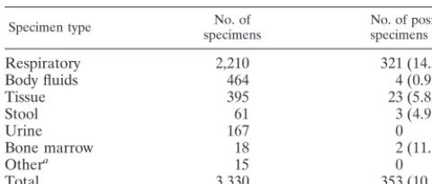
We compared 3,330 specimens collected from 2,346 patients treated at the six sites. By all culture systems, a total of 362 isolates of mycobacteria were recovered from 353 specimens, which had been collected from 247 patients. The majority of specimens tested (2,210 specimens [66%]) were from the re-spiratory tract. Overall, 353 (10.6%) of the specimens tested were positive for mycobacteria (Table 1). Among the respira-tory specimens, 321 (14.5%) were positive. Mycobacteria were also recovered from 2 (11.1%) of 18 bone marrow specimens, 23 (5.8%) of 395 tissue specimens, 3 (4.9%) of 61 stool spec-imens, and 4 (0.9%) of 464 body fluid specimens. Although 167 urine samples were tested, none were positive by any medium used in the study.
The numbers of isolates ofM. tuberculosiscomplex,
Myco-bacterium avium complex (MAC), and mycobacteria other
thanM. tuberculosis(MOTT, not inclusive of MAC) recovered
in each of the culture systems as well as the combined result of pairing each of the liquid media with solid media are listed in Table 2. Of the 362 isolates recovered during the study, there were 172 (47.5%) isolates of MAC, 132 (36.5%) isolates ofM.
tuberculosiscomplex, and 58 (16%) isolates of MOTT
recov-ered by all systems and media. As a single system, the BACTEC MGIT 960 detected the greatest number of myco-bacteria, 289 isolates (80%), followed by the BACTEC 460 TB System, with 271 isolates (75%), and the solid media, with 250 isolates (69%). When each of the broth medium systems was paired with the solid media and the combined recovery rates were compared, the same trends were noted. The BACTEC MGIT 960 plus solid media detected 335 mycobacteria (93%), as contrasted with 308 mycobacteria (85%) recovered in the BACTEC 460 TB system plus solid media.
As shown in Table 2, of the 132 isolates ofM. tuberculosis
complex recovered during the study, 119 (90%) were detected with the BACTEC 460 TB System, as contrasted with 105 (79%) detected with solid media and 102 (77%) detected with the BACTEC MGIT 960. When recovery on the combined solid media was paired with that on the broth systems, the BACTEC 460 TB System plus solid media detected 128 (97%) of the M. tuberculosis isolates as contrasted with 121 (92%) detected with the BACTEC MGIT 960 plus solid media.
[image:2.612.53.293.84.186.2]Of the 172 MAC isolates, more were recovered in the BACTEC MGIT 960 (147 isolates, or 86%) than in the BACTEC 460 TB system (123 isolates, or 72%) and with the combined solid media (106 isolates, or 62%). When the com-bined solid media were paired with the broth systems, the recovery of MAC was also greater with BACTEC MGIT 960
TABLE 1. Specimen types and recovery of mycobacteria
Specimen type specimensNo. of specimens (%)No. of positive
Respiratory 2,210 321 (14.5)
Body fluids 464 4 (0.9)
Tissue 395 23 (5.8)
Stool 61 3 (4.9)
Urine 167 0
Bone marrow 18 2 (11.1)
Othera 15 0
Total 3,330 353 (10.6)
[image:2.612.53.552.639.719.2]aSpecimen source not specified.
TABLE 2. Distribution of isolates recovered in each culture system and in combinations of solid and liquid systems
Organism
Total no. (%) of isolates recovered witha:
Total, all
media solid media9601 solid media460 TB1 Total,960 460 TBTotal, solid mediaTotal, 960 only 460 TBonly Solid mediaonly
M. tuberculosiscomplex 132 (100) 121 (92) 128 (97) 102 (77) 119 (90) 105 (79) 4 (3) 11 (8) 3 (2) MAC 172 (100) 160 (93) 136 (79) 147 (86) 123 (72) 106 (62) 36 (21) 12 (5) 3 (2)
MOTT 58 (100) 54 (93) 44 (76) 40 (69) 29 (50) 39 (67) 14 (24) 4 (7) 10 (17)
All 362 (100) 335 (93) 308 (85) 289 (80) 271 (75) 250 (69) 54 (15) 27 (7) 16 (4)
a960, BACTEC MGIT 960 system; 460 TB, BACTEC 460 TB system.
on May 15, 2020 by guest
http://jcm.asm.org/
plus solid media (160 isolates, or 93%) than with the BACTEC 460 TB system plus solid media (136 isolates, or 79%).
As a single system, the BACTEC MGIT 960 detected 40 (69%) of the 58 isolates of MOTT, whereas 39 (67%) of these isolates were detected with solid media and only 29 (50%) isolates were detected in the BACTEC 460 TB system. When the broth systems were paired with the solid media, the BACTEC MGIT 960 and solid media recovered 54 isolates (93%), whereas 44 (76%) isolates were found with the BACTEC 460 TB system and solid media.
As may be expected, there were several instances where one culture system was scored as contaminated while other media in the cohort were observed to be positive for AFB. In the present study, this occurred more often with the BACTEC MGIT 960 than with the other systems. There were 20 speci-mens for which mycobacteria were recovered from other co-hort media while only breakthrough contaminants were de-tected in the BACTEC MGIT 960 vial. In comparison, there were only eight specimens for which mycobacteria were recov-ered in other cohort media while only breakthrough contami-nants were detected in the BACTEC 460 TB System. During the study, when a culture vessel was found to be positive for breakthrough contamination, no further examination was per-formed on the contaminated medium while the other cohort media were followed to completion. As shown in Table 3, when these contaminated cohort specimens are excluded from the analysis, of 331 isolates recovered, the BACTEC MGIT 960 detected 281 (85%) isolates, followed by the BACTEC 460 TB System, with 251 isolates (76%), and the solid media, with 233 isolates (70%). When the broth media systems were paired with the solid media, the BACTEC MGIT 960 plus solid media detected 313 isolates (95%), and 282 isolates (85%) were re-covered in the BACTEC 460 TB system plus solid media. In this analysis, of the 118 isolates of M. tuberculosis complex recovered, 108 (92%) were detected with the BACTEC 460 TB System, as contrasted with 101 isolates (86%) detected with the BACTEC MGIT 960 and 95 isolates (81%) detected with solid media. When the combined solid media were paired with the broth systems, the BACTEC 460 TB System plus solid media
detected 114 (96%) of the M. tuberculosis isolates, and 111 isolates (94%) were detected with the BACTEC MGIT 960 plus solid media.
The TTD for M. tuberculosis complex and MAC in the BACTEC MGIT 960 and BACTEC 460 TB systems for the paired isolates (isolates recovered in both systems) and for the nonpaired isolates (total number of isolates recovered in each test system) are listed in Table 4. When all M. tuberculosis
complex isolates were included in the analysis, the TTD with the BACTEC MGIT 960 was 14.4 days and that with the BACTEC 460 TB system was 15.2 days. When specimens pos-itive forM. tuberculosis complex were paired, the TTD were 13.2 days with the BACTEC 460 TB system and 14.4 days with the BACTEC MGIT 960 (P50.055, not significant). The TTD for all MAC isolates in each system were 10.0 days with the BACTEC MGIT 960 and 10.4 days with the BACTEC 460 TB system. For the paired MAC isolates, the TTD were 9.8 days in the BACTEC MGIT 960 and 8.4 days in the BACTEC 460 TB System (P50.001).
In Table 5, the TTD for M. tuberculosis complex in the BACTEC MGIT 960 and that in the BACTEC 460 TB system for the paired and nonpaired isolates are grouped by whether the AFB smear result was negative or positive. Among both the nonpaired and the paired AFB smear-positive specimens, there was very little difference between the TTD for the BACTEC 460 TB system (10.8 days for nonpaired specimens and 10.3 days for paired specimens) and the BACTEC MGIT 960 (10.6 days for nonpaired specimens and 10.8 days for paired specimens). Notably, as can be seen in this table, the increased number of M. tuberculosiscomplex isolates recov-ered in the BACTEC 460 TB system was due almost exclu-sively to recovery from smear-negative samples. This may be a result of random inoculum distribution, particularly when small numbers of organisms are present in the original speci-men.
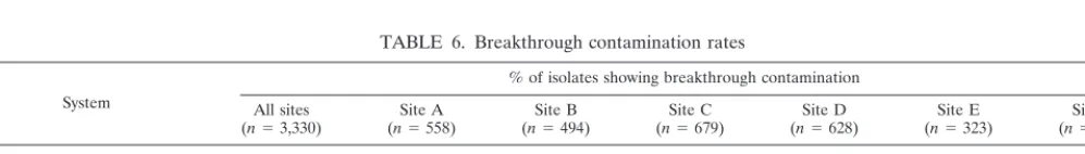
[image:3.612.54.554.83.163.2]Table 6 shows the breakthrough contamination rates for the BACTEC MGIT 960 and BACTEC 460 TB systems for spec-imens collected at all sites in the study. Breakthrough contam-ination was defined as the detection of any instrument-positive
TABLE 3. Distribution of isolates recovered in culture system and combinations with contaminated cohorts removed from analysis
Organism Total no. of isolates,all media
Total no. (%) of isolates recovered witha:
9601
solid media solid media460 TB1 Total, 960 Total, 460 TB solid mediaTotal,
M. tuberculosiscomplex 118 111 (94) 114 (97) 101 (86) 108 (92) 95 (81)
MAC 160 153 (96) 127 (79) 143 (89) 115 (72) 102 (64)
MOTT 53 49 (92) 41 (77) 37 (70) 28 (53) 36 (68)
All 331 313 (95) 282 (85) 281 (85) 251 (76) 233 (70)
a960, BACTEC MGIT 960 system; 460 TB, BACTEC 460 TB system.
TABLE 4. TTD ofM. tuberculosis, MAC, and all mycobacteria in BACTEC MGIT 960 and BACTEC 460 TB systems
Culture system
TTD, days (total no. of isolates detected) for:
M. tuberculosiscomplex MAC All mycobacteria
Nonpaireda Pairedb,c Nonpaireda Pairedb,d Nonpaireda Pairedb
BACTEC 960 14.4 (102) 14.4 (92) 10.0 (147) 9.8 (101) 11.8 (289) 11.1 (213)
BACTEC 460 15.2 (119) 13.2 (92) 10.4 (123) 8.4 (101) 12.5 (271) 11.3 (213)
aTotal for isolates recovered with either BACTEC MGIT 960 or BACTEC 460 TB system. bTotal for isolates recovered with both systems.
cP50.055, not significant. dP50.001.
on May 15, 2020 by guest
http://jcm.asm.org/
[image:3.612.53.551.627.696.2]tube that was AFB smear negative and subculture negative yet gram-stain smear positive for other (non-acid-fast) microor-ganisms. The contamination rate (percentage of isolates) noted with the BACTEC MGIT 960 was 8.1% (range, 1.8 to 14.6%), while that with the BACTEC 460 TB system was 4.9% overall (range, 0.9 to 9.2%). As shown in Table 6, at two of the trial sites, the contamination rates for the two systems were similar, while at the other four sites the contamination rate noted for the BACTEC MGIT 960 was higher than that noted for the BACTEC 460 TB.
The false-positive rate for the BACTEC MGIT 960 was 0.8% and was calculated as the number of BACTEC MGIT 960 vials which were instrument positive but were found to be smear negative and subculture negative for mycobacteria or other bacteria. During the study a total of 351 randomly se-lected BACTEC MGIT 960 instrument-negative vials (at the end of the 42-day protocol) were examined by AFB smear and subcultured to solid media. No false-negative samples from these random terminal subcultures of instrument-negative tubes were detected during the study.
DISCUSSION
Following the recent resurgence of tuberculosis in the United States, the rapid diagnosis of patients with active dis-ease has become a focus of much interest (1, 11). In addition, MAC has become the systemic bacterial pathogen most com-monly recovered from patients with AIDS and is an increas-ingly recognized pulmonary pathogen in nonimmunocompro-mised individuals as well (3). While amplified nucleic acid hybridization probe assays forM. tuberculosiscomplex provide an important adjunct to the diagnostic armamentarium, they have yet to displace traditional culture (2, 4). Among myco-bacterial culture detection systems, the BACTEC 460 TB system has long been the standard against which others are compared (5, 7). Newer methods that have recently been de-veloped, however, offer rapid TTD similar to that of the BACTEC 460 TB system while using fully automated, nonin-vasive, nonradiometric, self-contained incubator readers, each with a capacity of 240 to 383 mycobacterial culture vials (10, 13, 14). In addition to the automated instruments, the manual BBL MGIT system, which is a fluorescent indicator broth method, provides similar rapid performance characteristics,
but without the need for any instrumentation other than a UV light (15).
We evaluated the performance of the BACTEC MGIT 960, a high-capacity, fully automated, noninvasive system for the growth and detection of mycobacteria that requires neither radioactive media nor sharp implements. We compared the BACTEC MGIT 960 to the BACTEC 460 TB system and conventional solid media. Clearly, these data illustrate the su-perior performance characteristics of the two BACTEC broth systems as compared to solid media. Even though the LJ me-dium and two Middlebrook media were counted together as a single system, only 250 (69%) isolates of mycobacteria were recovered by using the combined solid media. Despite the advantages of the broth-based cultivation systems, traditional solid media still play a role in the recovery of mycobacteria from clinical samples and are recommended for use along with a liquid medium by the Centers for Disease Control (6). In the present study, no one system recovered all isolates of eitherM.
tuberculosis complex or MAC. The highest overall recovery
rates were noted when solid media were combined with a liquid medium system. It should be noted that 16 isolates (4%) were detected only in the solid media, including 3 isolates ofM.
tuberculosiscomplex. The variations in the concentrations of
organisms in the original specimens and the resulting random inoculum distribution among the multiple culture systems un-der testing likely account for some of these differences. Simi-larly, in a previous study of 1,184 respiratory specimens, no single medium tested provided the recovery of all isolates (12). In this study, among the three systems tested, the BACTEC 12B provided the highest percentage ofM. tuberculosis com-plex isolates recovered; the use of either 7H11 medium or LJ medium along with the 12B vial increased the recovery by 4 to 6%.
In the present study, the number of mycobacteria recovered in the BACTEC MGIT 960 system was greater than those recovered in the BACTEC 460 TB system and with solid me-dia. Among specimens collected at all sites, 48% of the isolates detected were MAC, 36% wereM. tuberculosiscomplex, and 16% were MOTT. In particular, the BACTEC MGIT 960 detected more isolates of MAC and MOTT than did the BACTEC 460 TB system. While a greater number ofM.
tu-berculosiscomplex isolates were detected with the BACTEC
460 TB than with the BACTEC MGIT 960, this advantage was almost exclusively seen in smear-negative samples and was greatly diminished when solid media were added to the anal-ysis. During the study, there were 11 specimens (10 of which were AFB smear negative) where the BACTEC MGIT 960 vial was found to have breakthrough contamination whileM.
tu-berculosiscomplex was recovered from the BACTEC 460 TB
[image:4.612.55.295.93.165.2]vial. Conversely, there was only one specimen (AFB smear negative) for which the BACTEC 460 TB vial was contami-nated while anM. tuberculosiscomplex isolate was recovered from the BACTEC MGIT 960 vial. When these specimens were removed from the analysis, as shown in Table 3, the recovery ofM. tuberculosiscomplex with the BACTEC MGIT
TABLE 5. TTD ofM. tuberculosisfor AFB smear-positive and smear-negative isolates in BACTEC 960 and 460 TB systems
Culture system TTD (range), no. of isolates AFB smear1 AFB smear2
BACTEC 960, total (n5102) 10.6 (5–24), 50 18.1 (7–43), 52 BACTEC 460, total (n5119) 10.8 (3–29), 51 18.4 (6–57), 68 BACTEC 960 (paired) 10.8, 47 18.1, 45 BACTEC 460 (paired) 10.3, 47 16.3, 45
TABLE 6. Breakthrough contamination rates
System
% of isolates showing breakthrough contamination All sites
(n53,330) (nSite A5558) (nSite B5494) (nSite C5679) (nSite D5628) (nSite E5323) (nSite F5648)
BACTEC MGIT 960 8.1 9.3 5.4 10.0 14.6 1.8 4.0
BACTEC 460 TB 4.9 5.0 0.6 9.2 7.1 0.9 3.7
Combined solid media 21.1 27.4 0.8 13.2 31.0 5.2 37.8
on May 15, 2020 by guest
http://jcm.asm.org/
[image:4.612.57.556.658.729.2]960 more closely approximated that of the BACTEC 460 TB system. On the other hand, when specimens from which MAC was recovered from a cohort vial but the other was contami-nated were removed from the analysis, the superiority of the BACTEC MGIT 960, either with or without solid media, re-mained significant.
In addition to overall recovery, the TTD is an important performance characteristic of mycobacterial detection systems. When all isolates recovered were included in the analysis, the TTD ofM. tuberculosiscomplex with the BACTEC MGIT 960 (14.4 days) was faster than that with the BACTEC 460 TB system (15.2 days). When only the 92 paired isolates (recov-ered in both broth systems) were included in the analysis, however, the TTD with the BACTEC 460 TB system (13.2 days) was faster than that with the BACTEC MGIT 960 (14.4 days). The reason for this is apparent in Table 5, where allM.
tuberculosiscomplex isolates recovered in each system are
sep-arated according to smear result. The higher recovery rate of these isolates in the BACTEC 460 TB system was almost exclusively due to recovery from smear-negative specimens, which may be expected to have fewer bacilli in the originating specimen and, concomitantly, a longer mean TTD. As seen in Table 4, among the total AFB-containing samples, the TTD recorded for the two BACTEC systems were essentially the same, regardless of AFB smear result.
As a whole, the rate of breakthrough contamination was found to be higher with the BACTEC MGIT 960 (8.1%) than with the BACTEC 460 TB system (4.9%). There was, however, a large variation in the contamination rate among the test sites for all the media tested. These wide variations may reflect the dissimilar conditions of specimen quality and transport time and conditions among the sites. Notably, at two of the six test sites there was little difference in the rate of contamination between the two systems, while at the other four sites, contam-ination rates were clearly higher with the BACTEC MGIT 960 than the BACTEC 460 TB system. This is similar to the expe-riences reported when the ESP (13), MB/BacT System (10), BACTEC 9000MB (14), and BBL MGIT (15) were compared to the BACTEC 460TB system. These higher rates of break-through contamination are likely a result of the fact that the media in the newer systems are more enriched than the BACTEC 460 TB media. The BACTEC 12B medium relies on the metabolic utilization of radiolabeled palmitic acid for the detection of14C-labeled CO
2, signaling the presence of
grow-ing mycobacteria. Other than the radiolabeled substrate, the 12B medium is very low in nutrient content and is not an optimal growth environment for most bacteria, which do not utilize the14C substrate and thus are not detected.
The amounts of labor and user interaction required for efficient operation are a substantial consideration in the selec-tion of any microbiology system. The BACTEC MGIT 960 system has very good performance characteristics, is easy to use, and readily fits into the customary mycobacteriology lab-oratory work flow. In large-volume laboratories, the 960-cul-ture tube capacity provides a decided advantage over the con-tinuously monitored instruments of lesser capacity. When a 42-day protocol is used in a laboratory with a 10% positivity rate, a single BACTEC MGIT 960 system could reasonably be expected to accommodate at least 8,000 specimens per year.
In summary, the newly introduced BACTEC MGIT 960 system is a dependable, high-capacity, compact, fully auto-mated continuous monitoring instrument for the recovery of mycobacteria from human clinical samples. When used in com-bination with solid media, the BACTEC MGIT 960 system shows performance comparable to that of the BACTEC 460 TB system for the detection ofM. tuberculosiscomplex, while providing greater recovery of MAC and MOTT.
ACKNOWLEDGMENT
This study was supported by Becton Dickinson Microbiology Sys-tems.
REFERENCES
1.Anhalt, J. P., F. G. Witebsky, and G. L. Woods.1993. College of American Pathologists position statement regarding rapid detection ofMycobacterium tuberculosis. Arch. Pathol. Lab. Med.117:873.
2.Eing, B. R., A. Becker, A. Sohns, and R. Ringelmann.1998. Comparison of Roche Cobas AmplicorMycobacterium tuberculosisassay with in-house PCR and culture for detection ofM. tuberculosis. J. Clin. Microbiol.36:2023–2029. 3.Epstein, M. D., C. P. Aranda, S. Bonk, B. A. Hanna, and W. Rom.1997. The significance ofMycobacterium aviumcomplex cultivation in the sputum of patients with pulmonary tuberculosis. Chest111:142–147.
4.Gamboa, F., G. Fernandez, E. Padilla, J. M. Manterola, J. Lonca, P. J. Cardona, L. Matas, and V. Ausina.1998. Comparative evaluation of initial and new versions of the Gen-Probe Amplified Mycobacterium Tuberculosis Direct Test for direct detection ofMycobacterium tuberculosisin respiratory and nonrespiratory specimens. J. Clin. Microbiol.36:684–689.
5.Huebner, R. E., R. C. Good, and J. I. Tokars.1993. Current practices in mycobacteriology: results of a survey of state public health laboratories. J. Clin. Microbiol.31:771–775.
6.Kent, P. T., and G. P. Kubica.1985. Public health mycobacteriology: a guide for the level III laboratory. Department of Health and Human Services, Centers for Disease Control, Atlanta, Ga.
7.Middlebrook, G., Z. Reggiardo, and W. D. Tigertt.1977. Automatic radio-metric detection of growth ofMycobacterium tuberculosisin selective media. Am. Rev. Respir. Dis.115:1066–1069.
8.Morgan, M. A., C. D. Horstmeier, D. R. DeYoung, and G. D. Roberts.1983. Comparison of a radiometric method (BACTEC) and conventional culture media for recovery of mycobacteria from smear-negative specimens. J. Clin. Microbiol.18:384–388.
9.Roberts, G. D., N. L. Goodman, L. Heifets, H. W. Larsh, T. H. Lindner, J. K. McClatchy, M. R. McGinnis, S. H. Siddiqi, and P. Wright.1983. Evaluation of the BACTEC radiometric method for recovery of mycobacteria and drug susceptibility testing of Mycobacterium tuberculosisfrom acid-fast smear-positive specimens. J. Clin. Microbiol.18:689–696.
10. Rohner, P., B. Ninet, C. Metral, S. Emler, and R. Auckenthaler.1997. Evaluation of the MB/BacT system and comparison to the BACTEC 460 system and solid media for isolation of mycobacteria from clinical specimens. J. Clin. Microbiol.35:3127–3131.
11. Schluger, N. W., and W. N. Rom.1994. Current approaches to the diagnosis of active pulmonary tuberculosis. Am. J. Respir. Crit. Care Med.149:264– 267.
12. Stager, C. E., J. P. Libonati, S. H. Siddiqi, J. R. Davis, N. M. Hooper, J. F. Baker, and M. E. Carter.1991. Role of solid media when used in conjunction with the BACTEC system for mycobacterial isolation and identification. J. Clin. Microbiol.29:154–157.
13. Tortoli, E., P. Cichero, M. G. Chirillo, M. R. Gismondo, L. Bono, G. Gesu, M. T. Simonetti, G. Volpe, G. Nardi, and P. Marone.1998. Multicenter comparison of ESP Culture System II with BACTEC 460TB and with Lo-wenstein-Jensen medium for recovery of mycobacteria from different clinical specimens, including blood. J. Clin. Microbiol.36:1378–1381.
14. Zanetti, S, F. Ardito, L. Sechi, M. Sanguinetti, P. Molicotti, G. Delogu, M. P. Pinna, A. Nacci, and G. Fadda.1997. Evaluation of a nonradiometric system (BACTEC 9000MB) for detection of mycobacteria in human clinical sam-ples. J. Clin. Microbiol.35:2072–2075.
15. Zuhre Badak, F., D. L. Kiska, S. Setterquist, C. Hartley, M. A. O’Connell, and R. L. Hopfer.1996. Comparison of Mycobacteria Growth Indicator Tube with BACTEC 460 for detection and recovery of mycobacteria from clinical specimens. J. Clin. Microbiol.34:2236–2239.