Copyright © 1999, American Society for Microbiology. All Rights Reserved.
Fluorescent Oligonucleotide Probes for Clinical and Environmental
Detection of
Acanthamoeba
and the T4 18S rRNA
Gene Sequence Type
DIANE R. STOTHARD,1† JOHN HAY,2JILL M. SCHROEDER-DIEDRICH,1†
DAVID V. SEAL,2ANDTHOMAS J. BYERS1*
Department of Molecular Genetics, The Ohio State University, Columbus, Ohio 43210,1and Tennent Institute of Ophthalmology, Western Infirmary, Glasgow University, Glasgow, United Kingdom2
Received 30 November 1998/Returned for modification 18 February 1999/Accepted 28 April 1999
The first genus- and subgenus-specific fluorescent oligonucleotide probes for in situ staining of
Acantha-moebaare described. Sequences of these phylogeny-based probes complement the 18S rRNA and the gene
encoding it (18S rDNA). The genus-specific probe (GSP) is a fluorescein-labeled 22-mer specific for
Acantha-moebaas shown here by its hybridization to growing trophozoites of all 12 knownAcanthamoeba18S rDNA
sequence types and by its failure to hybridize with amoebae of two other genera (Hartmannella vermiformisand
Balamuthia mandrillaris), two human cell lines, and two bacteria (Pseudomonas aeruginosaandEscherichia coli).
The sequence type T4-specific probe (ST4P) is a rhodamine-labeled 30-mer specific for Acanthamoeba18S
rDNA sequence type T4, as shown here in hybridization tests with trophozoites of all 12 sequence types. T4 is
the subgenus group associated most closely with Acanthamoeba keratitis (AK). GSP also was tested with
corneal scrapings from 17 patients with a high index of clinical suspicion of AK plus 5 patient controls. GSP stained both trophozoites and cysts, although nonspecific cyst wall autofluorescence also was observed. Results could be obtained with GSP in 1 to 2 days, and based on results from cell culture tests, the probe correctly
detected the presence or absence ofAcanthamoebain 21 of 24 specimens from the 22 patients. The use of GSP
with cultured trophozoites and cysts from corneal scrapings has illustrated the suitability of using fluorescent
oligonucleotide probes for identification of the genusAcanthamoebain both environmental and clinical
sam-ples. In addition, the use of ST4P with cultured amoebae has indicated the potential of oligonucleotide probes for use in subgenus classification.
The genus Acanthamoeba consists of small, ubiquitous amoebae that exhibit a biphasic life cycle consisting of a veg-etative trophozoite stage and a physiologically static cyst stage. These amoebae have been isolated from a variety of environ-mental sources (7) and are associated with human infection (17, 24–26, 37, 39). In immunocompromised patients, Acantha-moebainfections include granulomatous amoebic encephalitis (GAE), which is a fatal brain disease, and disseminated infec-tions of various other tissues. In otherwise healthy individuals, the prominent disease isAcanthamoebakeratitis (AK), a po-tentially sight-threatening eye infection. This disease was dis-covered in 1973 (19, 27). More than 500 cases of AK had been reported in the United States by 1993 (31), and another 200 cases had been reported by 1997 (25). AK is most prevalent among contact lens (CL) wearers (34). A recent prospective population-based study estimated that the incidence of AK among soft-CL wearers in Scotland was 1 in 6,710 (32). In other countries—for example, India—AK usually is associated with non-CL-associated ocular trauma (33).
Identification of acanthamoebae in ocular and other tissues can be difficult and time-consuming, even for trained micros-copists. In histological preparations, these amoebae look very similar to keratoplasts as well as neutrophils and monocytes,
and this often leads to false-negative and/or false-positive re-sults. It has been estimated that up to 70% of clinical AK cases are misdiagnosed as viral keratitis (2, 12, 18). Thus, many patients are initially treated with inappropriate drug therapies. Early detection ofAcanthamoebainfection is important be-cause trophozoites, which predominate in the initial stages of infection, are more susceptible to treatment than the sub-sequent cysts. Calcafluor white (42) and similar fluorescent stains, which often are used to detectAcanthamoeba, stain cyst walls and fail to detect trophozoites. The mean time to diag-nosis of AK can average 2.5 weeks longer for non-CL wearers than for CL users (6). This lag time may hamper disease resolution, since several studies have found that diagnosis and treatment within 1 month of onset results in lower morbidity and a better visual outcome (1, 2, 38). Thus, the availability of a rapid, accurate, and relatively simple diagnostic test would significantly enhance the initiation of appropriate chemother-apy.
Several RNA- and/or DNA-based methods are being devel-oped to aid in the detection and accurate identification of Acanthamoeba in clinical and environmental settings. These include PCR with and without DNA sequencing (16, 23, 35, 40), restriction fragment length polymorphism analysis (5, 36), and nucleic acid blotting methods (9). Although these tech-niques are important diagnostic aids, they involve indirect eval-uation of samples. They are unable to distinguish different developmental stages ofAcanthamoebaspp. and lack precision in the localization of the organisms in situ. These methods cannot determine whether the majority of amoebae in a sam-ple are cysts or trophozoites; they also cannot discern the density of the infection or the depth of corneal penetration of
* Corresponding author. Mailing address: Department of Molecular Genetics, The Ohio State University, 484 W. 12th Ave., Columbus, OH 43210-1292. Phone: (614) 292-5963. Fax: (614) 292-4466. E-mail: byers [email protected].
† Present address: Division of Infectious Diseases, Department of Medicine, Indiana University School of Medicine, Indianapolis, IN 46202.
2687
on May 15, 2020 by guest
http://jcm.asm.org/
the amoebae. Traditional histological techniques, in trained hands, often can identify amoebae in situ, but this depends on morphology or immunoreactivity. Furthermore, structural characteristics for identification of acanthamoebae at the sub-genus level are unreliable.
The most specific and sensitive technique devised to date for in situ staining is phylogenetic staining, first used by DeLong and coworkers (8) for identification of single cells in samples of mixed species. This technique uses oligonucleotide probes complementary to phylogenetically informative segments of rRNA. Different probes can provide accurate identification of an organism at various levels of taxonomic organization. In addition, specific probes labeled with different chromophores can be used in fluorescent in situ hybridization (FISH) to differentiate the different species or other taxonomic levels in a mixed sample.
The present article reports the development and use of two fluorescent probes. The first identifies all members of the ge-nusAcanthamoeba(genus-specific probe [GSP]); the second is specific for the subgenus group that is most commonly iden-tified in AK infections (sequence type T4-specific probe [ST4P]). We have used these probes for identification of Acan-thamoebain laboratory cultures and in human corneal scrap-ings from patients presumed to have AK based on traditional methods of microscopy and follow-up cultures.
MATERIALS AND METHODS
Cell cultures and corneal scrapings.The in situ hybridization of the modified universal probe (MUP), GSP, and ST4P described below was tested with cul-tured trophozoites from 21 strains (Table 1) representing the 12 different Acan-thamoeba18S rDNA sequence types previously described (35). Cultures were maintained at Ohio State University (OSU) as described previously (4, 10). GSP also was tested with 24 human corneal scraping specimens obtained in a study of microbial keratitis in Scotland (32). Acanthamoeba terricolawas a gift from Frederick Schuster, Brooklyn College, Brooklyn, N.Y. Human U937 lympho-cytes and human HeLa cells were provided by the OSU laboratories of Ing-ming Chiu and Mark Muller, respectively. The amoebaBalamuthia mandrillarisand theAcanthamoebastrains prefixed by CDC in Table 1 were gifts from Govinda S. Visvesvara of the U.S. Public Health Service’s Centers for Disease Control and Prevention, Atlanta, Ga.Hartmannella vermiformisandAcanthamoeba palesti-nensis, originally from the Cambridge Collection of Algae and Protozoa (see
Table 2), were provided by Peter H. H. Weekers, University of Nijmegen, Nijmegen, The Netherlands. The remainder of theAcanthamoebacultures (see Table 2) were obtained from Thomas Nerad at the American Type Culture Collection, Manassas, Va. Bacterial specimens were from laboratory cultures at OSU.
The corneal scrapings examined here were isolated by two of us (J.H. and D.V.S.) during a population-based longitudinal study of microbial keratitis in western Scotland (32). Scrapings were placed in sterile isotonic saline after being obtained from the eye by an ophthalmologist. Unstained aliquots were examined by bright-field or phase-contrast microscopy. This was followed by culture of samples forAcanthamoebain a medium described previously (15). One to 19 months after the initial specimen collection, portions of the original scraping samples were delivered to OSU, where they were retested for growth in culture and analyzed with the GSP probe described in this paper.
Pretreatment and fixation of amoebae and other cells for FISH.All solutions, with the exception of culture media, were prepared with diethylpyrocarbonate-treated double-distilled water (DEPC water). Portions of the human corneal scrapings (see Table 2) that had been suspended in sterile saline and stored at 4°C for 1 to 19 months were fixed. One milliliter of a laboratory cell culture or 100l of a scraping specimen was spun in a Microcentrifuge (Costar model 10) for 15 s to loosely pellet the cells. The medium was pipetted off, and the cells were resuspended by gentle shaking in 1⫻phosphate-buffered saline (PBS; 0.13 M NaCl, 7 mM Na2HPO4, and 3 mM NaH2PO4 in DEPC water; pH 7.2). The
cells were then loosely pelleted again by centrifugation for 15 s. The wash buffer was pipetted off, and the cells were gently resuspended in 1 ml of cold (4°C) 12% freshly depolymerized paraformaldehyde (PFA). The PFA was made by dissolv-ing 6 g of paraformaldehyde in 40 ml of 1⫻PBS for 10 min at 60°C with stirring. Then 10 ml of 0.1 M NaOH in DEPC water was added to clear the solution, and the mixture was chilled before being used. The cells were fixed at 4°C for at least 5 h and to a maximum of overnight. After being fixed, the cells were loosely pelleted by a 15-s centrifugation. The PFA was pipetted off, and the cells were washed in 1 ml of DEPC water with gentle shaking. The cells were loosely pelleted again by a 15-s centrifugation. The DEPC water was pipetted off, and the cells from cultures or corneal scrapings were resuspended in⬃100 or⬃30l, respectively, of 70% ethanol and stored at 4°C indefinitely.
Probes.Three oligonucleotide probes containing a fluorochrome at the 5⬘end were synthesized by Amitof Biotech, Inc. (Boston, Mass.). The 21-base MUP (5⬘-rhodamine-GWATTACCGCGGCTGCTGGCA-3⬘) was complementary to positions 653 to 633 in the 18S rRNA ofAcanthamoeba castellaniiNeff (13). The 22-base GSP (5-fluorescein-TTCACGGTAAACGATCTGGGCC-3⬘) was com-plementary to positions 957 to 936 in the Neff strain rRNA. The 30-base ST4P (5⬘-rhodamine-GCTGCCAAAACCAACTGAAAATAGGAGGAC-3⬘) was complementary to positions 1066 to 1037 in the Neff strain rRNA. MUP is based on the universal probe designed by Giovannoni and coworkers (11), with three modifications: base 14 was changed from K to T, a G between bases 16 and 17 was removed, and a GCA triplet was added to the 3⬘end. Probes were resus-pended in DEPC water to a final concentration of 1g/ml and stored at⫺20°C. Immediately before hybridization, probes were diluted to 30 ng/ml in DEPC water.
Whole-cell hybridization.Cells fixed in suspension were pipetted onto poly(L
-lysine)-coated microscope slides (Sigma, St. Louis, Mo.) in 2- to 5-l aliquots and air dried. The slides were then incubated in a methanol-formalin mixture (9:1) at room temperature for 20 min. The slides were then rinsed briefly in DEPC water and allowed to air dry. Thirty microliters of a hybridization mixture containing 60 ng of each probe in hybridization buffer (0.9 M NaCl, 20 mM Tris [pH 7.8], 5 mM EDTA, 0.01% sodium dodecyl sulfate) was pipetted onto each dry slide and covered with a 24- by 50-mm coverslip. The slides were immediately trans-ferred to a dark chamber humidified by a sponge soaked in DEPC water and incubated at 50 to 52°C for no more than 2 h of hybridization. Temperatures above 52°C led to a reduction in the fluorescence signal for all probes used.
After hybridization, the coverslips were floated off in DEPC water at room temperature and the slides were washed in wash buffer (30 mM NaCl, 4 mM Tris [pH 7.8], 1 mM EDTA) at 50°C for 20 min in the dark. The slides were rinsed quickly in DEPC water at room temperature and then counterstained with 4⬘,6-diamidino-2-phenylindole (DAPI; 1g/ml) for 10 min at room temperature in the dark. The slides were next washed briefly in DEPC water and allowed to air dry in the dark. Coverslips were mounted on dry slides by using 1 drop of Anti-fade Light mounting medium (Molecular Probes, Eugene, Oreg.) and perma-nently sealed with nail polish. The slides were stored indefinitely in the dark at 4°C. The preparations were examined with an inverted microscope (Zeiss Axio-scope, Oberkochen, Germany) fitted for epifluorescence detection with a high-pressure mercury bulb and filter sets CZ902 (for DAPI), CZ 915 (for rhoda-mine), and CZ 909 (for fluorescein).
RESULTS
Use of MUP with trophozoites.MUP was used here as a
[image:2.612.52.295.90.300.2]control that would stain all organisms tested in the FISH anal-yses. It was derived from the universal probe, designed by Giovannoni et al. (11), which originally was made to hybridize
TABLE 1. Acanthamoebastrains used in testing the genus- and T4-specific probesa
Strain Source sequence type18S rDNA
Acanthamoebaspecies strain V006a CDC0981:V006 T1
A. palestinensisGE 3aa ATCC 50252 T2
A. palestinensis1501/3ca CCAP 1501/3c T2
A. griffiniS7 ATCC 30731 T3
A. griffiniPanola Mt.a ATCC 30487 T3
A. castellaniiNeff ATCC 50373 T4
A. castellaniiV042 ATCC 50493 T4
A. castellaniiV014 ATCC 50492 T4
A. castellaniiMa ATCC 50370 T4
Acanthamoebaspecies strain Diamonda CDC Diamond T4
A. polyphagaV029 CDC0884:V029 T4
A. rhysodesSingh ATCC 30976 T4
Acanthamoebaspecies strain 88-2-37 ATCC 50497 T4
A. lenticulataPD2S ATCC 30841 T5
A. palestinensis2802 ATCC 50708 T6
A. astronyxisRay & Hayes ATCC 30137 T7
A. tubiashiOC-15c ATCC 30867 T8
A. terricolaFS F. Schuster T9
A. culbertsoniLilly A1 ATCC 30137 T10
A. hatchettiBH-2 T. Sawyer T11
A. heayliV013 CDC1283:V013 T12 aSpecies name has been revised according to the nomenclature of Stothard
et al. (35).
on May 15, 2020 by guest
http://jcm.asm.org/
to all three domains of life: archaebacteria, eubacteria, and eukaryotes. In the present study, however, the probe of Giov-annoni and coworkers did not provide a satisfactory signal due to the higher-stringency conditions used. Modification of two sites to be more consistent with the eubacteria and eukaryotes
used in the present study and addition of 3 bases at the 3⬘end led to a 100% match between MUP and targets and an unam-biguously positive red fluorescent signal from all cells tested. For example, hybridization of MUP withB. mandrillarisand A. castellaniiis illustrated by the red stain in Fig. 1.
FIG. 1–12. In situ hybridization of MUP, GSP, and ST4P with seven sequence types ofAcanthamoebaand with its closest relative, the genusBalamuthia. FIG. 1. One amoeba ofA. castellaniiV014 (T4) (upper right) and two amoebae of the largerB. mandrillarisV139 are stained red by MUP.
FIG. 2. Same field as Fig. 1, but onlyA. castellaniiV014 is stained green with GSP. FIG. 3. GSP stained amoebae ofAcanthamoebaspecies strain V006 (T1) green. FIG. 4.A. palestinensis1501/3c (T2) amoebae were stained green by GSP.
FIG. 5. GSP stained amoebae ofA. griffiniPanola Mt (T3) (bottom amoeba) andA. castellaniiV014 (T4) (top amoeba) green.
FIG. 6. Same field as Fig. 5, but onlyA. castellaniiV014 (T4) is stained with ST4P. Amoebae stained with this probe appear red because it has the same fluorochrome as MUP.
FIG. 7. GSP stainedA. lenticulataPD2S (T5) amoebae.
FIG. 8. A. tubiashiOV-15c (T8) amoebae were stained by GSP. FIG. 9. GSP stainedAcanthamoebaspecies cysts from a corneal scraping.
FIG. 10. Amoebae ofA. terricolaFS (T9) (the three large amoebae) andA. castellaniiV014 (T4) (the single smaller amoeba) were stained by GSP. FIG. 11. Same field as Fig. 10, but onlyA. castellaniiV014 (T4) is stained red with ST4P.
FIG. 12. Two sectioned cysts ofA. castellaniistained with GSP and showing green amoebae within yellow cyst walls.
on May 15, 2020 by guest
http://jcm.asm.org/
Use of GSP with trophozoites.Trophozoites double stained with MUP and GSP were used in tests for determining the specificity of GSP. Representatives from all 12 previously iden-tified sequence types of Acanthamoeba (35) (Table 1), two amoebae relatively closely related to the genusAcanthamoeba (H. vermiformis[41] andB. mandrillaris), bacteria ( Pseudomo-nas aeruginosaand Escherichia coli), and human cells (U937 and HeLa) were tested for hybridization with these two probes. As indicated above, MUP hybridized withAcanthamoebaand all control cells. GSP hybridized with all acanthamoebae tested but with no control organisms. The differential staining of MUP and GSP is illustrated in Fig. 1 and 2, which include a mixture of one relatively small acanthamoeba of sequence type T4 and two largerBalamuthiaamoebae. With the rhodamine filter (Fig. 1), all three amoebae are red, indicating hybridiza-tion with MUP. With the fluorescein filter (Fig. 2), only the smaller amoeba is green, indicating that GSP hybridized spe-cifically to A. castellanii. Evidence that GSP hybridizes with divergent sequence types ofAcanthamoebais illustrated by the green staining of amoebae of seven sequence types that had been hybridized with this probe, as shown in Fig. 3 (T1), 4 (T2), 5 (T3 and T4), 7 (T5), 8 (T8), and 10 (T9 and T4). See below for identification of T4 amoebae in Fig. 6 and 11.
Use of ST4P with trophozoites.The specificity of ST4P was
tested by using cells doubly stained with GSP and ST4P. Amoebae from each of the 12 described 18S rRNA gene (rDNA) sequence types of Acanthamoeba(Table 1) plus the bacteria, human cells, and other amoebae described above were used. ST4P successfully hybridized with all acanthamoe-bae of the T4 sequence type, but not with acanthamoeacanthamoe-bae of any of the other sequence types or with control cells. The degree of specificity is best observed in Fig. 5 and 6, which illustrate differential staining of the two most closely related sequence types, T3 (A. griffiniPanola Mt.) and T4 (A. castel-laniiV014). In Fig. 5, amoebae of both sequence types appear green with the fluorescein filter, indicating that both types have hybridized with GSP. In Fig. 6, however, only the amoeba of sequence type T4 appears red with the rhodamine filter, indi-cating that it alone has hybridized with ST4P. The specificity of ST4P also is seen in Fig. 10 and 11, in which staining with this probe and with GSP is compared for T4 (A. castellaniiV014) and a representative of a more distantly related sequence type, T9 (A. terricolaFS). Both amoebae are green in Fig. 10, indi-cating that both hybridized with GSP, but only the smaller A. castellaniiis red in Fig. 11 and, thus, was able to hybridize with ST4P.
Use of GSP with cysts from corneal scrapings.Hybridization
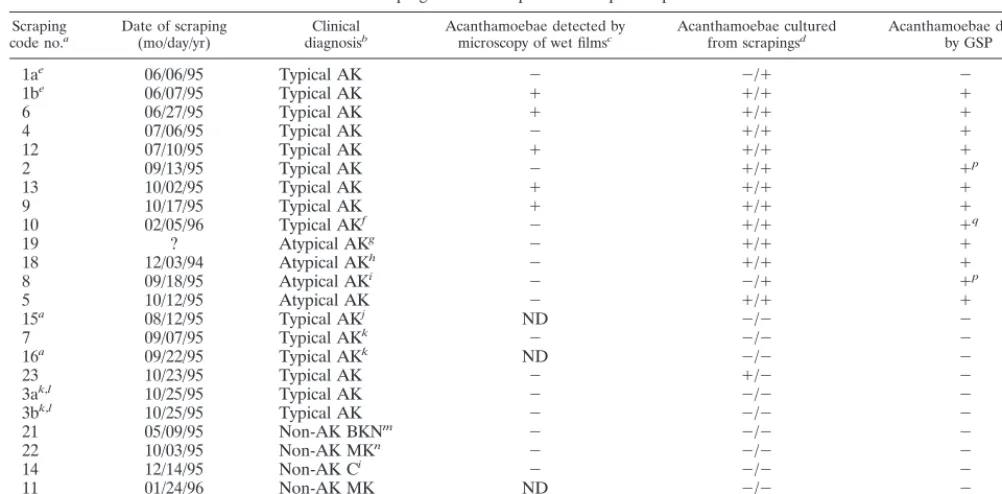
of GSP with intact cysts from a corneal scraping is illustrated by the green stain in Fig. 9. In addition, Acanthamoebacyst walls autofluoresced either in the presence or in the absence of hybridization conditions. Attempts to quench the fluorescence with a number of different agents failed. Nevertheless, the fluo-rescein filter set did distinguish between the yellow autofluo-rescence of the cyst walls and the green fluoautofluo-rescence attribut-able to hybridization of the fluorescein-labeled GSP to the rRNA and rDNA of the encysted amoebae. This distinction is best seen in sectioned cysts from the human cornea (Fig. 12). To test the usefulness of GSP with corneal scrapings, we employed 24 specimens obtained from 22 patients, 17 with either confirmed AK or a high index of suspicion of AK and 5 with non-Acanthamoebakeratitis or conjunctivitis (Table 2). Bright-field and phase-contrast microscopy were used with wet films to evaluate the specimens. Fifteen scrapings produced positive Acanthamoebacultures at Tennent Institute of Oph-thalmology (TIO), OSU, or both institutions. It was observed,
however, that wet-film microscopy detected acanthamoebae in only 6 (40%) of the 15 culture-positive specimens.
Microscopy using FISH with GSP was then used to evaluate the corneal scraping specimens. Acanthamoebae were detect-ed in 12 (80%) of the 15 culture-positive samples. Overall, FISH results were consistent with the positive or negative culture results for 21 (88%) of the 24 specimens. The only discrepan-cies were three FISH-negative specimens (no. 1a, 17, and 23) that were considered culture positive even though positive results were obtained at only one of the two institutions (see Discussion).
DISCUSSION
The major advantage of using fluorescent oligonucleotide probes and FISH for identification of Acanthamoeba is that such probes have the potential to simultaneously detect and classify amoebae in situ. FISH provides a means of rapid, unequivocal identification ofAcanthamoebain cases of AK for which clinical diagnosis is putative and the appropriate exper-tise for identification by bright-field and/or phase-contrast mi-croscopy is unavailable. As we have seen here, even when that expertise is available, the number of false negatives can be relatively high in the absence of FISH unless the observations are backed up by tests for culture growth. However, culture may take a week or more to yield satisfactory division of amoeba, especially if the cornea has been exposed to a range of antimicrobial or antiviral drugs prior to the procurement of the scraping specimen.
Although the examples of GSP and ST4P staining described in this paper have used amoebae growing in culture or in corneal scrapings, we also have been able to detect Acantha-moebain sectioned human cornea (Fig. 12) (20).
In these studies, successful culture of a specimen at both TIO and OSU provided the highest level of confidence that viable acanthamoebae were present in an original scraping. However, successful culture at either one of the two institu-tions also was considered strong evidence that amoebae were originally present in the scraping. This is because the amoebae in the scrapings we examined were encysted. In our experience, negative culture results due to failure of excystment are rela-tively common whereas positive culture results due to acciden-tal contamination with acanthamoebae are relatively rare. Thus, in the present study, the observance of an occasional failure of culture growth for scrapings that included cysts was not surprising. However, obtaining positive cultures from scrapings that did not originally include amoebae was highly unlikely. Therefore, in our analysis, all specimens giving rise to cultures at TIO, OSU, or both institutions were considered to have included acanthamoebae in the original scrapings. In some cases, positive cultures might have been obtained from samples with very few amoebae. Because FISH was performed on the original samples, small numbers of cells could have resulted in negative FISH results. On this basis, FISH with GSP detected Acanthamoeba in 80% of the culture-positive samples, and FISH and culture results were consistent for 88% of all samples. The comparable values are slightly higher if the FISH results, which were all obtained at OSU, are separately compared with culture results from each of the two laborato-ries. In this case, FISH detected amoebae in 86% (12 of 14) of culture-positive samples, and FISH and culture results were consistent for 92% (22 of 24) of all samples tested at OSU. At TIO, the values were 92% (11 of 12) and 92% (22 of 24), respectively. If comparisons of FISH and cell culture results are limited to the 20 specimens for which positive or negative culture results were identical at TIO and OSU, there was 100%
on May 15, 2020 by guest
http://jcm.asm.org/
agreement. Thus, FISH results were most consistent with cul-ture results when there was no ambiguity in the latter.
The data indicate that FISH with GSP clearly improves the accuracy of microscopy for detection ofAcanthamoeba. This method produced no false positives and only three false-neg-ative results among the 24 scrapings. The most likely explana-tion for the false negatives is that viable amoebae, although present, were too sparse to be picked up by FISH. This prob-lem probably could have been overcome by the use of a more effective method of cell concentration. The FISH result for specimen 1a (Table 2) appears to be a false negative since amoebae were successfully cultured at OSU and because spec-imen 1b, obtained from the same eye on the next day, was culture positive by all methods used, including FISH.
The FISH results for scraping specimens 17 and 23 also appear to be false negatives because amoebae were cultured successfully at either TIO or OSU and because PCR results to be discussed elsewhere (31a) indicated the presence of acan-thamoebae in both scrapings. Specimen 17 is a special case. The clinical diagnosis was bacterial keratitis, and the acan-thamoebae detected by wet-film microcopy and cell culture
were identified as transients present in the scraping. This result emphasizes that detection ofAcanthamoebashould not be the only criterion used for a diagnosis of AK. Finally, the FISH result for scraping specimen 8 appears to be a true positive because the presence of acanthamoebae also is supported by positive cell culture and PCR results at OSU.
Two important advantages of FISH are the unambiguous identification of the organisms and the relative rapidity with which results can be obtained. Identification with FISH can be accomplished in 1 to 2 days, whereas cell culture time is vari-able and can take up to several weeks. It is recommended, however, that all scrapings, especially those that give negative FISH results, be cultured. Any amoebae that grow can then be tested by FISH with GSP to determine whether they are acan-thamoebae.
[image:5.612.51.552.91.338.2]The availability of a large number of DNA sequences for the nuclear small-subunit rRNA gene (35) has made it possible to design cytological stains that are specific for the genus Acan-thamoebaand for separate lineages within this genus. We plan to develop a set of probes that could be used to identify the amoebae at the subgenus level. ST4P, which identifies 18S
TABLE 2. Comparison of bright-field and phase-contrast microscopy, culture growth, and FISH evaluations of corneal scrapings as follow-up to clinical presumption of AK
Scraping
code no.a Date of scraping(mo/day/yr) diagnosisClinicalb Acanthamoebae detected bymicroscopy of wet filmsc Acanthamoebae culturedfrom scrapingsd Acanthamoebae detectedby GSP
1ae 06/06/95 Typical AK ⫺ ⫺/⫹ ⫺
1be 06/07/95 Typical AK ⫹ ⫹/⫹ ⫹
6 06/27/95 Typical AK ⫹ ⫹/⫹ ⫹
4 07/06/95 Typical AK ⫺ ⫹/⫹ ⫹
12 07/10/95 Typical AK ⫹ ⫹/⫹ ⫹
2 09/13/95 Typical AK ⫺ ⫹/⫹ ⫹p
13 10/02/95 Typical AK ⫹ ⫹/⫹ ⫹
9 10/17/95 Typical AK ⫹ ⫹/⫹ ⫹
10 02/05/96 Typical AKf ⫺ ⫹/⫹ ⫹q
19 ? Atypical AKg ⫺ ⫹/⫹ ⫹
18 12/03/94 Atypical AKh ⫺ ⫹/⫹ ⫹
8 09/18/95 Atypical AKi ⫺ ⫺/⫹ ⫹p
5 10/12/95 Atypical AK ⫺ ⫹/⫹ ⫹
15a 08/12/95 Typical AKj ND ⫺/⫺ ⫺
7 09/07/95 Typical AKk ⫺ ⫺/⫺ ⫺
16a 09/22/95 Typical AKk ND ⫺/⫺ ⫺
23 10/23/95 Typical AK ⫺ ⫹/⫺ ⫺
3ak,l 10/25/95 Typical AK ⫺ ⫺/⫺ ⫺
3bk,l 10/25/95 Typical AK ⫺ ⫺/⫺ ⫺
21 05/09/95 Non-AK BKNm ⫺ ⫺/⫺ ⫺
22 10/03/95 Non-AK MKn ⫺ ⫺/⫺ ⫺
14 12/14/95 Non-AK Ci ⫺ ⫺/⫺ ⫺
11 01/24/96 Non-AK MK ND ⫺/⫺ ⫺
17 05/24/96 Non-AK BKSo ⫹ ⫺/⫹ ⫺
aAll samples except no. 15 and 16 were fresh corneal scrapings transferred to sterile isotonic saline. The exceptions are scrapings originally spread on non-nutrient
agar plates and then recovered from plate washings.
bC, conjunctivitis; MK, presumed microbial keratitis of bacterial origin; BKN, bacterial keratitis due toNeisseria meningitidis; BKS, bacterial keratitis due to
Staphylococcus aureus.
cBright-field or phase-contrast microscopy of original wet films.⫹, acanthamoebae detected;⫺, acanthamoebae not detected; ND, not done. dCulture results at TIO/OSU.
eScrapings 1a and b were from the same patient. fSevere AK after 3 weeks on chlorhexidine therapy. gTrachoma and acanthamoebae in a non-CL wearer (30).
hLarge epithelial defect, sporadic diffuse corneal endotheliitis thought to be due to herpes simplex virus in a non-CL wearer. iCL-associated conjunctival inflammation without keratitis.
jPatient was on Brolene and neomycin therapy at time of scraping; acanthamoebae found in CL storage case. kPatient was on topical 0.02% chlorhexidine therapy at time of scraping; acanthamoebae found in CL storage case. lBoth eyes of same patient; acanthamoebae found in CL storage case.
mBasement membrane dystrophy.
nRed eye, pain, and photophobia in a CL wearer.
oPeripheral ulcer in a CL wearer due toS. aureus, with transient acanthamoebae in the scraping sample (28). pOne cyst seen.
qTwo cysts seen.
on May 15, 2020 by guest
http://jcm.asm.org/
rDNA sequence type T4, is the first of these probes to become available. The specificity of this probe has been demonstrated for trophozoites in this study but remains to be demonstrated for cysts. We focused on this sequence type for the design of a probe mostly because of its apparent importance in AK. Our previous sequence studies (35) plus 18S rDNA sequences ob-tained for corneal scrapings used in the present study (31a) indicate that 29 of the 30 AK isolates we have examined to date have T4 sequences. The only exception is a T3 isolate (22), and this sequence type is very closely related to T4. Another reason for concentrating on the T4 probe is because we have 18S rDNA sequence information for many more T4 strains than for any other sequence type. The greater amount of information available about sequence variation for this lineage permitted the design of a more robust probe. ST4P hybridizes to a region of the 18S rRNA gene that differs among the sequence types. Thus, this region may be a good target for other probes de-signed to be specific for other sequence types. However, more information on sequence variation in most of these other types is required before a complete set of robust sequence type-specific probes can be developed.
The FISH protocol was designed to be both specific and sensitive. The cells tested included axenic cultures of tropho-zoites and corneal scraping specimens that mostly included cysts. GSP worked very well with either developmental stage, but ST4P was used only with multiplying amoebae. Amoebae actively growing in cultures gave the brightest signal, most likely because of a high rRNA content in the cytoplasm. How-ever, amoebae that have been stored in isotonic saline for 1 to 19 months, as was the case for some of the corneal scraping specimens, also were detectable. The fact that this technique works on stored samples is important because there can often be some delay between the collection of a scraping and its analysis in a specialized laboratory. The success of FISH with older stored samples is not surprising because the amoebae were present as cysts. It previously has been shown that en-cysted acanthamoebae can remain viable in CL saline for 14 to 90 days (3). In the present study, however, cysts remained viable during refrigeration in isotonic saline for at least 19 months, as shown, for example, by scraping sample 18 (Table 2), which was collected 3 December 1994 and still produced a culture at OSU in early July 1996.
The probes were designed to hybridize with 18S rRNA in addition to the large number of rDNA copies in the nucleolus in order to maximize the number of intracellular targets. Probe specificity at the level of sequence type was tested only with trophozoites because interpretation of results from cysts might be complicated by the autofluorescence of the cyst walls. It should be possible to alleviate this problem by using colori-metric detection of a nonfluorescent tag such as digoxigenin, which has been used with probes forAcanthamoeba26S rRNA (21).
The probes have a number of potential applications in ad-dition to those tested in the present study. For example, they can detect amoebae in deparaffinized tissue sections (Fig. 12). Also, Paillasson and coworkers (29) have recently developed a procedure for in situ hybridization of FISH probes to RNA in living human cells. The cells were permeabilized with strepto-lysin O and probed without fixation. They then were detected with a fluorescence-activated cell sorter. We recently proved a linkage between a patient with AK, the CL storage case con-tents, and the patient’s home water supply (22). Thus, it might be possible to use FISH probes in a fluorescence-activated cell sorter format for prophylactic studies of water from treatment plants, home water supplies, or other sources.
Although this report has focused on AK,Acanthamoebaalso
is responsible for GAE. Two of the specimens tested in this study,Acanthamoeba healyiV013 andA. castellaniiV006, are isolates from patients with GAE. This disease has been in-creasing in incidence due to its association with AIDS (14). Hawley et al. (14) recently reported the ability to identify Acanthamoebain cerebrospinal fluid via culturing. Nearly all reported cases of GAE have been fatal, but early diagnosis, possibly using FISH on cerebrospinal fluid, might improve the odds of successful treatment.
The level of specificity required in probes will depend on their use. In some cases, it may only be necessary to distinguish between organisms that are known to cause a particular infec-tion and those that do not. For studies of biodiversity, a more varied set of probes might be needed. The large collection of 18S rDNA sequences now available at GenBank plus the se-quence alignments available on the Internet (3a) should be consulted by those wishing to design other probes. In addition, since cysts can be the major component of clinical samples, methods of suppressing the autofluorescence of cyst walls or the use of nonfluorescent markers for the genus- and sequence type-specific probes will be explored.
ACKNOWLEDGMENTS
The molecular biology studies described in this report were funded by Public Health Service grant EY09073 from the National Eye Insti-tute.
We thank our colleague Gregory Booton for helpful discussions and assistance with the figures.
REFERENCES
1.Bacon, A. S., J. K. Dart, L. A. Ficker, M. M. Matheson, and P. Wright.1993.
Acanthamoebakeratitis. The value of early diagnosis. Ophthalmology100:
1238–1243.
2.Bacon, A. S., D. G. Frazer, J. K. Dart, M. Matheson, L. A. Ficker, and P. A. Wright.1993. A review of 72 consecutive cases ofAcanthamoebakeratitis, 1984–1992. Eye7:719–725.
3.Brandt, F. H., D. A. Ware, and G. S. Visvesvara.1989. Viability of Acantha-moebacysts in ophthalmic solutions. Appl. Environ. Microbiol.55:1144– 1146.
3a.Byers, T. J.1 April 1998, posting date. 18S rDNA sequence alignments. [Online.] http://www.biosci.ohio-state.edu/⬃tbyers/byers.htm.
4.Byers, T. J., R. A. Akins, B. J. Maynard, R. A. Lefken, and S. M. Martin.
1980. Rapid growth ofAcanthamoebain defined media; induction of encyst-ment by glucose-acetate starvation. J. Protozool.27:216–219.
5.Chung, D. I., H.-S. Yu, M.-Y. Hwang, T.-H. Kim, T.-O. Kim, H.-C. Yun, and H.-H. Kong.1998. Subgenus classification ofAcanthamoebaby riboprinting. Korean J. Parasitol.36:69–80.
6.Chynn, E. W., M. A. Lopez, D. Pavan-Langston, and J. H. Talamo.1995.
Acanthamoebakeratitis. Contact lens and noncontact lens characteristics. Ophthalmology102:1369–1373.
7.De Jonckheere, J. F.1991. Ecology ofAcanthamoeba. Rev. Infect. Dis.
13(Suppl. 5):S385–S387.
8.DeLong, E. F., G. S. Wickham, and N. R. Pace.1989. Phylogenetic stains: ribosomal RNA-based probes for the identification of single cells. Science
243:1360–1363.
9.Gast, R. J., and T. J. Byers.1995. Genus- and subgenus-specific oligonucle-otide probes forAcanthamoeba. Mol. Biochem. Parasitol.71:255–260. 10. Gast, R. J., D. R. Ledee, P. A. Fuerst, and T. J. Byers.1996. Subgenus
systematics ofAcanthamoeba: four nuclear 18S rDNA sequence types. J. Eukaryot. Microbiol.43:498–504.
11. Giovannoni, S. J., E. F. DeLong, G. J. Olsen, and N. R. Pace.1988. Phylo-genetic group-specific oligodeoxynucleotide probes for identification of sin-gle microbial cells. J. Bacteriol.170:720–726.
12. Goodall, K., A. Brahma, and A. Ridgeway.1996.Acanthamoebakeratitis: masquerading as adenovirus keratitis. Eye10:643–644.
13. Gunderson, J. H., and M. L. Sogin.1986. Length variation in eukaryotic rRNAs: small-subunit rRNAs from the protistsAcanthamoeba castellaniiand
Euglena gracilis. Gene44:63–70.
14. Hawley, H. B., J. S. Czachor, V. Malhotra, J. W. Funkhouser, and G. S. Visvesvara.1997.Acanthamoebaencephalitis in patients with AIDS. AIDS Reader7:137–144.
15. Hay, J., C. M. Kirkness, D. V. Seal, and P. Wright.1994. Drug resistance and
Acanthamoebakeratitis: the quest for alternative antiprotozoal chemother-apy. Eye8:555–563.
16. Howe, D. K., M. H. Vodkin, R. J. Novak, G. Visvesvara, and G. L.
on May 15, 2020 by guest
http://jcm.asm.org/
lin.1997. Identification of two genetic markers that distinguish pathogenic and nonpathogenic strains ofAcanthamoeba. Parasitol. Res.83:345–348. 17.John, D. T.1993. Opportunistically pathogenic free-living ameba, p. 143–
246. InJ. R. Baker and J. P. Kreier (ed.), Parasitic protozoa, 2nd ed. Academic Press, New York, N.Y.
18. Johns, K. J., D. M. O’Day, W. S. Head, R. J. Neff, and J. H. Elliott.1987. Herpes simplex masquerade syndrome:Acanthamoebakeratitis. Curr. Eye Res.6:207–212.
19. Jones, D. B., G. S. Visvesvara, and N. M. Robinson.1975.Acanthamoeba polyphagakeratitis andAcanthamoebauveitis associated with fatal menin-goencephalitis. Trans. Ophthalmol. Soc. U.K.95:221–232.
20. Kinnear, F., J. Hay, J. M. Schroeder-Diedrich, and T. J. Byers.Unpublished data.
21. Lai, S., M. Asgari, and H. R. Henney, Jr.1994. Non-radioactive DNA probe and polymerase chain reaction procedures for the specific detection of Acan-thamoeba. Mol. Cell. Probes8:81–89.
22. Ledee, D. R., J. Hay, T. J. Byers, D. V. Seal, and C. M. Kirkness.1996.
Acanthamoeba griffini. Molecular characterization of a new corneal patho-gen. Invest. Ophthalmol. Vis. Sci.37:544–550.
23. Lehmann, M. O., S. M. Green, N. Morlet, M. F. Keys, M. M. Matheson, J. K. G. Dart, J. I. McGill, and P. J. Watt.1998. Polymerase chain reaction analysis of corneal epithelial and tear samples in the diagnosis of Acantha-moebakeratitis. Invest. Ophthalmol. Vis. Sci.39:1261–1265.
24. Ma, P., G. S. Visvesvara, A. J. Martinez, F. H. Theodore, P.-M. Daggett, and T. K. Sawyer.1990.NaegleriaandAcanthamoebainfections: a review. Rev. Infect. Dis.12:490–513.
25. Martinez, A. J., and G. S. Visvesvara.1997. Free-living amphizoic and opportunistic amebas. Brain Pathol.7:583–598.
26. Murakawa, G. J., T. McCalmont, J. Altman, G. H. Telang, M. D. Hoffman, G. R. Kanter, and T. G. Berger.1995. Disseminated acanthamebiasis in patients with AIDS. A report of five cases and a review of the literature. Arch. Dermatol.131:1291–1296.
27. Nagington, J., P. G. Watson, and T. J. Playfair.1974. Amoebic infection of the eye. Lancetii:1547–1550.
28. Newman, W., J. Hay, B. Brown, and D. V. Seal.1997.Acanthamoebaas a “transient” in the corneal scrape of a poorly compliant soft contact lens wearer with peripheral keratitis. Eye11:937–939.
29. Paillasson, S., M. Van de Corput, R. W. Dirks, H. J. Tanke, M. Robert-Nicoud, and X. Ronot.1997. In situ hybridization in living cells: detection of RNA molecules. Exp. Cell Res.231:226–233.
30. Pyott, A., J. Hay, and D. V. Seal.1996.Acanthamoebakeratitis: first recorded
case from a Palestinian patient with trachoma. Br. J. Ophthalmol.80:849. 31. Rivasi, F., L. Longanesi, C. Casolari, G. P. Croppo, G. Pierini, E. Zunarelli,
and G. S. Visvesvara.1995. Cytologic diagnosis ofAcanthamoebakeratitis. Report of a case with correlative study with indirect immunofluorescence and scanning electron microscopy. Acta Cytol.39:821–826.
31a.Schroeder-Diedrich, J. M.Unpublished data.
32. Seal, D. V., C. M. Kirkness, H. G. B. Bennett, M. Peterson, et al. Population-based cohort study of microbial keratitis in Scotland: incidence and features. Submitted for publication.
33. Sharma, S., M. Srinivassan, and C. George.1990.Acanthamoebakeratitis in non-contact lens wearers. Arch. Ophthalmol.108:676–678.
34. Stehr-Green, J. K., T. M. Bailey, and G. S. Visvesvara.1989. The epidemi-ology ofAcanthamoebakeratitis in the United States. Am. J. Ophthalmol.
107:331–336.
35. Stothard, D. R., J. M. Schroeder-Diedrich, M. H. Awwad, R. J. Gast, D. R. Ledee, S. Rodriguez-Zaragoza, C. L. Dean, P. A. Fuerst, and T. J. Byers.
1998. The evolutionary history of the genusAcanthamoebaand the identi-fication of eight new 18s rRNA gene sequence types. J. Eukaryot. Microbiol.
45:45–54.
36. Sze´nası´, Z., T. Endo, K. Yagita, and E. Nagy.1998. Isolation, identification and increasing importance of “free-living” amoebae causing human disease. J. Med. Microbiol.47:5–16.
37. Tan, B., C. M. Weldon-Linne, D. P. Rhone, C. L. Penning, and G. S. Vis-vesvara.1993.Acanthamoebainfection presenting as skin lesions in patients with the acquired immunodeficiency syndrome. Arch. Pathol. Lab. Med.117:
1043–1046.
38. Tay-Kearney, M. L., C. N. McGhee, G. J. Crawford, and K. Trown.1993.
Acanthamoebakeratitis: a masquerade of presentation in six cases. Aust. N. Z. J. Ophthalmol.21:237–245.
39. Visvesvara, G. S., and J. K. Stehr-Green.1990. Epidemiology of free-living ameba infections. J. Protozool.37:25S–33S.
40. Vodkin, M. H., D. K. Howe, G. S. Visvesvara, and G. L. McLaughlin.1992. Identification ofAcanthamoebaat the generic and specific levels using the polymerase chain reaction. J. Protozool.39:378–385.
41. Weekers, P. H. H., R. J. Gast, P. A. Fuerst, and T. J. Byers.1994. Sequence variation in small-subunit ribosomal RNAs ofHartmannella vermiformisand the phylogenetic implications. Mol. Biol. Evol.11:684–690.
42. Wilhelmus, K. R., M. S. Osato, R. I. Font, N. M. Robinson, and D. M. Jones.
Rapid diagnosis ofAcanthamoebausing calcafluor white. Arch. Ophthalmol.
104:1309–1312.