0095-1137/06/$08.00⫹0 doi:10.1128/JCM.44.2.334–339.2006
Copyright © 2006, American Society for Microbiology. All Rights Reserved.
Evaluation of the New GenoType Mycobacterium Assay for
Identification of Mycobacterial Species
Cristina Russo,
1Enrico Tortoli,
2* and Donato Menichella
1Microbiology Laboratory, Bambino Gesu` Hospital, Rome,1and Regional Reference
Center for Mycobacteria, Microbiology and Virology Laboratory, Careggi Hospital, Florence,2Italy
Received 12 August 2005/Returned for modification 29 September 2005/Accepted 6 December 2005
The new commercial assay GenoType Mycobacterium, intended for the identification of mycobacteria, was evaluated with a panel of 197 strains belonging to 86 different taxa. The system, which relies on solid-phase reverse hybridization, is composed of two different kits: GenoType CM for the identification of more frequently detected mycobacterial species and GenoType AS for less common species. The sensitivity and specificity were 97.9% and 92.4% for the CM kit and 99.3% and 99.4% for the AS kit. Our results show that the system may represent a useful tool to substantially enlarge the number of mycobacteria that can be reliably identified in clinical laboratories.
Procedures for the identification of mycobacteria have greatly changed in the last years, and conventional biochemical and cultural methods are nowadays used only by a few labo-ratories worldwide. Even the methods based on analysis of cell wall lipids, which have played a primary role in the last 20 years of the 20th century (2, 8, 11), now seem to be in trouble because of the steady increase in the number of species within the genusMycobacterium(15).
As expected, the enormous progress in genetic knowledge has brought molecular methods into the limelight. Commercial DNA probes, first marketed by Gen-Probe (San Diego, Calif.), immediately turned out to be a handy and reliable tool, suit-able for accurate identification of the most common mycobac-terial species in many laboratories. The sensitivity and speci-ficity of such DNA probes are very high (6), and the only real limit of the system is the low number of species it is able to identify.
Several years ago, an important innovation was represented by the development of the reverse hybridization line probe assay. Such a system relies on paper strips on which multiple probes, each characterized by a different specificity, are immo-bilized. The amplified target sequence of the organism to be identified binds to the specific probe, and hybridization is re-vealed by the development of a colored line, while its position within the strip identifies the species.
The number of line probes in such commercial strips has risen over the last few years, and very recently a system using two different strips has been commercialized in Europe (not submitted to the U.S. FDA so far). Such a DNA probe system (GenoType Mycobacterium; Hain, Nehren, Germany) is avail-able in two different kits, named CM (for “common mycobac-teria”), which includes the most frequently encountered my-cobacteria, and AS (for “additional species”), which includes mycobacteria rarely isolated from clinical samples (Fig. 1).
The aim of this study was to investigate the specificity and sensitivity of this method using several reference strains and several clinical isolates.
MATERIALS AND METHODS
The 197 strains used to test the DNA probe system included both clinical isolates from our laboratory collections and reference strains from various in-ternational collections and covered a very wide spectrum of mycobacterial spe-cies. For several taxa, intraspecific variants were also included. This was the case forMycobacterium gordonae, for which, in addition to a number of typical strains, other strains were investigated, such as: isolates lacking pigmentation, isolates characterized by the uncommon high-performance liquid chromatography pro-file ii (3), isolates belonging to each of the three genotypes defined by micro-heterogeneity within the 16S rRNA gene (7), and isolates for which, in our hands, hybridization had failed with the commercial line probe assay INNO LiPA Mycobacteria (LiPA) (Innogenetics, Ghent, Belgium). Even in the case of My-cobacterium kansasii, of which five genetic variants have been reported (1, 12), strains belonging to the three most frequent genotypes (i, ii, and iii) were included in the study. Furthermore, two strains ofMycobacterium fortuitumnot hybridizing with any of the LiPA probes (E. Tortoli, unpublished data) were investigated. There were 151 clinical mycobacterial isolates, 37 reference strains, and 9 strains belonging to genera other thanMycobacteriumused in this study (Table 1).
In two-thirds of the cases, the identification of non-reference strains had been previously performed using biochemical and cultural tests (18) in combination with high-performance liquid chromatography (2), and, in the remaining cases, with LiPA (16). Moreover, on the occasion of the present study, all non-refer-ence strains were further checked by genetic sequencing of the first 500 bp of the 16S rRNA gene (4). For 11 of these strains, genetic sequencing revealed that the first identification was not correct mainly because of the inadequacy of pheno-typic tests to discriminate between species presenting similar features.
All of the isolates were processed after subcultivation on Middlebrook 7H11 agar. Only two strains ofMycobacterium genavensewere processed directly from liquid culture.
One hundred ninety-seven strains were assayed with GenoType Mycobacte-rium CM (CM), whereas only 167 of them were tested with GenoType Myco-bacterium AS (AS). In fact, several isolates belonging to species represented by a large number of strains were left out from the assay, as their testing with AS had only the aim of checking possible cross-reactivity.
16S rRNA gene sequencing.Amplification and sequencing were carried out
with MicroSeq 500 16S rRNA gene bacterial identification kit (Applied Biosys-tems, Foster City, Calif.) on the DNA extracted by using PrepMan Ultra (Ap-plied Biosystems). The identification of the sequences was obtained by compar-ison with the GenBank database.
GenoType.The GenoType assay was performed according to the
manufactur-er’s instructions: DNA extraction by the ultrasonic method was followed by PCR * Corresponding author. Mailing address: Centro Regionale di
Rife-rimento per la Diagnostica dei Micobatteri, Piastra dei Servizi, Osped-ale di Careggi, viOsped-ale Morgagni 85, 50134 Firenze, Italy. Phone: 39-055-7949199. Fax: 39-055-7949010. E-mail: e.tortoli@libero.it.
334
on May 16, 2020 by guest
http://jcm.asm.org/
amplification of a trait of the 23S rRNA gene, as recommended. Reverse hy-bridization and detection were carried out on a shaking water bath (TwinCuba-tor; Hain). The final identification was obtained by comparison of line probe patterns with the provided evaluation sheet (Fig. 1).
For calculation of sensitivity and specificity at the species level, the identifi-cations of all strains belonging to species compatible with the ones proposed by the system were considered as correct both when these identifications were unequivocal and when they were not. For the species not included in the strips, the identification was considered as correct when only the genus-specific probe was positive.
RESULTS
The genus Mycobacterium-specific probe in the CM strip correctly identified 186 out of 188 mycobacteria (sensitivity, 98.9%), and that in the AS strip identified 157 out of 158 mycobacteria (sensitivity, 99.4%). Specificity was 100% for the AS strip and 88.9% for the CM strip, since one out of nine nonmycobacterial strains (Tsukamurellaspp.) was weakly pos-itive with theMycobacteriumgenus probe (Table 2).
FIG. 1. Hybridization patterns produced in GenoType CM and AS with different mycobacterial species. 1, conjugate control; 2, universal probe; 3, genusMycobacteriumprobe; 4 to 17, “species”-specific probes.
on May 16, 2020 by guest
http://jcm.asm.org/
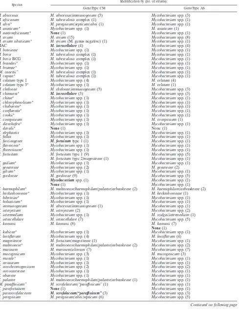
TABLE 1. Results of strains tested with GenoType CM and ASa
Species
Identification by (no. of strains):
GenoType CM GenoType AS
M. abscessus M. abscessus/immunogenum(5) Mycobacteriumspp. (3) M. africanum M. tuberculosiscomplex (1) Mycobacteriumspp. (1) M. alvei* M. peregrinum/septicum/alvei(1) Mycobacteriumspp. (1) M. asiaticum* Mycobacteriumspp. (1) M. asiaticum(1)
M. austroafricanum* None(1) Mycobacteriumspp. (1)
M. avium M. avium(15) Mycobacteriumspp. (8)
M. avium silvaticum* M. avium(M. genus negative) (1) Mycobacteriumspp. (1)
MAC M. intracellulare(4) Mycobacteriumspp. (4)
M. botniense Mycobacteriumspp. (1) Mycobacteriumspp. (1) M. bovis M. tuberculosiscomplex (1) Mycobacteriumspp. (1) M. bovisBCG M. tuberculosiscomplex (1) Mycobacteriumspp. (1) M. branderi* Mycobacteriumspp. (1) Mycobacteriumspp. (1)
M. brumae* Mycobacteriumspp. (1) Mycobacteriumspp. (1)
“M. canettii” M. tuberculosiscomplex (1) Mycobacteriumspp. (1) M. caprae* M. tuberculosiscomplex (1) Mycobacteriumspp. (1) M. celatumtype 1 Mycobacteriumspp. (4) M. celatum(4) M. celatumtype 3* Mycobacteriumspp. (1) M. celatum(1) M. chelonae M. chelonae/immunogenum(5) Mycobacteriumspp. (3) M. chimaera* M. intracellulare(3) Mycobacteriumspp. (3)
M. chitae* Mycobacteriumspp. (1) Mycobacteriumspp. (1)
M. chlorophenolicum* Mycobacteriumspp. (1) Mycobacteriumspp. (1) M. chubuense* Mycobacteriumspp. (1) Mycobacteriumspp. (1) M. confluentis* Mycobacteriumspp. (1) Mycobacteriumspp. (1)
M. cookii* Mycobacteriumspp. (1) Mycobacteriumspp. (1)
M. conspicuum Mycobacteriumspp. (1) M. conspicuum(1)
M. diernhoferi* Mycobacteriumspp. (1) Mycobacteriumspp. (1)
M. duvalii* None(1) None (1)
M. elephantis Mycobacteriumspp. (1) Mycobacteriumspp. (1)
M. fallax Mycobacteriumspp. (1) Mycobacteriumspp. (1)
M. farcinogenes M. fortuitumtype 1 (1) Mycobacteriumspp. (1) M. flavescens* Mycobacteriumspp. (1) Mycobacteriumspp. (1) M. florentinum* Mycobacteriumspp. (1) Mycobacteriumspp. (1) M. fortuitum M. fortuitumtype 1 (9) Mycobacteriumspp. (5) M. fortuitumtype 2/mageritense(1) Mycobacteriumspp. (1)
M. gadium* Mycobacteriumspp. (1) Mycobacteriumspp. (1)
M. genavense Mycobacteriumspp. (2) M. genavense(2)
M. gilvum* Mycobacteriumspp. (1) Mycobacteriumspp. (1)
M. gordonae M. gordonae(9) Mycobacteriumspp. (5)
Mycobacteriumspp. (1) Mycobacteriumspp. (1)
None(1) Mycobacteriumspp. (1)
M. haemophilum* M. malmoense/haemophilum/palustre/nebraskense(2) M. haemophilum/nebraskense(2) M. heckeshornense* Mycobacteriumspp. (1) M. heckeshornense(1)
M. hiberniae* Mycobacteriumspp. (1) Mycobacteriumspp. (1) M. holsaticum* Mycobacteriumspp. (1) Mycobacteriumspp. (1) M. immunogenum M. abscessus/immunogenum(1) Mycobacteriumspp. (1)
M. interjectum M. interjectum(2) Mycobacteriumspp. (2)
M. intermedium Mycobacteriumspp. (1) M. szulgai/intermedium(1) M. intracellulare M. intracellulare(7) Mycobacteriumspp. (5)
M. kansasii M. kansasii(8) M. kansasii(7)
None(1)
M. kubicae* Mycobacteriumspp. (1) Mycobacteriumspp. (1) M. lentiflavum Mycobacteriumspp. (4) M. lentiflavum(4) M. mageritense M. fortuitum/mageritense(1) Mycobacteriumspp. (1) M. malmoense* M. malmoense/haemophilum/palustre/nebraskense(2) Mycobacteriumspp. (2)
M. marinum M. marinum/ulcerans(7) Mycobacteriumspp. (7)
M. mucogenicum Mycobacteriumspp. (3) M. mucogenicum(3)
M. murale* Mycobacteriumspp. (1) Mycobacteriumspp. (1)
M. neoaurum Mycobacteriumspp. (1) Mycobacteriumspp. (1)
M. nonchromogenicum Mycobacteriumspp. (2) Mycobacteriumspp. (2) M. novocastrense Mycobacteriumspp. (1) Mycobacteriumspp. (1)
M. obuense Mycobacteriumspp. (1) Mycobacteriumspp. (1)
M. palustre M. malmoense/haemophilum/palustre/nebraskense(1) Mycobacteriumspp. (1) “M. paraffinicum” M. scrofulaceum/“paraffinicum”(1) Mycobacteriumspp. (1)
M. parafortuitum None(1) Mycobacteriumspp. (1)
M. parascrofulaceum M. scrofulaceum/“paraffinicum”(3) Mycobacteriumspp. (3) M. peregrinum M. peregrinum/alvei/septicum(6) Mycobacteriumspp. (5)
Continued on following page
on May 16, 2020 by guest
http://jcm.asm.org/
One hundred thirty-eight strains belonging to 33 of the 37 taxa covered by the GenoType were correctly identified. For 97 of them, the identification was unambiguous, while for 41 it fell within a group including two or more alternatives. All but one of the nonmycobacterial strains were nonreactive with the ge-nus- and species-specific probe. The strain of Tsukamurella positive by the genus-specific probe also presented the pattern ofMycobacterium peregrinumin the CM strip. The nonmyco-bacterial strains, belonging to species more closely related to theMycobacteriumgenus, hybridized as expected with the uni-versal probe present in both strips.
With the CM strip, misidentification occurred in 12 cases (four strains belonging to theMycobacterium avium complex [MAC], threeMycobacterium chimaerastrains, three Mycobac-terium parascrofulaceumstrains, and one strain each of Myco-bacterium farcinogenes, Mycobacterium porcinum, and Myco-bacterium senegalense), while identification was missed in four cases, in which the hybridization pattern did not match any of the expected ones. The sensitivity and specificity were 97.9% and 92.4%, respectively (Table 2).
With the AS strip, there was one case of a non-sense pattern
for one strain ofM. kansasii (out of eight) and one case of missed identification at the genus level (Mycobacterium duva-lii). Sensitivity was 99.3%, and specificity was 99.4% (Table 2).
DISCUSSION
The GenoType Mycobacterium assay is able to identify up to 37Mycobacteriumspecies, using two strips. The technology is based on reverse-phase hybridization aimed at revealing an amplification product of a proprietary fragment of approxi-mately 230 bp within the 23S rRNA gene.
Hybridization patterns may be characterized either by hy-bridization of a single probe or by hyhy-bridization of multiple probes simultaneously (Fig. 1), thus suggesting the presence of more polymorphic traits scattered within the target.
The system represents the evolution of a previous kit, based on a single strip, which practically overlaps the present CM. The most evident improvement is represented by the introduc-tion of the AS strip, but another important aspect of the new system is the introduction of the genusMycobacterium-specific probe, which has been added to the already present universal probe. The combined results of such probes allow us to distin-guish, among the strains not recognized at the species level, the ones belonging to the genusMycobacteriumand those belong-ing to genetically related genera (Nocardia, Gordonia, etc.) from the other species.
Assayed with a large number of species, the system proved very reliable. The values of sensitivity and specificity, which turned out to be good with CM (98 and 92%), were excellent with AS (99% for both).
A limit of the system is represented, however, by patterns shared by more than one species (up to four; Fig. 1). In addi-TABLE 2. Sensitivity and specificity of GenoType assay
Assay % Sensitivity % Specificity
Genus-specific probe
CM strip 98.9 88.9
AS strip 99.4 100
Species-specific probes
CM strip 97.9 92.4
[image:4.585.46.542.74.334.2]AS strip 99.3 99.4
TABLE 1—Continued
Species
Identification by (no. of strains):
GenoType CM GenoType AS
M. phlei* Mycobacteriumspp. (1) M. phlei(1)
M. porcinum* M. fortuitumtype 1 (1) Mycobacteriumspp. (1) M. poriferae* Mycobacteriumspp. (1) Mycobacteriumspp. (1) M. rhodesiae* Mycobacteriumspp. (2) Mycobacteriumspp. (2) M. scrofulaceum M. scrofulaceum/“paraffinicum”(2) Mycobacteriumspp. (2) M. senegalense M. fortuitumtype 2/mageritense(1) Mycobacteriumspp. (1)
M. shimoidei Mycobacteriumspp. (1) M. shimoidei(1)
M. simiae Mycobacteriumspp. (5) M. simiae(5)
M. smegmatis Mycobacteriumspp. (2) M. smegmatis(2)
M. szulgai Mycobacteriumspp. (3) M. szulgai/intermedium(3)
M. terrae Mycobacteriumspp. (2) Mycobacteriumspp. (2)
M. thermoresistibile Mycobacteriumspp. (1) Mycobacteriumspp. (1) M. tokaiense Mycobacteriumspp. (1) Mycobacteriumspp. (1)
M. triplex Mycobacteriumspp. (1) Mycobacteriumspp. (1)
M. triviale Mycobacteriumspp. (1) Mycobacteriumspp. (1)
M. tuberculosis M. tuberculosiscomplex (5) Mycobacteriumspp. (5)
M. tusciae Mycobacteriumspp. (1) Mycobacteriumspp. (1)
M. xenopi M. xenopi(10) Mycobacteriumspp. (6)
None(1)
Tsukamurellaspp. M. peregrinum/septicum/alvei(1) Organism with high guanidine-cytosine content (1)
Legionella pneumophila None (1) None (1)
Rhodococcus equi Organism with high guanidine-cytosine content (2) Organism with high guanidine-cytosine content (2) Gordoniaspp. Organism with high guanidine-cytosine content (2) Organism with high guanidine-cytosine content (2) Nocardia asteroides Organism with high guanidine-cytosine content (3) Organism with high guanidine-cytosine content (3)
a
Discrepant results are in boldface; asterisked species include one reference strain.
on May 16, 2020 by guest
http://jcm.asm.org/
[image:4.585.42.284.639.725.2]tion to the ones declared by the manufacturer (seven on the CM strip and two on the AS strip), several ambiguous patterns, almost all concerning the CM strip, emerged from our inves-tigation. In particular, the following species turned out not to be distinguishable:M. parascrofulaceumfromM. scrofulaceum; M. chimaeraand several intermediate MAC spp. from Myco-bacterium intracellulare; andM. farcinogenes,M. porcinum, and M. senegalensefromM. fortuitum.The differentiation of slowly growing species, hybridizing with the same probe, may be easily achieved by investigating few simple phenotypic features (Ta-ble 3), whereas it seems to be more pro(Ta-blematic for rapid growers, which may need definitive speciation by genetic se-quencing.
Published evaluations of GenoType are available only for the previous version, overlapping the present CM assay. The relevant specificity and sensitivity results are substantially in agreement with our results (9, 13, 14). The use of a larger panel, however, allowed us to add further information, such as the misidentification of species not tested before (M. farcino-genes,M. chimaera,M. porcinum, and M. senegalense). Differ-ently from others, we did not detect any strain that could not be amplified, while, in our hands, a few strains failed to hy-bridize with the genus-specific probe or produced a pattern not overlapping any of those provided by the system. We are, however, surprised by an aspect which had never been pointed out before (5, 10, 17): the identification, either asM. aviumor asM. intracellulare, of all the strains belonging to the MAC, including the ones genetically distinct from bothM. aviumand M. intracellulare.
The only other commercial method with characteristics sim-ilar to GenoType is LiPA (16). In addition to the 18 taxa identified by both systems, the combination of CM and AS is suitable for identifying 15 further species. On the other hand, with LiPA, different from GenoType, all but one the hybrid-ization patterns were unique to single species. GenoType showed a better sensitivity in identifying the speciesM. fortui-tum: all 10 strains tested were in fact correctly identified, while two of them had been missed by LiPA (E. Tortoli, unpublished data). Some limits of specificity of the M. fortuitum probe (cross-reactivity with M. senegalenseand M. mageritense), re-ported for LiPA (16), were also present in GenoType.
One of the unique features of GenoType is the differentia-tion of two types within the speciesM. fortuitum. No clinical difference related to such variants is known so far, and the distinction appears at present to have only epidemiological value.
We would propose some suggestions in order to improve the GenoType system. The inclusion in the CM strip of a new line probe, combining all the specificities present in AS, would allow us to target the use of the AS strip only for the strains it is able to identify. Moreover, the development of new, more specific, probes for M. avium and M. intracellulare and the combination in a new line probe (targeting the whole MAC) of the presentM. aviumandM. intracellulare probes would sub-stantially increase the reliability of identification of the strains included in this complex.
In conclusion, the new GenoType assay appears to be reli-able and suitreli-able to identify almost all of the most frequently isolated mycobacteria (apart from Mycobacterium terrae and Mycobacterium nonchromogenicum), thus reducing the number of strains to be submitted to reference centers. The high num-ber of overlapping patterns in the CM strip may be a problem. It should be noted, however, that most of them concern rarely detected species.
REFERENCES
1.Alcaide, F., I. Richter, C. Bernasconi, B. Springer, C. Hagenau, R.
Schulze-Ro¨bbecke, E. Tortoli, R. Martı´n, E. Bo¨ttger, and A. Telenti.1997.
Hetero-geneity and clonality among isolates ofMycobacterium kansasii: implications for epidemiological and pathogenicity studies. J. Clin. Microbiol.35:1959– 1964.
2.Butler, W. R., and J. O. Kilburn.1988. Identification of major slowly growing
pathogenic mycobacteria andMycobacterium gordonaeby high-performance liquid chromatography of their mycolic acids. J. Clin. Microbiol.26:50–53.
3.Cage, G. D.1992. High-performance liquid chromatography patterns of
Mycobacterium gordonaemycolic acids. J. Clin. Microbiol.30:2402–2407.
4.Cloud, J. L., H. Neal, R. Rosenberry, C. Y. Turenne, M. Jama, D. R. Hillyard,
and K. C. Carroll.2002. Identification ofMycobacteriumspp. by using a
commercial 16S ribosomal DNA sequencing kit and additional sequencing libraries. J. Clin. Microbiol.40:400–406.
5.Frothingham, R., and K. H. Wilson.1993. Sequence-based differentiation of
strains in theMycobacterium aviumcomplex. J. Bacteriol.175:2818–2825.
6.Gonzales, R., and B. A. Hanna.1987. Evaluation of Gen-Probe DNA
hy-bridization system for the identification ofMycobacterium tuberculosisand
Mycobacterium avium-intracellulare. Diagn. Microbiol. Infect. Dis.8:69–77.
7.Kirschner, P., and E. C. Bo¨ttger.1992. Microheterogeneity within rRNA of
Mycobacterium gordonae. J. Clin. Microbiol.30:1049–1050. (Letter.)
[image:5.585.40.539.76.248.2]8.Lambert, M. A., C. W. Moss, V.A. Silcox, and R. C. Good.1986. Analysis of
TABLE 3. Distinctive features suitable to differentiate slowly growing species which share an identical GenoType pattern
Species Characteristic(s)
M. szulgai...No growth at 41°C M. intermedium...Growth at 41°C
M. haemophilum...No growth on LJa
M. nebraskense...Growth on LJ
M. scrofulaceum...Tween 80 negative “M. paraffinicum”...Tween 80 positive
M. malmoense...Growth on LJ, nonchromogenic, no growth at 41°C M. haemophilum...No growth on LJ, nonchromogenic, no growth at 41°C M. palustre...Growth on LJ, scotochromogenic, growth at 41°C M. nebraskense...Growth on LJ, scotochromogenic, no growth at 41°C
M. ulcerans...Identified by AS M. marinum...Not identified by AS
aLJ, Lowenstein-Jensen medium.
on May 16, 2020 by guest
http://jcm.asm.org/
mycolic acid cleavage products and cellular fatty acids ofMycobacterium
species by capillary gas chromatography. J. Clin. Microbiol.23:731–736.
9.Ma¨kinen, J., A. Sarkola, M. Marjama¨ki, M. K. Viljanen, and H. Soini.2002.
Evaluation of GenoType and LiPA MYCOBACTERIA assays for identifi-cation of Finnish mycobacterial isolates. J. Clin. Microbiol.40:3478–3481.
10.Mijs, W., P. de Haas, R. Rossau, T. van der Laan, L. Rigouts, F. Portaels,
and D. van Soolingen.2002. Molecular evidence to support a proposal to
reserve the designationMycobacterium avium subsp. avium for bird-type isolates and ’M. aviumsubsp.hominissuis’ for the human/porcine type ofM. avium. Int. J. Syst. Evol. Microbiol.52:1505–1518.
11.Minnikin, D. E., L. Al-Shamaony, and M. Goodfellow.1975. Differentiation
ofMycobacterium,Nocardia, and related taxa by thin-layer chromatographic analysis of whole-organism methanolysates. J. Gen. Microbiol.88:200–204.
12.Picardeau, M., G. Prod’Hom, L. Raskine, M. P. LePennec, and V. Vincent.
1997. Genotypic characterization of five subspecies ofMycobacterium kan-sasii. J. Clin. Microbiol.35:25–32.
13.Ruiz, P., J. Gutierrez, F. J. Zerolo, and M. Casal.2002. GenoType
Myco-bacterium assay for identification of mycobacterial species isolated from human clinical samples by using liquid medium. J. Clin. Microbiol.40:3076– 3078.
14.Sarkola, A., J. Makinen, M. Marjamaki, H. J. Marttila, M. K. Viljanen, and
H. Soini.2004. Prospective evaluation of the GenoType assay for routine
identification of mycobacteria. Eur. J. Clin. Microbiol. Infect. Dis.23:642– 645.
15.Tortoli, E.2003. Impact of genotypic studies on mycobacterial taxonomy: the
new mycobacteria of the 1990s. Clin. Microbiol. Rev.16:319–354.
16.Tortoli, E., A. Mariottini, and G. Mazzarelli.2003. Evaluation of
INNO-LiPA MYCOBACTERIA v2: improved reverse hybridization multiple DNA probe assay for mycobacterial identification. J. Clin. Microbiol.41:4418– 4420.
17.Tortoli, E., L. Rindi, M. J. Garcia, P. Chiaradonna, R. Dei, C. Garzelli, R. M.
Kroppenstedt, N. Lari, R. Mattei, A. Mariottini, G. Mazzarelli, M. I.
Mur-cia, A. Nanetti, P. Piccoli, and C. Scarparo.2004. Proposal to elevate the
genetic variant MAC-A, included in theMycobacterium aviumcomplex, to species rank asMycobacterium chimaerasp. nov. Int. J. Syst. Evol. Microbiol.
54:1277–1285.
18.Vincent, V., B. A. Brown-Elliott, K. C. Jost, Jr., and R. J. Wallace, Jr.2003.
Mycobacterium: phenotypic and genotypic identification, p. 560–584.InP. R. Murray, E. J. Baron, J. H. Jorgensen, M. A. Pfaller, and R. H. Yolken (ed.), Manual of clinical microbiology, 8th ed., vol. 1. ASM Press, Washington, D.C.