2017 International Conference on Computer, Electronics and Communication Engineering (CECE 2017) ISBN: 978-1-60595-476-9
A Method for Quickly and Exactly Extracting Hepatic Vessel
Yue-jing QIAN
Collage of Information and communications, Zhejiang Industry & Trade Vocational College, Wenzhou, Zhejiang, 325003, China
Keywords: Hessian matrix, Region growing, Blood vessel segmentation.
Abstract. It is of vital importance that providing detailed and accurate information about hepatic vessel for liver surgery planning, such as pre-operative planning of living donor liver transplantation (LDLT). Due to the different blood flow rate of intra-hepatic vascular systems and the restrictions of CT scan, it is common that hepatic vessels are filled with contrast medium during the scan and in high intensity in the hepatic venous phase images. As a result, the hepatic vessel segmentation result obtained from the hepatic venous phase images is always contaminated which makes accurate hepatic vessel modeling difficult. In this paper, we proposed a method for quick and accurate hepatic vessel extraction. Firstly, the Hessian method is used to enhance the vessel areas. Then, a region growing vessel segmentation algorithm can obtain the hepatic vessel. Experiments show that the proposed method can segment the liver vessels accurately with a certain practicability and operability.
Introduction
Liver is the largest parenchyma in human body and part of the digestive system that performs many tasks including metabolic activities, storage and filtration of blood, excretory function, and repelling wastes. Liver cancer is considered as one of the major diseases of liver, and it is one of the main death factors world-wide. To remove cancerous tumors with the least damage to liver vessels, which is the most frequent treatment for this disease, a physician has to know the approximate positions of the tumors and their distances to the major vessels [1]. There are four vascular structures in liver: hepatic artery, hepatic vein, hepatic portal vein, and bile ducts. Imaging modalities are used as non-invasive tools for diagnosis of liver diseases, treatment planning, evaluation of treatment and training. CT-scanners are used more frequently than other modalities for liver imaging [2]. Manual delineation of the vessels in hundreds of slices is a time-consuming and very difficult task. Due to the significant effect of visualization and the quantitative analysis of hepatic vessels [3], several researches have been devoted to develop enhancement filters for tubular structures and medial-axes.
Usually, image enhancement processing is needed before doing vessel segmentation. Image enhancement is one of the most commonly used in the field of image processing technology. According to the processing size of image region, image enhancement technologies can be divided into global enhancement and local enhancement. Global enhancement is to enhance the overall information of the image, however, this might easily lead to the local details missing, and so is not suitable for the segmentation of liver vessels processing. The traditional local enhancement methods include local histogram equalization, the noise removal method based on local statistical characteristics, adaptive histogram equalization with limited contrast], and so on. As the gray level of vessels in the liver area is relatively close the gray level of vessels surrounding tissues, the traditional local enhancement methods cannot obtain satisfactory enhancement effect. Tomohiro Takagi [6] proposed the mathematical method to construct fuzzy model, m. Wilscy [7] used the model for color image sharpening enhancing and gained good results. Based on this, this paper proposes a local enhancement algorithm based on fuzzy method for liver image enhancement processing.
In this paper, a new filter based on the Hessian is designed for enhancement of liver vessel. The proposed filter is applied to synthetic and clinical images of 2D/3D dimensions. Then we discuss how to select the seed point automatically. The most last step, using region growing algorithm to segment vessel. In “Discussion”, the results are discussed, and “Conclusion and future works” concludes the paper and describes future works.
Vascular Enhancement Based on the Hessian Matrix Eigenvalue
Hessian method is a kind of method which uses the high order differential direction to extract image features. The direction and measures of the tubular structure are gained by the analysis of characteristics of 3d gray-scale image Hessian matrix H:
xx xy xz
xy yy yz
xz yz zz
I I I
H I I I
I I I
(1)
We can suppose that 1, 2, 3 are the three eigenvalues of H with corresponding eigenvectors
1
, 2, 3, and their absolute values meet the conditions: 1 2 3 . For a tubular structure,
1
vrepresents the direction with the minimum amount of gray level change, which is the direction of blood vessels. As tubular structures with different diameters only show the tube character under the scale of the specific scale, so it’s necessary to calculate the vascular vector field under the framework of multi-scale to extract the blood vessels with different diameter. Multi-scale vector field is first to calculate the eigenvalue and eigenvector of a Hessian matrix under various scales, and then choose the largest measure as the final vector. We assume original image I, scale factor σwith a range of [a1, a2], iteration step-size Step, and according to the papers, we can set the range of σ as [1,10], and set the iteration step-size as 2.The basic steps of blood vessels enhance are as follows:
Step1: Input image (gray image), and then get the pixel matrix I. Step2: For each pixel Ix y z, , in I , repeat the steps from Step3to Step10. Step3: Initialize the space scaleσ=a1,and the enhance factor zmax 0 Step4: If the space scale σ meets the stop condition, do the step 9
Step5: Calculate the second order differential convolution of the element Ix y z, , and Gaussian function.
2 3
2 2 2
2 2 2
0 0 0
( , , , )
1 exp exp 1 exp
2 2 2
i a b
or
I x y z R R s
c
el se (2)
1 2
2
3 2 3
, ,
a b j
j
R R S
(3)
max max( max, ( , ))i
z Z I V
(4) According to the papers, we set the values of , as 0.5, and set C as half of the biggest Hessian matrix norm.
Step8: Change the value of σ with a specify step and return to step 4 to do iteration.
Step9: When the iteration ends, the enhance factor which is the biggest enhance filter output value
max
z and the corresponding eigenvalues 1, 2, 3 and eigenvectors v1, v2, v3of Hessian matrix H. Step10: Judge whether the pixel is belonging to blood vessel based on the gained value ofzmax. That is the bigger value ofzmax, the more possible of the pixel being a part of blood vessel.
[image:3.612.92.519.67.207.2]The characteristics of Hessian matrix eigenvalues are showing in the following Table 1.
Table 1. The Hessian matrix eigenvalues characteristics.
2D 3D
Direction of the pattern
λ1 λ2 λ1 λ2 λ3
N N N N N noise image
L L H- The disk structure(bright)
L L H+ The disk structure(dark)
L H- L H- H- Tubular structure(bright)
L H+ L H+ H+ Tubular structure(dark)
H- H- H- H- H- Globular structure(bright)
H+ H+ H+ H+ H+ Globular structure(dark)
annotation: H is high, L is low, N is noise, +/- is the characteristic value of +/-
Vascular Segmentation Based on Region Growing Algorithm
The proposed algorithm adaptively builds and segments successive local cubes, one by one. And, for the segmentation in each local cube, a region growing technique is adopted. First, to determine the initial local cube, the vessel area is selected around the user-given seed point at the slice, by using a simple threshold-based method. Then, the center of the initial local cube is placed at the user-given point and the length of every edge is set to double the diameter of the selected vessel area. At each procedure, after a local cube of the proper size is determined, vessel segmentation and branch detection are performed in it. Then, the positions of next local cubes are estimated based on connected component labeling, which will be described in detail in the next subsection. Here, each connected component is regarded as a seed area of each next local cube. Among the detected next local cubes, one local cube is chosen as a successor and the others are put to the branch queue for later processing. If no next local cube is found in the current one, the processing for the current branch stops and the processing for a new branch begins by popping a local cube from the branch queue. The whole procedure finishes when no more local cubes remain in the branch queue.
approximated by connecting the center positions of local cubes, and can be used in various applications, such as automatic flyway generation in a virtual angioscopy system.
The Simulation Results
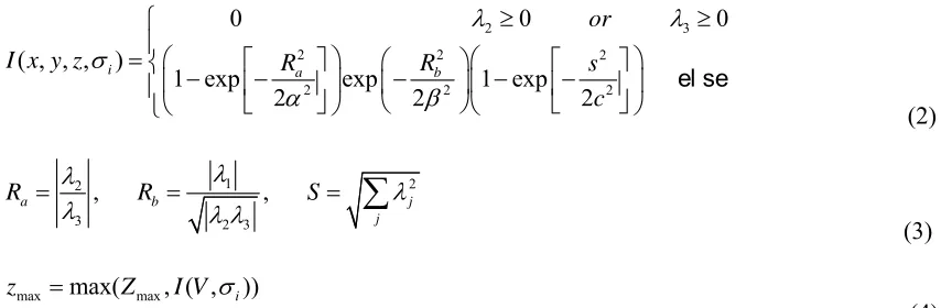
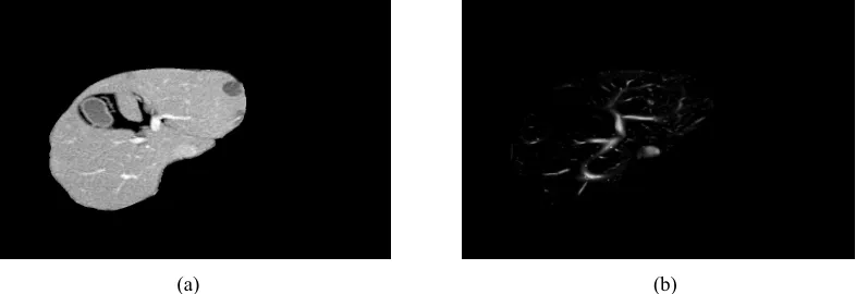
Experimental hardware equipment is Intel dual-core processor 3.2 G and 8GB memory. The software and its version is matlab2014. Experiments are conducted to select one of the first image contrast, and then through the 3D imaging software to show the blood vessels segmentation effect. Figure 1.a is the original liver image, Figure 1.b is Hessian enhancement liver images, after comparing two images, we can find that the liver vascular contrast with other regions in the enhanced image. Figure 2.a is the finally 3D reconstruction result of vessel structure, Figure 2.b is the 3D reconstruction vessel structure in the liver.
(a) (b)
Figure 1. The original liver image and the fuzzy enhancement liver image.
[image:4.612.110.503.229.364.2]
(a) (b)
Figure 2. The 3D reconstruction result of vessel structure.
Conclusion
Local enhancement algorithm based on Hessian matrix eigenvalue, the area can be used to extract blood vessels. Then we propose a vessel segmentation algorithm based on local cube tracking. The algorithm segments the vessel in the subsequent local cubes iteratively, starting from the initial local cube determined by a user-selected point. Thereby, it provides prospective results even for a long vessel structure with slowly varying intensity. Contrary to existing algorithms, the proposed algorithm
Acknowledgement
References
[1] Marieb E.N., Hoehn K.N. Human Anatomy and Physiology [J]. Benjamin/cummings Pub Co, 2015, 3(2): 82-86.
[2] Lee, Ming J. Grant's atlas of anatomy [M]. Williams & Wilkins, 1991.
[3] Barnes H.A. Segmental anatomy of the liver: poor correlation with CT. [J]. Radiology, 1998, 206(1): 151-6.
[4] Su Zhang, Xuesong Lu, Yuanyuan Shen, et al. A new approach for automatic segmentation of LSCM blood vessel images of time sequence based on region growing [J]. IEEE/ICME International Conference on Complex Medical Engineering. 2007, 689-693.
[5] Fetita C., Lucidarme O. CT hepatic venography: 3D vascular segmentation for preoperative evaluation [C]//8th International Conference on Medical Image Computing and Computer-Assisted Intervention, 2005, 3750: 830-837.
[6] R. Adams and L. Bischof, “Seeded region growing,” IEEE Transactions on Pattern Analysis and Machine Intelligence, vol. 16, no. 6, pp. 641–647, 1994.
[7] Preetha, “Image segmentation using seeded region growing,” in Proceedings of the International Conference on Computing, Electronics and Electrical Technologies (ICCEET ’12), pp. 576–583, March 2012.
[8] Takagi, T., Sugeno, M.: Fuzzy identification of systems and its applications to modeling and control. IEEE Trans. Syst., Man, Cybern. SMC-15(1), 116–132 (1985).