Fc-fusion mimetics
Hanieh Khalili
1,2, Peng T Khaw
2and Steve Brocchini
1,21
UCL School of Pharmacy, University College London, 29-39 Brunswick
Square, London WC1N 1AX, UK
2
NIHR Biomedical Research Centre, Moorfields Eye Hospital and UCL Institute
Abstract
To achieve effective bivalency and high affinity, the two Fabs in an IgG antibody are mobile and are linked together as if each Fab (or protein) is bound at the end of linear molecule (Figure 1). FpFs 1 are IgG antibody mimetics (Figure 1) designed to have enhanced stability and binding properties compared to IgGs. They are prepared from PEG-di(mono-sulfone) 3 and two antibody fragments (Fabs).1 Fc-based fusion proteins2 (Figure 1) are also capable of exploiting the therapeutic advantages of bivalency that are displayed by IgGs. Several Fc-fusion proteins are registered for clinical use3 and they will continue to offer considerable clinical potential because of Fc recycling, but they can be difficult to produce during early preclinical research and to scale for production.4 Fc-fusion proteins are also often prone to aggregation during downstream processing5 and have similar stability limitations as IgGs. There are therapeutic applications where the Fc is not needed or can cause problems.6 One area of interest is the use of antibody based medicines in organ specific applications such as the eye. In such cases, Fc recycling does not occur and effector function can be deleterious, especially in the treatment of inflammatory conditions. Improved stability is important to formulate more concentrated solutions to decrease the frequency of dose administration and improved binding properties such as slower dissociation rates are important for organ specific targeting. In an effort to further explore the potential of antibody-based mimetics that are made using the PEG-di(mono-sulfone) 3 (Scheme 1A, Figure 1) we describe an Fc-fusion mimetic that we call RpR 2, for receptor binding region-PEG-receptor binding region.
ether bonds in a rebridged disulfide are more stable than the original disulfide bond. To make the desired RpR 2 we first had to obtain the VEGFR1-VEGFR2 fragment 4 by proteolytic digestion of aflibercept to remove the Fc domain (Scheme 1B).
It was first confirmed that aflibercept migrated to an approximate molecular weight of about 115 kDa by SDS PAGE (Figure 2, lane 1). Aflibercept was then treated with dithiothreitol (DTT) to reduce the accessible disulfides thought to exist in an hinge like region between the binding domain (VEGFR1-VEGFR2) and the Fc domain. A broad band appeared at ~55-60 kDa by SDS-PAGE (Figure 2, lane 2). We believe this band corresponds to the monomeric VEGFR1-VEGFR2-Fc 5 (Scheme 1B). Glycosylation is usually somewhat heterogeneous in therapeutic proteins, so we inferred that the broadness of the band at ~55-60 kDa was due to glycosylation heterogeneity. DDT was then removed using a PD-10 column and the reduced aflibercept solution was incubated with Ellman’s reagent which indicated the presence of 4 accessible cysteine thiols in aflibercept (Figure 1S, Table 1S, ESI). This suggested that there are 2 cysteines in each VEGFR1-VEGFR2-Fc 5 monomer which can form two disulfides in aflibercept analogous to what is found in the hinge region of IgG antibodies. Hence it was thought possible that an RpR 2 derived from aflibercept could be prepared using the PEG-di(mono-sulfone) 3. If only one cysteine had been present in the VEGFR1-VEGFR2-Fc 5 monomer, there are stable, mono-thiol conjugation linkers available9 that would have been utilised in a bifunctional reagent analogous to PEG-di(mono-sulfone) 3.
Proteolytic digestion of aflibercept was then examined in an effort to obtain the monomeric VEGFR1-VEGFR2 fragment 4 (Scheme 1B). It was necessary that the cleavage should in aflibercept should occur to give the monomeric VEGFR1-VEGFR2 fragment 4 to include the cysteines that form the accessible disulfides in aflibercept. Preliminary digestion studies of aflibercept using immobilised papain yielded only difficult to characterise small peptide fragments. We had previously used papain to digest IgGs to obtain Fabs to make FpFs,1, 10 but recognised that proteolytic digestion of different antibody subclasses and motifs can be difficult to control.11
Genovis). This provided a purified a non-Fc containing fragment at 60-70 kDa (Figure 2, lane 4) which was thought to be the VEGFR1-VEGFR2 dimer 6 (Scheme 1B).
Incubation of VEGFR1-VEGFR2 dimer 6 with DTT caused this fragment to disappear to give 2 lower molecular weight fragments (Figure 2, lane 5). These fragments are thought to be the desired VEGFR1-VEGFR2 monomer 4 (Scheme 1). Two bands are often observed after reduction of Fabs that are obtained by proteolytic digestion. This is often thought to be due to miscleavage reactions during proteolysis. This may be exacerbated with aflibercept due to structural differences with IgGs and aflibercept glycosylation. There are five N-glycosylation sites on each monomeric VEGFR1-VEGFR2-Fc fragment 5 which may be partially or completely glycosylated. There may also be additional heterogeneity caused by differences in saccharide structure in the glycosylated protein.
To prepare the RpR 2, the VEGFR1-VEGFR2 dimer 6 was first incubated with DTT for 30 minutes to give the VEGFR1-VEGFR2 monomer 4 (Scheme 1B). The reaction mixture was carefully eluted over a PD-10 column to remove the DTT while avoiding disulfide reformation, and then the PEG di(mono-sulfone) reagent 3 (derived from a 10 kDa PEG precursor) was added to the solution of the monomeric VEGFR1 -VEGFR24. Incubation of the reaction mixture for 3 h (Figure 2S, lane 1, ESI) was then followed by purification by size exclusion chromatography (Figure 2S, lanes 2-10, ESI) to give the purified RpR 2 which appeared in a band at approximately 70 kDa (Figure 2, lanes 6 and 7). Two detection dyes were used, first coomassie blue to detect protein (lane 6) and then barium iodide to detect the PEG (lane 7) being conjugated to the protein. Starting from 0.8 mg (in 1.0 mL) of VEGFR1-VEGFR2 dimer 6, approximately 0.16 mg (in 0.5 mL) of RpR 2 was obtained (~ 20 % yield).
At 25˚C the purified RpR 2 displayed a solution size of 10.7 ± 0.5 nm (Pd, 0.7 ± 0.1 nm), which is similar to the starting aflibercept (10.2 ± 0.7 nm; Pd, 0.6 ± 0.1 nm). The FpF antibody mimetics 1 were also a similar solution size to the corresponding IgG.1 This is in stark contrast to when PEG is conjugated only at one terminus to a single protein where the solution size of a PEG-protein conjugate is dominated by the random coil nature of PEG.13 When only one terminus of PEG is conjugated to a protein, the other PEG terminus has considerable freedom to allow the PEG to maintain a large solution structure.
nm) which is similar to both aflibercept and RpR 2. Interestingly, when the VEGFR1 -VEGFR2 fragment 6 was treated with DTT and the cysteine thiols were blocked with iodoacetamide, the cysteine thiol-capped monomeric VEGFR1-VEGFR2 fragment 7 (Scheme 1B) displayed a solution size of 7.2 ± 0.4 nm (Pd, 0.7 ± 0.1 nm). Although the dimer 6 is twice the molecular weight of the monomer 7, its solution size is only about 40% larger suggesting that there is intramolecular association of the VEGFR1 -VEGFR2 domains within the dimer 6.
The binding properties of the RpR 2 and aflibercept were then evaluated by surface plasmon resonance (Biacore) to determine the affinity (KD), and the rate constants of association (ka) and dissociation (kd) (Table 1). Vascular endothelial growth factor-165 (VEGF165), which is a ligand for aflibercept, was immobilised at a density to minimise or prevent rebinding events (91 RU).1, 15 The dissociation rate (kd) for the RpR 2 was slower than what was observed with aflibercept. Interestingly, the ka appeared to be slightly faster in RpR 2 compared to aflibercept. This is in contrast to what was previously observed for anti-VEGF FpF which had a slower association rate than the precursor IgG antibody.1 However it was the decreased kd of RpR 2 that appeared to be the dominating factor to cause the improved affinity of RpR 2 compared to aflibercept (Table 1). Representative fitting curves for aflibercept and RpR 2 are shown in the ESI (Figure S3, ESI).
Exploiting reduced dissociation rates may be a viable strategy to increase efficacy by increasing the residence time and mode of action within specific tissue.16 Although the reduction in kd for FpF 1 is also slower than the parent IgG,1 there appears to be a greater relative reduction in kd for the RpR 2 compared to its parent Fc-fusion (i.e. aflibercept). During initial dissociation steps from the ligand of one of the two VEGFR1-VEGFR2 domains in the RpR 2, PEG conformational flexibility may be more efficient for rebinding than the polypeptide linking the Fc domain to the VEGFR1 -VEGFR2 domain in aflibercept. This suggests there is less flexibility in the bivalent binding moieties in the Fc-fusion protein (aflibercept) than there is in an IgG (e.g. bevacizumab).
the aflibercept disulfides to better optimise dissociation rates would be expected to make aflibercept less stable. Such an added polypeptide sequence to increase the flexibility of the VEGFR1-VEGFR2 receptor domains would invariably lack secondary structure in a similar way to the hinge region of IgG antibodies. While the hinge region in IgG antibodies provides the flexibility needed for cooperative and bivalent binding of both Fabs,17 the IgG hinge region is also vulnerable to degradation and disulfide scrambling.18 The stable conjugation imparted by PEG-di(mono-sulfone) 3 and use of a PEG scaffold provides enough flexibility of the VEGFR1-VEGFR2 binding moieties to potentially maximise both association and dissociation rates that could be important in the development of new therapeutics.
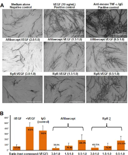
RpR 2 was then evaluated in vitro using a human umbilical vein endothelial cell (HUVEC) co-culture (Figure 3). This assay measures the migration and the formation of an anastomosing network that is characterised by tubule and junction formation during HUVEC proliferation. These processes are characteristic for angiogenesis and are often a good in vitro measurement for angiogenesis.19 RpR 2 and aflibercept were incubated with VEGF165 at different molar ratios of 3.0, 1.5, 0.5 for 2 hours at 370C prior to incubation with HUVECs. VEGF165 and anti-mouse TNF-α IgG were used for positive controls. Images were obtained after fixing HUVECs with an anti-CD31 antibody to differentiate between the endothelial tubular network and non-endothelial structures of similar apparent morphology (Figure 3A). These images suggest that both aflibercept and the RpR 2 have similar anti-angiogenic properties. Quantification of tubule (Figure S5, ESI) and junction formation (Figure 3B) (AngioSys Image Analysis Software, TCS Cellworks Ltd.) showed that the formation of these structures were similarly inhibited in a concentration dependent manner by both RpR 2 and aflibercept.
References
1. H. Khalili, A. Godwin, J. W. Choi, R. Lever, P. T. Khaw, and S. Brocchini,
Bioconjugate Chem., 2013, 24, 1870-1882.
2. (a) S. M. Chamow, T. Ryll, H. B. Lowman, and D. Farson, eds. Therapeutic Fc-Fusion Proteins. Vol. 10. 2013, Wiley Blackwell: Weinheim. 419; (b) D. M. Czajkowsky, J. Hu, Z. Shao, and R. J. Pleass, EMBO Mol. Med., 2012, 4, 1015-1028.
3. W. R. Strohl, BioDrugs 2015, 29, 215-239.
4. (a) A. B. V. Spriel, H. H. V. Ojik, and J. G. J. Winkel, Immunol. Today 2000, 21, 391-398; (b) H. R. Hoogenboom, Nat. Biotechnol., 1997, 15, 125-126.
5. (a) A. A. Cordes, C. W. Platt, J. F. Carpenter, and T. W. Randolph, J. Pharm. Sci., 2012, 101, 1400-1409; (b) M. C. Manning, D. K. Chou, B. M. Murphy, R. W. Payne, and D. S. Katayama, Pharm. Res., 2010, 27, 544-575.
6. (a) M. Kolfschoten, J. Schuurman, M. Losen, W. K. Bleeker, P. Martinez, E. Vermeulen, T. H. D. Bleker, D. Wiegman, T. Vink, L. A. Aarden, M. H. D. Baets, J. Winkel, R. C. Aalberse, and P. Parren, Science 2007, 13, 1554-1557; (b) S. Murinello, R. F. Mullins, A. J. Lotery, V. H. Perry, and J. L. Teeling, Invest. Ophthalmol. Vis. Sci., 2014, 55, 247-258; (c) A. L. Nelson, MAbs, 2010, 2, 77-83; (d) A. Chapman, P. Antoniw, M. Spitali, S. West, S. Stephens, and D. King,
Nat. Biotechnol., 1999, 17, 780-783.
7. (a) EMA (European Medicines Agency), Assessment report for aflibercept (EMA/646256/2012), European Medicines Agency Committee for Medicinal Products for Human Use, Editor. 2012. p. 83; (b) K. K. Ciombor, J. Berlin, and E. Chan, Clin. Cancer. Res., 2013, 19, 1920-1925; (c) J. Holash, S. Davis, N. Papadopoulos, S. D. Croll, L. Ho, M. Russell, P. Boland, R. Leidich, D. Hylton, E. Burova, E. Ioffe, T. Huang, C. Radziejewski, K. Bailey, J. P. Fandl, T. Daly, S. J. Wiegand, G. D. Yancopoulos, and J. S. Rudge, Proc. Natl. Acad. Sci. U S A, 2002, 99, 11393-11398.
8. (a) S. Shaunak, A. Godwin, J. W. Choi, S. Balan, E. Pedone, D. Vijayarangam, S. Heidelberger, I. Teo, M. Zloh, and S. Brocchini, Nat. Chem. Biol., 2006, 2, 312-313; (b) S. Balan, J. W. Choi , A. Godwin, I. Teo, C. M. Laborde, S. Heidelberger, M. Zloh, S. Shaunak, and S. Brocchini, Bioconjugate Chem., 2007, 18, 61-76.
9. G. Badescu, P. Bryant, J. Swierkosz, F. Khayrzad, E. Pawlisz, M. Farys, Y. Cong, M. Muroni, N. Rumpf, S. Brocchini, and A. Godwin, Bioconjugate Chem., 2014, 25, 460-469.
10. H. Khalili, A. Godwin, J. Choi, R. Lever, and S. Brocchini, Bioconjugate Chem., 2012, 23, 2262-2277.
11. (a) M. G. Mage, Methods Enzymol., 1980, 70, 142-150; (b) P. Parham, J. Immunol., 1983, 131, 2895-2902.
12. (a) B. P. Johansson, O. Shannon, and L. Bjorck, PLoS One, 2008, 3, 1-6; (b) P. Åkesson, L. Moritz, M. Truedsson, B. Christensson, and U. V. Pawel-Rammingen, Infection and Immunity 2006, 74, 497–503.
13. C. Fee, Biotechnol. Bioeng., 2007, 98, 725-731.
14. (a) F. Lo Verso and C. N. Likos, Polymer, 2008, 49, 1425-1434; (b) A. N. Semenov, J. F. Joanny, and A. R. Khokhlov, Macromolecules, 1995, 28, 1066-1075.
15. J. Yang, X. Wang, G. Fuh, L. Yu, E. Wakshull, M. Khosraviani, E. S. Day, B. Demeule, J. Liu, S. J. Shire, N. Ferrara, and S. Yadav, Mol. Pharm., 2014, 11, 3421-3430.
16. G. Vauquelin and S. J. Charlton, Br. J. Pharmacol., 2010, 161, 488-508. 17. (a) W. Paul, ed. Fundamental immunology. 2012, Lippincott Williams and
Mcpherson, Immunol. Rev. , 1998, 163, 35-43; (c) X. Wang, S. Kumar, and S. Singh, Pharm. Res., 2011, 28, 3128-3143.
18. (a) J. Vlasak and R. Ionescu, MAbs, 2011, 3, 253-263; (b) B. Yan, D. Boyd, T. Kaschak, J. Tsukuda, A. Shen, Y. Lin, S. Chung, P. Gupta, A. Kamath, A. Wong, J.-M. Vernes, G. Y. Meng, K. Totpal, G. Schaefer, G. Jiang, B. Nogal, C. Emery, M. Vanderlaan, P. Carter, R. Harris, and A. Amanullah, J. Biol. Chem., 2012, 287, 5891-5897.
Figure Legends
Figure 1. IgG and Fc fusion proteins and their respective mimetics, FpF 1 and RpR 2. Figure 2. SDS-PAGE gels of VEGFR1-VEGFR2 dimer 6 obtained by the proteolytic digestion of aflibercept and preparation of RpR 2. Novex Bis-Tris 4-12% gel stained with colloidal blue for protein and barium Iodide for PEG (lane 7). M: standard protein markers, Lane 1: aflibercept, Lane 2: aflibercept treated with DTT to give VEGF1 -VEGF2-Fc monomer 5, Lane 3: aflibercept-Ides digestion mixture, Lane 4: VEGFR1 -VEGFR2 dimer 6, Lane 5: VEGFR1-VEGFR2 monomer 4, Lanes 6,7: purified RpR 2. Figure 3. (A) Representative images that were used for AngioSys analysis to quantitate tubule formation using a HUVEC-fibroblast angiogenesis assay. The dark structures (tubules) are indicative of angiogenesis and were analysed to determine the number of junctions. (B) Number of junctions observed for medium alone, medium + VEGF, Anti-mouse TNF-a IgG + VEGF, aflibercept + VEGF and RpR + VEGF. Ratios are the amount of test compound to VEGF. VEGF was present at a fixed concentration of 10 ng/mL. Data are expressed as the mean of three individual cultures per treatment environment.
Scheme Legend
Scheme 1.(A) Preparation RpR 2 from PEG-di(mono-sulfone) 3 and two equivalents of the VEGFR1-VEGFR2 monomer 4. (B) Use of aflibercept to obtain the monomeric VEGF1-VEGF2 4 and VEGF1-VEGF2-Fc 5 fragments. Proteolytic digestion of aflibercept with the IdeS enzyme results in the cleavage of the Fc to give the VEGFR1 -VEGFR2 dimer 6 that after treatment with DTT gives the VEGFR1-VEGFR2 monomer 4 which was used to make RpR 2. The VEGFR1-VEGFR2 monomer 4 was also incubated iodoacetamide to give the thiol capped VEGFR1-VEGFR2 fragment 7 for binding studies.
Table Legend
)LJXUHV
)LJXUH
)LJXUH
6FKHPH
7DEOH
6DPSOH ND
î0V N G
îV
.'
NGNDQ0
$IOLEHUFHSW
9(*)59(*)5