• No results found

Long term persistence of donor nuclei in a Duchenne muscular dystrophy patient receiving bone marrow transplantation

N/A
N/A
Protected

Academic year: 2020

Share "Long term persistence of donor nuclei in a Duchenne muscular dystrophy patient receiving bone marrow transplantation"

Copied!
9
0
0

Loading.... (view fulltext now)

Full text

(1)

Long-term persistence of donor nuclei in a

Duchenne muscular dystrophy patient receiving

bone marrow transplantation

Emanuela Gussoni, … , Louis M. Kunkel, Kenneth Weinberg

J Clin Invest. 2002;

110(6)

:807-814.

https://doi.org/10.1172/JCI16098

.

Duchenne muscular dystrophy (DMD) is a severe progressive muscle-wasting disorder

caused by mutations in the dystrophin gene. Studies have shown that bone marrow cells

transplanted into lethally irradiated mdx mice, the mouse model of DMD, can become part of

skeletal muscle myofibers. Whether human marrow cells also have this ability is unknown.

Here we report the analysis of muscle biopsies from a DMD patient (DMD-BMT1) who

received bone marrow transplantation at age 1 year for X-linked severe combined immune

deficiency and who was diagnosed with DMD at age 12 years. Analysis of muscle biopsies

from DMD-BMT1 revealed the presence of donor nuclei within a small number of muscle

myofibers (0.5-0.9%). The majority of the myofibers produce a truncated, in-frame isoform of

dystrophin lacking exons 44 and 45 (not wild-type). The presence of bone marrow-derived

donor nuclei in the muscle of this patient documents the ability of exogenous human bone

marrow cells to fuse into skeletal muscle and persist up to 13 years after transplantation.

Article

Stem cells

Find the latest version:

(2)

Introduction

Bone marrow transplantation (BMT) is used to treat a variety of human diseases such as severe combined immune deficiency (SCID) (1, 2), Fanconi anemia (3, 4), osteogenesis imperfecta (5, 6), and inborn errors of

metabolism or macrophage function such as lysosomal storage diseases or osteopetrosis (7, 8). Adult bone mar-row (BM) contains mesenchymal stem cell progenitors (MSCs), which can give rise to osteocytes, chondrocytes, adipocytes, and skeletal muscle (9, 10). Marrow also con-tains progenitors for neural (11, 12), hepatic (13, 14), cardiac (15, 16), and epithelial cells (17), indicating the potential of BMT to repair nonhematopoietic tissues. Engraftment of donor-derived cells in nonhemato-poietic tissues has been described in human BMT recip-ients (18, 19). However, recent findings demonstrated that BM cells fuse to and subsequently adapt the phe-notype of host cells in vitro, thus questioning the trans-differentiation abilities of BM cells in vivo (20).

Duchenne muscular dystrophy (DMD), caused by mutation of the dystrophingene, is the most common and severe form of muscular dystrophy, with loss of independent ambulation occurring at approximately 10

Long-term persistence of donor

nuclei in a Duchenne muscular

dystrophy patient receiving bone

marrow transplantation

Emanuela Gussoni,

1

Richard R. Bennett,

1

Kristina R. Muskiewicz,

1

Todd Meyerrose,

2

Jan A. Nolta,

2

Irene Gilgoff,

3

James Stein,

4

Yiu-mo Chan,

5

Hart G. Lidov,

1

Carsten G. Bönnemann,

6

Arpad von Moers,

7

Glenn E. Morris,

8

Johan T. den Dunnen,

9

Jeffrey S. Chamberlain,

10

Louis M. Kunkel,

1,11

and Kenneth Weinberg

2

1Division of Genetics, Children’s Hospital, Boston, Massachusetts, USA

2Division of Research Immunology and Bone Marrow Transplantation, Department of Pediatrics,

Children’s Hospital Los Angeles, Los Angeles, California, USA

3Department of Pediatrics, Rancho Los Amigos Medical Center, Downey, California, USA 4Department of Surgery, Children’s Hospital Los Angeles, Los Angeles, California, USA 5Geisinger Hospital, Weis Center for Research, Danville, Pennsylvania, USA

6Division of Neurology, Children’s Hospital of Philadelphia, and University of Pennsylvania, Philadelphia, Pennsylvania, USA 7University Children’s Hospital, Department of Neuropediatrics, Charité, Berlin, Germany

8North East Wales Institute, Wrexham, United Kingdom

9Center for Human and Clinical Genetics, Leiden University Medical Center, Leiden, The Netherlands 10Department of Neurology, University of Washington, Seattle, Washington, USA

11The Howard Hughes Medical Institute at Children’s Hospital, Boston, Massachusetts, USA

Duchenne muscular dystrophy (DMD) is a severe progressive muscle-wasting disorder caused by muta-tions in the dystrophingene. Studies have shown that bone marrow cells transplanted into lethally irra-diated mdx mice, the mouse model of DMD, can become part of skeletal muscle myofibers. Whether human marrow cells also have this ability is unknown. Here we report the analysis of muscle biopsies from a DMD patient (DMD-BMT1) who received bone marrow transplantation at age 1 year for X-linked severe combined immune deficiency and who was diagnosed with DMD at age 12 years. Analysis of muscle biopsies from DMD-BMT1 revealed the presence of donor nuclei within a small number of muscle myofibers (0.5–0.9%). The majority of the myofibers produce a truncated, in-frame isoform of dystrophin lacking exons 44 and 45 (not wild-type). The presence of bone marrow–derived donor nuclei in the muscle of this patient documents the ability of exogenous human bone marrow cells to fuse into skeletal muscle and persist up to 13 years after transplantation.

This article was published online in advance of the print edition. The date of publication is available from the JCI website, http://www.jci.org. J. Clin. Invest.110:807–814 (2002). doi:10.1172/JCI200216098.

Received for publication June 5, 2002, and accepted in revised form July 23, 2002.

Address correspondence to:Louis Kunkel, Division of Genetics, Howard Hughes Medical Institute, Children’s Hospital, 320 Longwood Avenue, Boston, Massachusetts 02115, USA. Phone: (617) 355-7576; Fax: (617) 277-0496;

E-mail: kunkel@enders.tch.harvard.edu.

Conflict of interest:No conflict of interest has been declared.

Nonstandard abbreviations used:bone marrow transplantation (BMT); severe combined immune deficiency (SCID); bone marrow (BM); mesenchymal stem cell progenitors (MSCs); Duchenne muscular dystrophy (DMD); graft-versus-host disease (GVHD); fluorescence in situ hybridization (FISH);

4′,6-diamidine-2-phenylindole dihydrochloride (DAPI).

(3)

years of age (21, 22). Loss of dystrophin protein is also observed in the mdxmouse, the mouse model of DMD (23). In mdxmice and DMD patients, dystrophin expres-sion is restored within revertant myofibers and results from skipping of exons surrounding the mutation, giv-ing rise to truncated in-frame transcripts (24–26).

Skeletal muscle myofibers are multinucleate syncytial structures that arise from fusion of mononuclear myoblasts (27). Besides myoblasts, other cells can partic-ipate in the formation of myotubes in vitro (28) or fuse to preexisting myofibers in vivo (29–33). Thus, for therapy of muscle disorders, fusion of wild-type cells to dys-trophic myofibers leads to the incorporation of normal nuclei capable of expressing the desired gene product (34). Studies in the mdxmouse have indicated that dif-ferent cell types can contribute to the repair of dystroph-ic myofibers in vivo, including myoblasts (35), fibroblasts (29), and BM cells after BMT (30–32). Thus, preclinical testing of BMT for therapy of muscular dystrophy is cur-rently ongoing. Here we describe a patient, DMD-BMT1, who received BMT at 1 year of age for X-linked SCID, was diagnosed with DMD at age 12 years, and has a very mild muscular dystrophy phenotype. This fortuitous case rep-resents a unique opportunity to study whether human bone marrow cells have the potential to engraft into dis-eased skeletal muscle, persist for 13 years after BMT, and contribute to the mild phenotype of the patient.

Methods

The studies were performed in accordance with a pro-tocol approved by the Committee on Clinical Investi-gations (Institutional Review Board) of Children’s Hos-pital Los Angeles, with the informed consent of the patient’s parents and the assent of the patient, referred to as DMD-BMT1.

Analysis of skin, marrow, and peripheral blood DNA samples. DMD-BMT1 skin fibroblasts and BM-derived MSCs were grown to confluence in DMEM 10% FCS or Dexter’s Original Medium (Life Technologies Inc., Grand Island, New York, USA). Peripheral blood lymphocytes were iso-lated by Ficoll separation (Amersham-Pharmacia Biotech Europe GmbH, Uppsala, Sweden), and FACS sorting was used to purify CD3+T lymphocytes and CD13/CD14+

myeloid cells, respectively. Genomic DNA was extracted using Puregene kit (Gentra Systems Inc., Minneapolis, Minnesota, USA). Dystrophinexons were amplified using a comprehensive set of primers (36). For each PCR reac-tion, 300 ng of forward or reverse primers were added to 200 ng of template genomic DNA. The sequences of exon 45 primers were as follows: 45F2 5′- GGTGTCTTTCT-GTCTTGTATCCTT-3′; 45R2 5′- CCTATTAGATCTGTCGCC-CTAC-3′. Red Hot Taq (ABgene North America, Rochester, New York, USA) was used at 1.5 U per reaction with 1.5 mM MgCl2. Genomic DNA was initially

dena-tured at 94°C for 3 minutes. Amplification was per-formed for 35 cycles under the following conditions: 94°C for 5 seconds, 50–56°C (depending on the primer pair) for 15 seconds, and 72°C for 30 seconds, with a sin-gle, final extension at 72°C lasting 3 minutes. PCR

product sequencing was performed on an ABI 373 fluo-rescent sequencer after purification using QIAquick PCR purification kit (QIAGEN Inc., Valencia, California, USA).

RT-PCR analysis of dystrophin transcripts. Total RNA was extracted using the RNeasy kit (QIAGEN Inc.), and cDNA was synthesized and amplified with the One Step RT-PCR kit (QIAGEN Inc.). Forward and reverse primers were as follows: CC42F-C48R (CC42F 5′-ATGCTCCTGACCTCTGTGCTAAG-3′; C48R 5′- ACC-ACAGCAGCAGATGATTTAACT-3′) or CC42F-CC45R (CC45R 5′-GGCTTCCCAATTTTTCCTGTAGAA-3′) or CC45F-CC47R (CC45F 5′- CAATCCTCAAAAACAGAT-GCCAGT-3′; C47R 5′-TTCTGGGCTTATGGGAGCACTTAC -3′). Reverse transcription was performed in a Hybaid thermocycler for 30 minutes at 50°C followed by 15 minutes at 95°C. Amplification was performed for 30 cycles under the following conditions: 94°C for 1 minute, 52°C for 40 seconds, and 72°C for 50 seconds, with a final extension at 72°C for 10 minutes. A second round of PCR amplification was performed using 5 µl of undiluted (DMD-BMT1) or diluted (1:100, unaffected control) PCR product from the first round of amplifica-tion. Two hundred nanograms of dystrophin primers were used (C43F-C48R; C43F-C47R; C43F-CC45R; CC45F-C46R) (C43F 5′- CGCCTGTGGAAAGGGTGAAGC-TAC-3′; C46R 5′-TTCAAGTGGGATACTAGCAATGT-3′) under identical amplification conditions. PCR products were concentrated in a speed vacuum and loaded on a 1.5% NuSieve agarose gel. Bands were gel purified (QIAGEN Inc.) and sequenced on an ABI 373 sequencer.

For real-time RT-PCR (Taqman; Applied Biosystems Inc., Foster City, California, USA), primers and probes were designed (ABI Software Inc., Foster City, California, USA) to specifically amplify full-length dystrophin (primers 44F 5′-TCCCCAGTTGCATTCAATGTT-3′; 45R 5′-ATCTTAAGGAACTCCAGGATGGC-3′; Taqman probe 5′-TGACAACAGTTTGCCGCTGCCC-3′); 44-46 isoform (out of frame, primers 44F 5′- TGACAAGATATTCTTTTG-TTCTTCTAGCCT-3′; 46R 5′- CAGTTTCTCAGAAAGACA-CAAATTCC-3′; Taqman probe 5′- AAGATACCATTTGTATT-TAGCATGTTCCCAATTCTCA-3′); 43-46 (in frame, primers 43F 5′-TCATTTAAATCTCTTTGAAATTCTGACAA-3′; 46R 5′-CCAATGGGAAAAAGTTAACAAAATGTA-3′; Taqman probe 5′-TCTTTTGTTCTTCTAGCCCTTGTCGGTCCTT-3′), and 44-48 (in frame, primers 44F 5′- CATGCTAAATA-CAAATGGTATCTTAAGGT-3′; 48R 5′- CAAGCTGCCCAAG-GTCTTTTAT-3′; Taqman probe 5′- TCCAGAGCTTTACCT-GAGAAACAAGGAGAAATTG-3′). Primers and probes were used in the Taqman Universal PCR master mix at a final concentration of 200 µM. PCR conditions were 2 min-utes at 50°C (once), 10 minmin-utes at 95°C (once) followed by 15 seconds at 95°C, and 1 minute at 60°C for 40 cycles (single-round PCR).

(4)

MANDYS102 (exon 43 epitope), MANEX 46B (exon 46 epitope) (24, 37), diluted 1:20; DYS1 and NCL-DYS2 (Novocastra Laboratories, Newcastle, United Kingdom), diluted 1:100. The affinity-purified rabbit polyclonal Ab CAP 6-10 directed to the dystrophin cDNA residues 6,181–9,544 (exon 42–64 protein epi-topes) was used at a final concentration of 0.3 µg/ml (diluted 1:1,000) (38). Cultured cells or tissue sections were incubated with the primary Ab overnight at 4°C, washed three times for 10 minutes in PBS at room tem-perature, and incubated at room temperature for 1 hour with either a FITC- or a Cy5-conjugated secondary Ab (diluted 1:100; Jackson Immunochemicals Inc., West Grove, Pennsylvania, USA). Sections were washed again in PBS, mounted in Vectashield (Vector Laboratories, Burlingame, California, USA), and examined using a Zeiss Axiophot microscope (Carl Zeiss Inc., Thornwood, New York, USA) (32). The same protocol was followed for simultaneous detection of dystrophin-positive myofibers and T cells using anti-dystrophin CAP 6-10 (diluted 1:1,000) and anti-human CD2 (diluted 1:100; DAKO Corp., Carpinteria, California, USA) overnight at 4°C, followed by incubations with anti-rabbit Cy5 and anti-mouse FITC (Jackson Immunochemicals Inc.), both diluted 1:100. For flow cytometry, CD5 (Leu-1) anti-body was purchased from Becton-Dickinson Immuno-cytometry Systems (San Jose, California, USA).

Fluorescence in-situ hybridization. Following immunohis-tochemistry, coverslips were removed, and muscle tissue sections were refixed in Histochoice (AMRESCO Inc., Solon, Ohio, USA) as described previously (39). The genomic phage clone λNN.1 or the cosmid clone cyD4.87 (40) containing genomic sequences flanking and includ-ing dystrophinexon 45 were separately labeled by nick translation with digoxigenin-11-dUTP (Roche Molecular Biochemicals, Indianapolis, Indiana, USA) using 1 µg of purified DNA (41). The X chromosome FITC-labeled centromeric probe was purchased (Rainbow Scientific Inc., Windsor, Connecticut, USA) and used according to the manufacturer’s instructions. Sections and probe were denatured at 70°C, and 200 ng of denatured, prean-nealed digoxigenin-labeled λNN.1 or CyD4.87 probe was added per slide. Sections were hybridized overnight at 37°C and processed as described previously (39).

Results

Clinical history. DMD-BMT1 is a 15-year-old male who was diagnosed with X-linked SCID at 6 months. There was a maternal family history of male infants who had died of opportunistic infections in infancy. DMD-BMT1 received conditioning with cyclophosphamide and anti-thymocyte globulin prior to receiving an HLA-haploidentical T cell–depleted BMT from his father. T cell depletion was accomplished by incubation of the donor marrow with anti-CD5 mAb and complement. Because the patient did not receive myeloablative conditioning, the expected out-come after BMT is that he would have donor-derived T lymphocytes, but that all other hematopoietic lineages would be host derived. After transplant, DMD-BMT1 had

grade 3 acute graft-versus-host disease (GVHD), which required immunosuppression with cyclosporine and steroids. DMD-BMT1’s immune system has shown per-sistent absence of humoral function, manifested by chron-ic recurrent middle ear infections. He has been treated with intravenous immunoglobin monthly for his hypogammaglobulinemia.

DMD-BMT1 walked at 2 years of age, the delay being attributed to his difficult post-BMT course. His parents reported that he had no interest in physical activities, but his intellectual development has been above aver-age. During a scheduled visit at age 12, findings consis-tent with muscular dystrophy were noted, including ele-vated serum creatine kinase value (8,000–9,000 IU/ml), history of falling at school, shortened Achilles tendons, limb muscle hypertrophy, and Gower’s sign (22). Analy-sis of blood leukocyte DNA revealed a deletion of dys-trophinexon 45, although a faint signal was present after long exposure of the autoradiogram. Currently, at age 15 years, DMD-BMT1 can still walk, but uses a wheel-chair approximately 50% of the time.

Molecular diagnosis of DMD-BMT1. To confirm the dele-tion of dystrophin exon 45 in DMD-BMT1, a skin fibroblast cell line was established from the patient. PCR amplification of the fibroblast-derived DNA was performed using primers to all 79 dystrophinexons. All exons amplified from an unaffected control, and only exon 45 failed to amplify from DMD-BMT1, thus agreeing with the original finding that the deletion is restricted to dystrophinexon 45 (Figure 1a).

To determine the contribution of donor cells to the peripheral blood of DMD-BMT1 after BMT, genomic DNA was extracted from DMD-BMT1’s T lymphocyte and myeloid cell fractions. Amplification by PCR using primers to dystrophinexons 40 and 45 revealed that the T lymphocytes contained cells of donor origin as expected, while only a very faint exon 45 band was seen in the myeloid cells, indicating their predominant host origin (Figure 1b). Amplification of exon 40 was unal-tered in all fractions.

To exclude the possibility that DMD-BMT1 is a somat-ic mosasomat-ic for a dystrophingene deletion, which would limit the ability to unequivocally detect donor-derived cells, his mother’s carrier status was ascertained by two independ-ent methods. PCR amplification and sequence analysis of

(5)

both X chromosomes (Figure 2a), while chromosome spreads from DMD-BMT1’s mother consistently showed signals on only one X chromosome (Figure 2b), proving her carrier status and essentially excluding the possibili-ty that DMD-BMT1 was a somatic mosaic for dystrophin

exon 45 deletion. Interphase nuclei from DMD-BMT1–cultured fibroblasts showed no hybridization with the dystrophinexon 45 probe, while an X chromo-some centromeric probe consistently showed one hybridization signal (Figure 2c).

To evaluate whether MSCs of donor origin were still present in DMD-BMT1 13 years after transplantation, a marrow aspirate was performed and the adherent cells were grown in vitro. The MSC culture was greater than 99% positive for CD105, a MSC marker (10, 42) (Figure 2d). Simultaneous FISH and immunofluorescence analyses demonstrated the presence of donor nuclei within CD105+MSCs at a frequency of 9 in 1,165 nuclei

(0.77%) (Figure 2e). Recent reports have indicated that mouse BM cells can fuse spontaneously to other mononuclear cell types and subsequently convert to a large tetraploid cell upon nuclear fusion (20). To deter-mine if the donor cells in DMD-BMT1 MSC cultures were diploid and not fused to host cells we examined interphase nuclei by FISH using an X chromosome

centromeric probe (green) and a dystrophinexon 45 DNA probe (red), simultaneously. Donor nuclei appeared to have single red and green hybridization signals (Figure 2, f–g), while nuclei from an unaffected female revealed the presence of double green and red hybridization sig-nals in greater than 90% of the nuclei, as expected (Fig-ure 2, h–i). These results indicate that donor-derived MSC nuclei present in DMD-BMT1 marrow over a decade after BMT are diploid, not fused with host nuclei.

Muscle biopsy analyses. To ascertain engraftment of donor cells and expression of normal dystrophin in DMD-BMT1’s skeletal muscle, biopsies were surgically removed from the left calf and right quadriceps. Muscle biopsies from an unaffected individual, as well as two unrelated DMD patients (D1 and D2), were performed for clinical indications independent of this study and were provided from other institutions as control tissues. DMD patients D1 and D2 had a confirmed single-exon deletion of dystrophinexon 45, but did not receive BMT (data not shown).

[image:5.576.98.501.55.340.2]

Immunofluorescence and FISH analyses. For immunoflu-orescence analysis, frozen muscle biopsies from DMD-BMT1 and from control DMD patients D1 and D2 were serially sectioned and individually incubated with Ab’s recognizing different dystrophin epitopes. Upon

Figure 1

(6)

examination, clusters of dystrophin-positive myofibers were observed in all patients, with no apparent difference between sections from DMD-BMT1 (Figure 3, a–c) and nontransplanted DMD patient D1 (Figure 3, d–f) and D2 (data not shown). Sections stained with MANDYS102 recognizing a protein epitope within dystrophin exon 43 (43, 44) (Figure 3, a and d) or CAP6-10 recognizing pro-tein epitopes within dystrophinexons 42–64 (38) (Figure 3, c and f) revealed faint dystrophin reactivity in most myofibers and strong reactivity in interspersed, clustered myofibers that also appeared to be positive with MANEX46B, an Ab specific to a protein epitope within

dystrophinexon 46 (Figure 3, b and e). There was no

detectable difference in dystrophin staining between non-transplanted controls and DMD-BMT1.

[image:6.576.253.537.55.327.2]

FISH analysis of immunostained DMD-BMT1 tissue sections using simultaneously a dystrophin exon 45 DNA probe (red) and a X chromosome centromeric probe (green) revealed the presence of diploid donor nuclei fused within dystrophin-positive myofibers (magenta) with single green and red hybridization sig-nals (Figure 3, g and h). Tissue sections from an unaf-fected female revealed the expected double green and red hybridization signals in 63% of nuclei (Figure 3i). FISH analysis on muscle tissue sections from D1 or D2 failed to detect dystrophinexon 45 (data not shown).

Figure 2

Detection of dystrophinexon 45 deletion in DMD-BMT1 and his mother by FISH analysis. (a) Unaffected female. Chromosomes are counterstained in blue by 4′ ,6-diami-dine-2-phenylindole dihydrochloride (DAPI) and both X chromosomes have red hybridizations signals using a dystrophinexon 45–specific probe. (b) DMD-BMT1’s mother’s chromosome spreads. Only one X chromo-some hybridized with the dystrophinexon 45-specific probe, thus confirming her DMD carrier status for dys-trophinexon 45 deletion. (c) DMD-BMT1 fibroblast nuclei hybridized with an X-centromeric probe (green) and a dystrophinexon 45 probe (red), which failed to hybridize. (dand e) Detection of donor-derived nuclei in MSC cultures of DMD-BMT1. (d) By immunofluo-rescence, more than 95% of cultured MSCs from DMD-BMT1 are positive for CD105 (green). (e) FISH analysis using a dystrophinexon 45 cosmid probe (red) shows presence of donor-derived MSCs. (fand g) Simultane-ous hybridization of DMD-BMT1 MSC nuclei with an X-centromeric DNA probe (green) and dystrophinexon 45 probe (red) by FISH. Donor nuclei in DMD-BMT1 MSCs are diploid. (hand i) Nuclei from an unaffected female hybridized by FISH with the X-centromeric and dystrophinexon 45 FISH probes. Two green and red hybridization signals are expected in each nucleus. Nuclei are counterstained in blue by DAPI.

Figure 3

[image:6.576.53.536.524.739.2]
(7)

Since DMD-BMT1 is a hematopoietic chimera in which the T and NK lymphocytes are of donor origin, it was important to establish whether the donor-derived nuclei detected within dystrophin-positive myofibers were due to artifactual overlay of T lymphocytes or incorporation of donor nuclei in a host myofiber. Simul-taneous immunostaining of dystrophin and the T cell–specific protein CD2 did not show evidence that donor T lymphocytes had fused with the host myofibers. Instead, T lymphocytes were detected only interstitially in two separate series of serial sections spanning a total length of 700 µm (Figure 4, a and b). FISH analysis of 11 of these consecutive sections using a dystrophinexon 45 cosmid probe revealed eight interstitial T lymphocytes of donor origin (Figure 4c), eight donor nuclei fused with-in dystrophwith-in-positive myofibers lackwith-ing the T lympho-cyte marker CD2 (Figure 4, d–f), and two donor nuclei fused in dystrophin-negative myofibers.

RT-PCR analyses of mRNA transcripts. To investigate at the molecular level the type(s) of dystrophin transcripts expressed in the muscle of DMD-BMT1, nested RT-PCR analyses were performed. First-round cDNA amplifica-tion using primers specific for dystrophin exons 42–48 revealed the presence of a band of predicted size from an unaffected control and a band of reduced size from DMD-BMT1, D1, and D2 (data not shown). Reamplifi-cation of diluted PCR products using the nested primers

C43F-C48R or C43F-C47R revealed the presence of mul-tiple bands (Figure 5a), which were purified and sequenced. The largest band corresponded to the tran-script lacking exon 45 (out of frame), while the smaller bands were consistent with transcripts either lacking exons 44 and 45 or exons 45, 46, and 47, respectively (Figure 5b). These transcripts are predicted to express in-frame, truncated dystrophin proteins 43–46 and 44–48 (24, 45). Multiple faint bands were also detected near the expected molecular weight of full-length dystrophin in DMD-BMT1 (Figure 5a). These bands were also purified, but sequence analysis failed to determine whether these transcripts corresponded to full-length dystrophin.

To detect wild-type dystrophin transcripts in DMD-BMT1, a first round of PCR amplification was performed using primers CC42F-CC45R or CC45F-C47R, followed by nested PCR amplification using primers C43F-CC45R or CC45F-C46R. Bands of expected size containing exon 45 were amplified from the unaffected control and DMD-BMT1 (Figure 5c). In contrast, amplification of cDNA from D1 and D2 using primers CC42F-CC45R failed to amplify a band, confirming that dystrophinexon 45 is deleted in these patients (data not shown).

To estimate the relative amounts of dystrophin tran-scripts present in DMD-BMT1 muscle, we performed real-time quantitative RT-PCR analysis (Figure 5d). The most abundant dystrophin cDNA in DMD-BMT1’s muscle is the exon 45–deleted isoform, followed by the 43–46 and 44–48 truncated in-frame isoforms (Figure 5d). There is detectable full-length dystrophin in DMD-BMT1 at an estimated amount of 0.0005% rela-tive to normal (Figure 5d). A similar amount of normal dystrophin transcripts was detected in cDNA derived from peripheral blood lymphocytes of an unaffected individual (data not shown), thus making it difficult to rule out whether wild-type dystrophin transcripts in DMD-BMT1 are produced by fused donor nuclei or by interstitial donor-derived T lymphocytes. No wild-type dystrophin transcripts were amplified from D1 or D2 muscle-derived cDNA (data not shown).

Discussion

Studies in rodents have demonstrated that BM contains progenitors for hematopoietic and nonhematopoietic lineages (9, 11, 12, 13, 46), including skeletal muscle (30–32). Similarly, human BM cells can give rise to hepa-tocytes (18, 47, 48), cardiomyocytes (19), osteocytes (5, 6), and epithelial cells in vivo after BMT (18).

[image:7.576.71.271.52.278.2]

DMD-BMT1 has a very mild DMD clinical course, which prompted us to investigate whether the BMT he received for the treatment of X-SCID also slowed the progression of DMD symptoms through fusion of donor cells into dystrophic myofibers, resulting in expression of wild-type dystrophin. Our analyses demonstrate that the relatively mild clinical course of DMD-BMT1 is not likely due to BMT, but to exon skip-ping and expression of truncated dystrophin protein. Molecular analyses had indicated that DMD-BMT1 inherited a dystrophinexon 45 deletion from his mother

Figure 4

(8)

and that he is not a somatic mosaic for the deletion. Thus, FISH analysis using dystrophinexon 45 probes would unequivocally detect the presence of donor-derived nuclei in tissues obtained from DMD–BMT1. In contrast, an X chromosome centromeric probe would detect donor and host-derived nuclei. FISH analysis of muscle biopsies from DMD-BMT1 revealed that donor-derived nuclei had fused to dystrophin-positive myofibers. These nuclei were diploid and did not become tetraploid in vivo by fusion with host nuclei (20). Codetection of dystrophinexon 45 by FISH and the T cell–specific marker CD2 demonstrated that most of the T cells within the muscle of DMD-BMT1 are of donor origin, as expected from the BMT. This analysis also sug-gests that donor nuclei fused within dystrophin-positive myofibers are unlikely to be incidental T cells lying on the host myofiber since they were not expressing CD2. However, these results do not completely exclude the possibility that a donor-derived T lymphocyte fused to a host myofiber had subsequently downregulated the expression of CD2. The presence of fused nuclei that are derived from the transplanted donor BM cells suggests the existence within adult human BM of a precursor cell that has the potential to fuse into mature skeletal mus-cle in vivo. Persistence of donor cells was also detected in MSC cultures 13 years after BMT.

Protein analysis by immunofluorescence revealed that 0.2–0.5% of DMD-BMT1’s myofibers brightly stain using a panel of different dystrophin Ab’s. A

similar percentage of dystrophin-pos-itive myofibers was also detected in two other DMD patients who had not received BMT, suggesting that the BM transplant in DMD-BMT1 did not significantly increase the proportion of dystrophin-positive myofibers. Quantitative RT-PCR analysis of dys-trophin transcripts expressed in DMD-BMT1’s muscle indicated that the most abundant in-frame dys-trophin transcript is 43-46, followed by 44-48, and then full-length dys-trophin. In light of this, it is not pos-sible to conclude that the mild disease course observed in DMD-BMT1 is due to the presence of the full-length dystrophin derived from donor cells, but is more likely the result of high levels of truncated dystrophin. Other reports have described expression of truncated dystrophin isoforms in DMD patients deleted for dystrophin

exon 45, although the resulting phe-notype varies from mild (49–54) to extremely severe (45). Since the first analysis of DMD-BMT1’s muscle was performed 13 years after BMT, it remains unclear whether at earlier time points a higher proportion of donor BM cells had engrafted into host myofibers and produced full-length dystrophin.

Because the aim of the haploidentical BM transplant for DMD-BMT1 was treatment of X-SCID, the condi-tions for transplantation may not have favored donor cell engraftment into host muscle. For example, to achieve engraftment in DMD-BMT1 marrow without inducing fatal GVHD, purging of the donor marrow with an anti-CD5 Ab and complement was used to remove mature T lymphocytes, which may have result-ed in nonspecific losses of myogenic precursors. Con-versely, the engraftment that was observed may reflect the profound defect in cell-mediated immunity due to SCID. Previous analyses of long-term engraftment of donor-derived muscle progenitors have used immun-odeficient hosts (30, 32) and the role of immune responsiveness in determining the ability of marrow-derived muscle progenitors to engraft recipient skele-tal muscle remains to be characterized.

[image:8.576.61.354.55.256.2]

In summary, this study has demonstrated that adult human-derived BM cells can become part of skeletal muscle myofibers. Donor-derived nuclei were identi-fied in two separate muscle biopsies of a DMD patient 13 years after BMT. The patient described here is the result of the occurrence of DMD in a patient undergo-ing BMT for an established indication. The application of BMT for the treatment of DMD will require exten-sive preclinical studies to establish ideal progenitor cells and conditions for transplantation.

Figure 5

(9)

Acknowledgments

The authors would like to thank Kristine O’Brien for crit-ical review of this manuscript and helpful discussions. Neena Kapoor, Ami Shah, Robertson Parkman, Donald Kohn, and Gay Crooks provided invaluable help in both the care of DMD-BMT1 and the clinical studies described in this paper. The authors are grateful to DMD-BMT1 and his family for their interest, patience, and willingness to participate in the studies. This work was supported by grants from the Charles Hood Foundation (E. Gussoni), the Muscular Dystrophy Association, USA (E. Gussoni, L.M. Kunkel), and the NIH (P50 HL-54850, J.A. Nolta, K. Weinberg; M01 RR00043, K. Weinberg; 1PO1NS40828-01, L.M. Kunkel, E. Gussoni). L.M. Kunkel is an Investi-gator of the Howard Hughes Medical Institute. The clin-ical care of the patient was provided at the Children’s Hospital Los Angeles satellite of the General Clinical Research Center at the University of California.

1. Gatti, R.A., Meuwissen, H.J., Allen, H.D., Hong, R., and Good, R.A. 1968. Immunological reconstitution of sex-linked lymphopenic immunological deficiency. Lancet. 2:1366–1369.

2. Buckley, R.H., et al. 1999. Hematopoietic stem-cell transplantation for the treatment of severe combined immunodeficiency. N. Engl. J. Med. 340:508–516.

3. Kohli-Kumar, M., et al. 1994. Bone marrow transplantation in Fanconi ane-mia using matched sibling donors. Blood. 84:2050–2054.

4. Gluckman, E., et al. 1995. Bone marrow transplantation for Fanconi ane-mia. Blood. 86:2856–2862.

5. Horwitz, E.M., et al. 1999. Transplantability and therapeutic effects of bone marrow-derived mesenchymal cells in children with osteogenesis imper-fecta. Nat. Med. 5:309–313.

6. Horwitz, E.M., et al. 2001. Clinical responses to bone marrow transplanta-tion in children with severe osteogenesis imperfecta. Blood. 97:1227–1231. 7. Parkman, R., Crooks, G., Kohn, D.B., Lenarsky, C., and Weinberg, K. 1995. Bone marrow transplantation for metabolic diseases. Cancer Treat. Res.

76:87–96.

8. McMahon, C., et al. 2001. Bone marrow transplantation corrects osteopet-rosis in the carbonic anhydrase II deficiency syndrome. Blood.

97:1947–1950.

9. Prockop, D.J. 1997. Marrow stromal cells as stem cells for nonhematopoi-etic tissues. Science. 276:71–74.

10. Pittenger, M.F., et al. 1999. Multilineage potential of adult human mes-enchymal stem cells. Science. 284:143–147.

11. Brazelton, T.R., Rossi, F.M., Keshet, G.I., and Blau, H.M. 2000. From mar-row to brain: expression of neuronal phenotypes in adult mice. Science. 290:1775–1779.

12. Mezey, E., Chandross, K.J., Harta, G., Maki, R.A., and McKercher, S.R. 2000. Turning blood into brain: cells bearing neuronal antigens generated in vivo from bone marrow. Science. 290:1779–1782.

13. Lagasse, E., et al. 2000. Purified hematopoietic stem cells can differentiate into hepatocytes in vivo. Nat. Med. 6:1229–1234.

14. Theise, N.D., et al. 2000. Derivation of hepatocytes from bone marrow cells in mice after radiation-induced myeloablation. Hepatology. 31:235–240. 15. Kocher, A.A., et al. 2001. Neovascularization of ischemic myocardium by human bone-marrow-derived angioblasts prevents cardiomyocyte apopto-sis, reduces remodeling and improves cardiac function. Nat. Med.7:430–436. 16. Orlic, D., et al. 2001. Bone marrow cells regenerate infarcted myocardium.

Nature. 410:701–705.

17. Krause, D.S., et al. 2001. Multi-organ, multi-lineage engraftment by a sin-gle bone marrow-derived stem cell. Cell. 105:369–377.

18. Korbling, M., et al. 2002. Hepatocytes and epithelial cells of donor origin in recipients of peripheral-blood stem cells. N. Engl. J. Med.346:738–746. 19. Quaini, F., et al. 2002. Chimerism of the transplanted heart.N. Engl. J. Med.

346:5–15.

20. Terada, N., et al. 2002. Bone marrow cells adopt the phenotype of other cells by spontaneous cell fusion. Nature. 416:542–545.

21. Emery, A. 1988. Duchenne muscular dystrophy. Oxford University Press. New York, New York, USA.

22. Engel, A.G., Yamamoto, M., and Fischbeck, K.H. 1994. Dystrophinopathies. In Myology. 2nd ed. Volume 2. A.G. Engel and C. Franzini-Armstrong, edi-tors. McGraw Hill. New York, New York, USA. 1130–1188.

23. Sicinski, P., et al. 1989. The molecular basis of muscular dystrophy in the

mdxmouse: a point mutation. Science. 244:1578–1580.

24. Thanh, L.T., Nguyen, T.M., Helliwell, T.R., and Morris, G.E. 1995. Charac-terization of revertant muscle fibers in Duchenne muscular dystrophy, using exon-specific monoclonal antibodies against dystrophin. Am. J. Hum. Genet. 56:725–731.

25. Wilton, S.D., Dye, D.E., and Laing, N.G. 1997. Dystrophin gene transcripts skipping the mdxmutation. Muscle Nerve. 20:728–734.

26. Lu, Q.L., et al. 2000. Massive idiosyncratic exon skipping corrects the non-sense mutation in dystrophic mouse muscle and produces functional rever-tant fibers by clonal expansion. J. Cell Biol. 148:985–996.

27. Buckingham, M. 2001. Skeletal muscle formation in vertebrates. Curr. Opin. Genet. Dev. 11:440–448.

28. Chaudhari, N., Delay, R., and Beam, K.G. 1989. Restoration of normal func-tion in genetically defective myotubes by spontaneous fusion with fibrob-lasts. Nature. 341:445–447.

29. Gibson, A.J., et al. 1995. Dermal fibroblasts convert to a myogenic lineage in mdxmouse muscle. J. Cell Sci.108:207–214.

30. Ferrari, G., et al. 1998. Muscle regeneration by bone marrow-derived myo-genic progenitors. Science.279:1528–1530.

31. Bittner, R.E., et al. 1999. Recruitment of bone-marrow-derived cells by skele-tal and cardiac muscle in adult dystrophic mdxmice. Anat. Embryol. (Berl.). 199:391–396.

32. Gussoni, E., et al. 1999. Dystrophin expression in the mdxmouse restored by stem cell transplantation. Nature.401:390–394.

33. Pye, D., and Watt, D.J. 2001. Dermal fibroblasts participate in the forma-tion of new muscle fibres when implanted into regenerating normal mouse muscle. J. Anat. 198:163–173.

34. Partridge, T.A. 1991. Invited review: myoblast transfer: a possible therapy for inherited myopathies? Muscle Nerve.14:197–212.

35. Blau, H.M., and Springer, M.L. 1995. Muscle-mediated gene therapy.

N. Engl. J. Med.333:1554–1556.

36. Bennett, R.R., Dunnen, J., O’Brien, K.F., Darras, B.T., and Kunkel, L.M. 2001. Detection of mutations in the dystrophin gene via automated DHPLC screening and direct sequencing. BMC Genet.2:17. http://www. biomedcentral.com/1471-2156/2/17.

37. Le Thiet, T., et al. 1995. Characterization of genetic deletions in Becker mus-cular dystrophy using monoclonal antibodies against a deletion-prone region of dystrophin.Am. J. Med. Genet. 58:177–186.

38. Byers, T.J., Lidov, H.G.W., and Kunkel, L.M. 1993. An alternative dystrophin transcript specific to the peripheral nerve. Nat. Genet. 4:77–80.

39. Gussoni, E., et al. 1996. A method to codetect introduced genes and their products in gene therapy protocols.Nat. Biotechnol.14:1012–1016. 40. Rosenberg, C., et al. 1998. Clinical diagnosis of heterozygous dystrophin

gene deletions by fluorescence in situ hybridization. Neuromuscul. Disord. 8:447–452.

41. Lichter, P., et al. 1989. High-resolution mapping of human chromosome 11 by in situ hybridization with cosmid clones. Science. 247:64–69. 42. Cheifetz, S., et al. 1992. Endoglin is a component of the transforming

growth factor-beta receptor system in human endothelial cells. J. Biol. Chem.

267:19027–19030.

43. Nguyen, T.M., Ginjaar, I.B., van Ommen, G.J., and Morris, G.E. 1992. Mon-oclonal antibodies for dystrophin analysis. Epitope mapping and improved binding to SDS-treated muscle sections. Biochem. J.288:663–668. 44. Nguyen, T.M., and Morris, G.E. 1993. Use of epitope libraries to identify

exon-specific monoclonal antibodies for characterization of altered dys-trophins in muscular dystrophy. Am. J. Hum. Genet.52:1057–1066. 45. Prior, T.W., et al. 1997. Dystrophin expression in a Duchenne muscular

dys-trophy patient with a frame shift deletion. Neurology. 48:486–488. 46. Woodbury, D., Schwarz, E.J., Prockop, D.J., and Black, I.B. 2000. Adult rat

and human bone marrow stromal cells differentiate into neurons. J. Neu-rosci. Res. 61:364–370.

47. Alison, M.R., et al. 2000. Hepatocytes from non-hepatic adult stem cells.

Nature. 406:257.

48. Theise, N.D., et al. 2000. Liver from bone marrow in humans. Hepatology. 32:11–16.

49. Baumbach, L.L., Chamberlain, J.S., Ward, P.A., Farwell, N.J., and Caskey, C.T. 1989. Molecular and clinical correlations of deletions leading to Duchenne and Becker muscular dystrophies. Neurology. 39:465–474. 50. Gillard, E.F., et al. 1989. Molecular and phenotypic analysis of patients with

deletions within the deletion-rich region of the Duchenne muscular dys-trophy (DMD) gene. Am. J. Hum. Genet.45:507–520.

51. Hodgson, S., et al. 1989. Correlation of clinical and deletion data in Duchenne and Becker muscular dystrophy. J. Med. Genet.26:682–693. 52. Nicholson, L.V., et al. 1993. Integrated study of 100 patients with Xp21

linked muscular dystrophy using clinical, genetic, immunochemical, and histopathological data. Part 2. Correlations within individual patients.

J. Med. Genet. 30:737–744.

53. Winnard, A.V., et al. 1993. Characterization of translational frame excep-tion patients in Duchenne/Becker muscular dystrophy. Hum. Mol. Genet.

2:737–744.

Figure

Figure 1Deletion of dystrophin exon 45 in DMD-BMT1 genomic DNA by PCR. (a) DNA amplification performed using primers to dystrophin exons
Figure 2
Figure 4
Figure 5

References

Related documents

For all the tested operating conditions, the effect of CNG and diesel fuel injection pressure, together with the amount of fuel injected during the pilot injection, were analyzed

(2004) reported a limited randomised double-blinded, placebo-controlled trial investigating the use of phenytoin for the prevention of early post- traumatic seizures in children

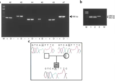
At the end the introduced assay with primer combination (A) is able to detect the point mutation in all samples of formalin-fixed, paraffin-embedded tissue (FFPE) and blood (see

AAP National Center of Medical Home Initiatives for Children With Special Needs (training programs and materials and other resources for pediatricians including materials to help

METHODS: In this study 100 patients of COPD coming to OPD as well as indoor cases in NCH, Surat from JANUARY 2016 to NOVEMBER 2016 for correlation of 6 Minutes Walk Test

Though the clearest sources of international law do not explicitly provide the right to free legal assistance for children in removal proceedings, trends in

The aim of this work was to evaluate the prevalence of Campylobacter jejuni (as affected by refrigerated storage) in chicken samples obtained from the wholesale poultry market in

In this study, because there was no association between periodontal disease-causing bacteria and the dietary intervention group, the improvement in peri- odontal disease may be