Adenosine kinase inhibition promotes survival
of fetal adenosine deaminase–deficient
thymocytes by blocking dATP accumulation
C. Justin Van De Wiele, … , Hong Jiang, Linda F. Thompson
J Clin Invest. 2002;
110(3)
:395-402.
https://doi.org/10.1172/JCI15683
.
Thymocyte development past the CD4
–CD8
–stage is markedly inhibited in adenosine
deaminase–deficient (ADA-deficient) murine fetal thymic organ cultures (FTOCs) due to the
accumulation of ADA substrates derived from thymocytes failing developmental
checkpoints. Such cultures can be rescued by overexpression of Bcl-2, suggesting that
apoptosis is an important component of the mechanism by which ADA deficiency impairs
thymocyte development. Consistent with this conclusion, ADA-deficient FTOCs were
partially rescued by a rearranged T cell receptor
b
transgene that permits virtually all
thymocytes to pass the
b
-selection checkpoint. ADA-deficient cultures were also rescued by
the adenosine kinase inhibitor 5
¢
-amino-5
¢
-deoxyadenosine (5
¢
A5
¢
dAdo), indicating that the
metabolite responsible for the inhibition of thymocyte development is not adenosine or
deoxyadenosine, but a phosphorylated derivative of an ADA substrate. Correction of
ADA-deficient FTOCs by 5
¢
A5
¢
dAdo correlated with reduced accumulation of dATP, implicating
this compound as the toxic metabolite. In ADA-inhibited FTOCs rescued with a Bcl-2
transgene, however, dATP levels were superelevated, suggesting that cells failing positive
and negative selection continued to contribute to the accumulation of ADA substrates. Our
data are consistent with dATP-induced mitochondrial cytochrome c release followed by
apoptosis as the mechanism by which ADA deficiency leads to reduced thymic T cell
production.
Article
Genetics
Find the latest version:
http://jci.me/15683/pdf
Introduction
Adenosine deaminase (ADA) catalyzes the irreversible deamination of adenosine and deoxyadenosine to inosine and deoxyinosine, respectively. Mutations in the ADA gene that result in loss of enzyme activity cause severe combined immunodeficiency (1). Biochemical
aberra-tions due to ADA deficiency have been delineated over the past 30 years, but it is still unclear why loss of this enzyme activity exhibits such profound effects on the immune system (reviewed in ref. 2). Adenosine and deoxyadeno-sine, the substrates of ADA, are generated in the microen-vironment of emerging thymocytes through normal mechanisms of lymphocyte selection. Thymocytes failing developmental checkpoints die and are degraded by thymic macrophages (3) generating adenosine and deoxyadenosine (4, 5). In a normal thymus, ADA catabo-lizes these metabolites, but in ADA deficiency they accu-mulate (6, 7) and exert lymphotoxic effects either direct-ly (2) or after conversion to phosphorylated derivatives such as AMP and dATP (2, 8–11). In an environment where up to 95% of the cells undergo programmed cell death, it is easy to visualize the potential of a cell to accu-mulate toxic levels of purine metabolites.
ADA-deficient murine fetal thymic organ culture (FTOC) is an excellent model of the human disease (12) because it exhibits many biochemical features of ADA-deficient patients, including ADA substrate and dATP accumulation as well as S-adenosylhomocys-teine (SAH) hydrolase inhibition. Furthermore, the yield of thymocytes from ADA-deficient cultures is 85–95% less than in control cultures, with thymocyte development becoming progressively more impaired
Adenosine kinase inhibition promotes survival
of fetal adenosine deaminase–deficient thymocytes
by blocking dATP accumulation
C. Justin Van De Wiele,
1,2James G. Vaughn,
1Michael R. Blackburn,
3Catherine A. Ledent,
4Marlene Jacobson,
5Hong Jiang,
1and Linda F. Thompson
1,21Immunobiology and Cancer Program, Oklahoma Medical Research Foundation, Oklahoma City, Oklahoma, USA 2Department of Microbiology and Immunology, University of Oklahoma, Oklahoma City, Oklahoma, USA 3Department of Biochemistry, University of Texas/Houston Medical School, Houston, Texas, USA 4Université Libre de Bruxelles, Brussels, Belgium
5Merck Research Laboratories, West Point, Pennsylvania, USA
Thymocyte development past the CD4–CD8– stage is markedly inhibited in adenosine
deaminase–deficient (ADA-deficient) murine fetal thymic organ cultures (FTOCs) due to the accu-mulation of ADA substrates derived from thymocytes failing developmental checkpoints. Such cul-tures can be rescued by overexpression of Bcl-2, suggesting that apoptosis is an important compo-nent of the mechanism by which ADA deficiency impairs thymocyte development. Consistent with this conclusion, ADA-deficient FTOCs were partially rescued by a rearranged T cell receptor β trans-gene that permits virtually all thymocytes to pass the β-selection checkpoint. ADA-deficient cultures were also rescued by the adenosine kinase inhibitor 5′-amino-5′-deoxyadenosine (5′A5′dAdo), indi-cating that the metabolite responsible for the inhibition of thymocyte development is not adenosine or deoxyadenosine, but a phosphorylated derivative of an ADA substrate. Correction of ADA-defi-cient FTOCs by 5′A5′dAdo correlated with reduced accumulation of dATP, implicating this com-pound as the toxic metabolite. In ADA-inhibited FTOCs rescued with a Bcl-2transgene, however, dATP levels were superelevated, suggesting that cells failing positive and negative selection contin-ued to contribute to the accumulation of ADA substrates. Our data are consistent with dATP-induced mitochondrial cytochrome crelease followed by apoptosis as the mechanism by which ADA defi-ciency leads to reduced thymic T cell production.
J. Clin. Invest.110:395–402 (2002). doi:10.1172/JCI200215683
Received for publication April 12, 2002, and accepted in revised form June 11, 2002.
Address correspondence to: Linda F. Thompson, Oklahoma Medical Research Foundation, 825 NE 13th Street, Oklahoma City, Oklahoma 73104, USA. Phone: (405) 271-7235;
Fax: (405) 271-8568; E-mail: Linda-Thompson@omrf.ouhsc.edu. C. Justin Van De Wiele’s present address is: Department of Surgery, College of Medicine, University of Oklahoma, Tulsa, Oklahoma, USA.
Hong Jiang’s present address is: Curatek Pharmaceuticals, Elk Grove Village, Illinois, USA.
Conflict of interest: No conflict of interest has been declared. Nonstandard abbreviations used: adenosine deaminase (ADA); fetal thymic organ culture (FTOC); S-adenosylhomocysteine (SAH); T cell receptor βtransgene (TCRβ); carbobenzoxy-Val-Ala-Asp-fluoromethyl ketone (z-VADfmk); apoptotic protease-activating factor-1 (Apaf-1); adenosine receptor 2a (A2aR); 2′-deoxycoformycin (17) (dCF); 5′-N-ethylcarboxamidoadenosine (NECA); xanthine amine congener (XAC); 5′-amino-5′
past the CD4–CD8–CD25+CD44– stage, the point where T cell receptor β(TCRβ) V→DJ gene rearrange-ments occur. This is presumably because of the accu-mulation of ADA substrates derived from thymocytes that fail to rearrange an in-frame TCRβchain. This conclusion is supported by rescue of ADA-inhibited FTOCs by the pan-caspase inhibitor carbobenzoxy-Val-Ala-Asp-fluoromethyl ketone (z-VADfmk). Inhibi-tion of caspase activity prevents the accumulaInhibi-tion of dATP, most likely by preventing the apoptotic death of thymocytes failing βselection. ADA-deficient FTOCs are also rescued by a Bcl-2 transgene or deletion of
apoptotic protease-activating factor-1(Apaf-1), providing
further evidence of the involvement of apoptosis in the inhibition of lymphocyte development observed in our cultures. However, whether these latter cultures were rescued by prevention of substrate accumulation or by direct intervention in the mechanism of toxicity medi-ated by ADA substrates was not determined.
Adenosine, deoxyadenosine, AMP, and dATP have all been implicated in causing the lymphopenia observed in ADA deficiency (2). Aberrant adenosine receptor engagement can induce thymocyte apoptosis in vitro. Inhibition of pyrimidine synthesis by AMP can starve developing cells of molecules required for both RNA and DNA production. Inhibition of SAH hydrolase by both adenosine and deoxyadenosine can result in feed-back inhibition of methylation reactions required for cell viability. Inhibition of ribonucleotide reductase by dATP can prevent production of DNA precursors and thus inhibit cell proliferation. Phosphorylation events coupled with dATP-mediated stimulation of adenine ribonucleotide catabolism can deplete cellular ATP. Finally, dATP can induce mitochondrial cytochrome c
release and subsequent apoptosis (11). We show here that normalization of dATP levels by inhibition of adenosine kinase abrogates the effects of ADA defi-ciency in FTOC, suggesting that dATP is the toxic metabolite. Furthermore, since our cultures can also be rescued by a Bcl-2transgene, our data are consistent with dATP-induced cytochrome crelease from mito-chondria followed by initiation of the apoptotic cas-cade as the mechanism of toxicity.
Methods
Mice. C57BL/6 mice (Taconic Farms, Germantown, New York, USA or The Jackson Laboratory, Bar Harbor, Maine, USA) and transgenic mice expressingBcl-2under the control of the lckproximal promoter (The Jackson Laboratory) (13) were purchased from the indicated sources. Transgenic mice expressing a rearranged OVA257–264–specific TCRβchain (14) were obtained from Francis Carbone (Monash University, Melbourne, Aus-tralia). ADA-deficient (7), adenosine receptor 2a–defi-cient (A2aR-defi2a–defi-cient) (15), and A3R-defi2a–defi-cient mice (16) have been described previously. The Adagene-targeted allele has been designated m1. Because Adam1/m1mice are not viable, Adam1/+ mice were bred to each other to obtain Adam1/m1fetal thymuses for FTOCs. Adam1/+and
A3R–/–mice were crossed to produce Adam1/+A3R–/–mice. These mice were bred to each other to obtain
Adam1/m1A3R–/–fetal thymuses for FTOC. All mice were bred and maintained in our animal facility under spe-cific pathogen-free conditions (Laboratory Animal Resource Center, Oklahoma City, Oklahoma, USA) in accordance with procedures outlined in “Guide to Care and Use of Laboratory Animals” (National Research Council, Washington, D.C., USA).
Drugs and reagents. The specific ADA inhibitor,
2′-deoxycoformycin (17) (dCF), was obtained from Parke Davis (Ann Arbor, Michigan, USA) or SuperGen (Dublin, California, USA). The general adenosine recep-tor agonist 5′-N-ethylcarboxamidoadenosine (NECA), the general adenosine receptor antagonist 8-[4-[[[[(2- aminoethyl)amino]carbonyl]methyl]oxy]phenyl]-1,3-dipropylxanthine (xanthine amine congener, or XAC), and the adenosine kinase inhibitor, 5′-amino-5′ -deoxyadenosine (18) (5′A5′dAdo) were obtained from Sigma-Aldrich (St. Louis, Missouri, USA).
FTOC. Thymuses were removed from fetuses derived from timed pregnant mice on day 14 or 15 of gestation and cultured in FTOC as described previously (12). FTOCs with ADA-deficient lobes were performed in serum-free medium, HyQ-CCM1 (HyClone Laboratories, Logan, Utah, USA), supplemented with glutamine and antibiotics. In experiments with C57BL/6 fetuses, thy-muses were separated into individual lobes, randomized, and cultured in the presence or absence of 5 µM dCF and/or test reagents. In FTOCs with transgenic or gene-targeted mice, one lobe from each thymus was cultured under control conditions and the other under experi-mental conditions. At harvest, lobes of the pertinent genotypes were pooled and pushed through 70-µm nylon screens to make single cell suspensions. Cells were then counted and characterized for cell surface phenotype.
Genotyping. ADA-deficient thymuses derived from
breeding either Adam1/+ or Adam1/+A3R–/– mice were identified by ADA enzyme assays (19) on the corre-sponding fetal livers. TCRβchain and Bcl-2transgenic fetuses were identified by PCR on DNA extracted from fetal livers by digestion overnight at 55°C in pro-teinase K, followed by phenol/chloroform extraction and ethanol precipitation. For PCR, the following conditions were used in a 20-µl reaction: 2.5 mM MgCl2, 200 µM dNTPs, 1 µM each primer, and 2 U of Taq polymerase (Roche Applied Science, Nutley, New Jersey, USA). The PCR primers were as follows: (a)
TCRβ, forward, 5′-TCCAGTCTCCAAGACACATAATC-3′ and reverse, 5′-GACCGAAGTACTGTTCATAATTG-3′and
(b)Bcl-2, forward, 5′-GTAGCCATTGCAGCTAGGTG-3′
and reverse, 5′- CTTTGTGGAACTGTACGGCCCCAGCAT-GCG-3′. The cycling parameters were: (a) TCRβ, 94°C for 7 minutes (94°C for 15 seconds, 60°C for 15 sec-onds, 72°C for 45 seconds) 33/72°C for 2 minutes and
(b)Bcl-2, 94°C for 7 minutes (94°C for 15 seconds,
Ab’s and immunofluorescent staining. Staining was per-formed with the following Ab’s as described previ-ously (20): FITC-rat anti-CD4 (PharMingen, San Diego, California, USA) and phycoerythrin-rat (PE) CD8α(Caltag Laboratories Inc., Burlingame, Cali-fornia, USA) using isotype-matched rat myeloma proteins as controls. Propidium iodide (PI) was added to all stains (5 µg/ml) to exclude dead cells from analysis. Data were collected on 20,000–50,000 cells using a Becton-Dickinson FACScan or FACS-Calibur (Becton Dickinson Immunocytometry Sys-tems, Mountain View, California, USA) and analyzed with CELLQuest software (Becton Dickinson Immunocytometry Systems).
Adenosine receptor expression. Adenosine receptor
expression was analyzed by RT-PCR with RNA pre-pared from day-15 fetal thymocytes according to stan-dard protocols. The PCR primers were: A1R, forward, 5′-AGGCACTTCGCGATGCTA-3′ and reverse, 5′
-CCTTTTTGTTGAGCTGCTTA-3′; A2aR, forward, 5′
-GGATCAACAGCAACCTGC-3′ and reverse, 5′- CTTC-CTTCTGCAGTGTGGA-3′; A2bR, forward, 5′- CAGCTA-GAGACGCAAGAC-3′ and reverse, 5′- GAGGACAG-CAGCTTTTATTC-3′; A3R, forward, 5′- GGTCAAGCT-GACAGTCAGAT-3′ and reverse, 5′- CAAACAAGAAGA-GAACCAGAAA-3′; and β-actin, forward, 5′- CCTAA-GGCCAACCGTGAAAAG-3′and reverse, 5′- TCTTCATG-GTGCTAGGAGCCA-3′. The PCR conditions for the
A1R, A2aR, A3R, and β-actinwere: 95°C for 5 minutes, 58°C for 1 minute, 72°C for 1 minute, once; 95°C for 1 minute, 58°C for 1 minute, 72°C for 1 minute, 40 times. The PCR conditions for the A2bR were identi-cal, except that the annealing temperature was 52°C. The PCR products were separated on agarose gels and visualized by EtBr staining.
dATP measurements. Single cell suspensions were
pre-pared at 4°C from 25–40 cultured thymic lobes. Aliquots were removed for cell counts and CD4/CD8 staining, and the remaining cells were pelleted by a 20-second spin at 16,000 gin a microcentrifuge. After aspi-ration of supernatant, the thymocytes were resuspend-ed with vigorous vortexing in 0.5 ml of 60% methanol and extracted overnight on dry ice. dATP levels were determined by HPLC as previously described (7).
SAH hydrolase enzyme assays. Cultured thymic lobes
were rinsed with cold PBS and then flash-frozen in liq-uid nitrogen and stored at –80°C prior to analysis. SAH hydrolase enzyme activities were determined by meas-uring the formation of SAH from homocysteine and radiolabeled adenosine as described previously (7).
Results
A transgenic TCRβchain partially corrects the effects of ADA
inhibition. ADA deficiency causes a block in thymocyte
development past the point of βselection (12) because of the accumulation of ADA substrates derived from apoptotic thymocytes failing this developmental check-point. This assertion is supported by the rescue of ADA-deficient FTOCs by the pan-caspase inhibitor,
[image:4.576.306.533.385.606.2]z-VADfmk, along with normalization of levels of dATP. We reasoned that a TCRβchain transgene should also correct the effects of ADA deficiency by preventing thy-mocyte death by neglect due to failure of βselection. As expected, dCF inhibited the development of thymocytes in 2-day FTOCs with control littermates of TCRβchain transgenic mice, as shown by both a decrease in the cell yield and in the percentages of CD4+CD8+thymocytes (Figure 1, a and b). As predicted, a TCRβchain transgene rescued thymocyte differentiation in ADA-inhibited cul-tures. TCRβtransgenic lobes treated with 5 µM dCF pro-duced 17-fold more CD4+CD8+ (double positive [DP]) thymocytes than similarly treated littermate control lobes (Figure 1, b and d). Rescue was only partial, how-ever, because dCF-treated transgenic cultures had just 72% of the number of thymocytes as transgenic media control cultures (Figure 1, c and d). These data suggest that ADA substrates derive not only from thymocytes that fail βselection but also from those that fail positive and negative selection. The correction observed in trans-genic cultures likely resulted from thymocyte expansion and differentiation during a period of delayed accumu-lation of toxic levels of ADA substrates. Expression of the transgene did not prevent dATP accumulation (data not shown), suggesting that this phenomenon occurs for only a very narrow window in time, perhaps because the cultures are not perfectly synchronized and apoptosis
Figure 1
A transgenic TCRβchain partially corrects the effects of ADA inhibi-tion. FTOCs were performed at day 14 of gestation with or without 5
due to failure at the positive/negative selection check-point may occur in some cells earlier than βselection in others. Further experiments revealed that the degree of rescue, defined as ( number of DP thymocytes per lobe from dCF-treated transgenic cultures)/(number of DP thymocytes per lobe from control transgenic cultures)
×100, was dependent upon the length of the cultures; rescue was essentially complete in 1-day cultures and declined as the culture period lengthened (Figure 1e). Rescue was also poor if cultures were initiated after a sig-nificant number of thymocytes had already passed the
βselection checkpoint (data not shown).
Inhibition of adenosine kinase corrects thymocyte
develop-ment within ADA-inhibited FTOC. To differentiate
between toxicity induced by nonphosphorylated ver-sus phosphorylated forms of ADA substrates, we eval-uated the ability of an adenosine kinase inhibitor to correct thymocyte development in ADA-deficient murine FTOCs. In the mouse, the conversion of adenosine and deoxyadenosine to AMP and dAMP, respectively, is dependent primarily upon the activity of adenosine kinase (21). Treatment of ADA-inhibit-ed lobes with the adenosine kinase inhibitor, 5′A5′dAdo, for 5 days resulted in a tenfold increase (range: 5.5 to tenfold) in cell recovery and a 44-fold increase (range: 27- to 47-fold) in the absolute num-bers of CD4+CD8+thymocytes per lobe (Figure 2, b and d). Five-day cultures of genetically ADA-deficient fetal thymic lobes (Adam1/m1) gave similar results, as did experiments with an alternate adenosine kinase inhibitor, 5′-iodotubercidin (data not shown). These findings demonstrate that inhibition of adenosine kinase largely abrogates the effects of ADA deficiency, as judged by thymocyte differentiation and prolifera-tion, and indicate that the inhibition of thymocyte development in ADA-deficient FTOC is due to a phos-phorylated form of an ADA substrate and not adeno-sine or deoxyadenoadeno-sine.
Neither aberrant adenosine receptor engagement nor SAH hydrolase inhibition are responsible for the inhibition of
thy-mocyte production in ADA-deficient FTOC. To confirm the
assertion that adenosine and deoxyadenosine are not directly responsible for the lymphotoxicity in ADA defi-ciency, we evaluated two mechanisms by which these molecules have been hypothesized to inhibit lympho-cyte development: aberrant engagement of adenosine receptors and inhibition of SAH hydrolase. RT-PCR revealed that A2aR, A2bR, and A3R were expressed in murine thymus at day 15 of gestation (Figure 3), the time when the FTOC cultures were initiated, validating the potential of these receptors as mediators of toxici-ty due to ADA inhibition.
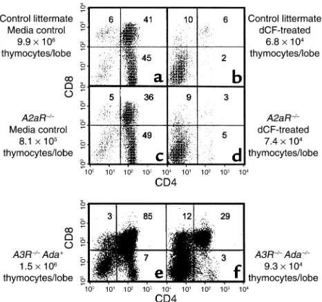
To address involvement of individual adenosine recep-tors in ADA deficiency, fetal thymuses from A2aR and A3R knockout mice were used in ADA-deficient FTOC. The absence of either receptor did not seem to be dele-terious to developing thymocytes because CD4/CD8 expression profiles were similar between receptor knockout (Figure 4, c and e) and wild-type fetal thy-muses (Figure 4a, and data not shown). An absence of either the A2aR or A3R failed to provide protection from the consequences of ADA deficiency. In dCF-treat-ed A2aR–/–FTOCs (Figure 4d) there was a 91% and greater than 99% inhibition in the absolute numbers of total and CD4+CD8+thymocytes per lobe, respectively, compared with control cultures (Figure 4c). Similarly, in FTOCs with Adam1/m1A3R–/–thymuses (Figure 4f), the yields of total and CD4+CD8+ thymocytes were de-creased by 96% and 98%, respectively, compared with FTOCs with Ada+A3R–/–thymuses (Figure 4e).
We next used the general adenosine receptor agonist, NECA, to evaluate the potential role of the A2bR as well as the possibility that multiple adenosine receptors need-ed to be engagneed-ed to see an effect on thymocyte
develop-Figure 2
[image:5.576.58.294.56.188.2]Inhibition of adenosine kinase rescues thymocyte development with-in ADA-with-inhibited FTOC. FTOCs were performed with C57BL/6 thy-muses at day 15 of gestation in media alone (a), 5 µM dCF (b), 1 µM 5′A5′dAdo (c), or 5 µM dCF plus 1 µM 5′A5′dAdo (d). After 5 days, thymocytes were harvested, counted, and stained with FITC-anti-CD4, PE-anti-CD8, and PI to exclude dead cells. The data are repre-sentative of one experiment out of four.
Figure 3
Adenosine receptor expression in murine fetal thymocytes. Adeno-sine receptor expression was analyzed by RT-PCR with RNA from day-15 fetal thymocytes (T). Brain tissue (B) was used as a positive control. RT-PCR was also performed with primers specific for
[image:5.576.334.495.488.647.2]ment. However, NECA, at concentrations up to 100 µM, did not prove deleterious as judged by CD4/CD8 expres-sion profiles and fetal thymic lobe cellularity (Figure 5, a and b). Likewise, the general adenosine receptor antag-onist, XAC, at 25 µM failed to protect developing thy-mocytes from the consequences of ADA deficiency (Fig-ure 5, e and f). FTOCs treated with XAC and dCF exhibited decreases in absolute numbers of total (86%) and CD4+CD8+thymocytes (94%) per lobe, similar to cultures treated with dCF alone (85% and 97%, respec-tively). Taken together, these data suggest that adenosine receptor engagement is not the mechanism by which ADA deficiency inhibits T cell development.
An additional consequence of ADA deficiency is inhi-bition of SAH hydrolase by adenosine or deoxyadeno-sine (22, 23), potentially leading to decreased cellular transmethylation reactions (24). In 3-day FTOCs, res-cue of ADA-deficient FTOCs by an adenosine kinase inhibitor (Figure 2) did not prevent inhibition of SAH hydrolase (Figure 6); enzyme activity was inhibited by approximately 85% in ADA-deficient cultures, irre-spective of adenosine kinase inhibition. Thus, inhibi-tion of SAH hydrolase is not the mechanism responsi-ble for the inhibition of thymocyte development in ADA-deficient FTOC.
Inhibition of adenosine kinase prevents the accumulation of
dATP in ADA-inhibited FTOC. The ability of 5′A5′dAdo to
correct thymocyte differentiation in ADA-deficient
cul-tures led to the conclusion that lymphotoxicity was caused by a phosphorylated ADA substrate. To confirm this, we measured dATP in ADA-inhibited cultures cor-rected by 5′A5′dAdo. The dATP accumulation was inhib-ited by 90% compared with cultures treated with dCF alone (Figure 7). Thus, rescue of thymocyte differentia-tion in ADA-deficient FTOC by adenosine kinase inhi-bition correlated with a decrease in dATP levels.
Overexpression of Bcl-2 corrects the effects of ADA inhibition but
does not prevent dATP accumulation. We reported previously
that ADA-deficient FTOCs could be rescued by overex-pression of a Bcl-2transgene (12). To determine whether Bcl-2 acted to prevent death by neglect of thymocytes fail-ing developmental checkpoints (thus limitfail-ing the avail-ability of ADA substrates) or to prevent apoptosis induced by ADA substrate accumulation, we measured dATP in dCF-treated FTOCs from Bcl-2transgenic mice and con-trol littermates (Figure 8). The dATP was elevated an aver-age of 2.5-fold in the Bcl-2cultures compared with those from control littermates. These data indicate that Bcl-2 does not prevent ADA substrate accumulation, but rather inhibits its effects, and are consistent with mitochondri-al-dependent apoptosis as the route by which thymocyte development is impaired by ADA deficiency.
Discussion
[image:6.576.60.293.55.274.2] [image:6.576.306.538.399.615.2]ADA-deficient murine FTOC was used to assess the bio-chemical mechanism(s) by which a loss of ADA enzyme
Figure 4
Targeted deletion of the adenosine A2aR or A3R does not correct the effects of ADA deficiency in FTOC. FTOCs were performed at day 15 of gestation with fetal thymuses from A2aR–/–and A3R–/–Adam1/m1
mice and appropriate controls. A2aR+/+(aand b) and A2aR–/–(cand
d) fetal thymic lobes were cultured for 8 days in the presence (b
and d) or absence (a and c) of 5 µM dCF. A3R–/–Ada+ (e) and
A3R–/–Adam1/m1(f) fetal thymic lobes were cultured for 5 days. At the
end of culture, thymocytes were harvested, counted, and stained with FITC-anti-CD4, PE-anti-CD8, and PI to exclude dead cells. The data are representative of one experiment out of two.
Figure 5
An adenosine receptor agonist does not mimic and an adenosine recep-tor antagonist does not prevent the effects of ADA deficiency in FTOC. FTOCs were performed with C57BL/6 thymuses at day 15 of gestation in the presence of (a) 0.1% DMSO (carrier for NECA), (b) 100 µM NECA, (c) 0.1 mM acetic acid (carrier for XAC), (d) 5 µM dCF, (e) 25
activity blocks the development of T cells. Previous work from our group (12) demonstrated that the earliest effects of ADA deficiency on thymocyte development were caused by the accumulation of ADA substrates derived from thymocytes undergoing apoptosis. The pan-caspase inhibitor, z-VADfmk, both corrected dif-ferentiation and prevented the accumulation of dATP, presumably by inhibiting the death of thymocytes that failed βselection. This hypothesis was further corrobo-rated in the present study by the ability of an in-frame
TCRβchain transgene to partially correct thymocyte dif-ferentiation in short-term ADA-deficient FTOCs. The transgene failed to provide substantial protection in cultures longer than 2 days, however, probably because of the accumulation of ADA substrates derived from thymocytes failing positive selection, because a trans-genic TCRβchain does not assure TCRs competent to recognize self peptide plus MHC when paired with endogenous rearranged TCRαchains. Indeed, dATP was quite elevated in 3.5-day ADA-inhibited FTOCs with
TCRβchain transgenic thymuses (data not shown). We next employed 5′A5′dAdo, a potent inhibitor of adenosine kinase, the primary enzyme responsible for the phosphorylation of both adenosine and deoxy-adenosine in murine thymus (21), to address the ques-tion of whether toxicity is mediated directly by ADA substrates or by phosphorylated derivatives. Correction of thymocyte differentiation with a concomitant 90% reduction in cellular dATP levels argues strongly against mechanisms of toxicity mediated by adenosine and deoxyadenosine and suggests that the culprit is dATP. To strengthen this assertion, various proposed
mechanisms of toxicity due to adenosine and deoxy-adenosine or their metabolites were evaluated.
One proposed mechanism of toxicity was adenosine receptor signaling, because engagement of these recep-tors induces apoptosis in thymocyte suspensions (25–28). However, ADA-deficient FTOCs performed with thy-muses from A2aRand A3Rknockout mice revealed no protective effect. Since the absence of a single adenosine receptor might not prevent toxicity, the general adeno-sine receptor antagonist XAC was tested for its ability to protect ADA-deficient FTOCs. Prevention of adenosine receptor engagement did not affect normal thymocyte differentiation nor did it protect developing thymocytes from the effects of ADA deficiency. In a parallel approach, FTOCs were performed with NECA, a general adenosine receptor agonist, to see if this agent would mimic the con-sequences of ADA deficiency. NECA, used at concentra-tions up to 1,000-fold higher than necessary for inducing apoptosis of thymocytes in suspension cultures, had no observable deleterious effects on thymocyte differentia-tion or proliferadifferentia-tion. The reason why NECA induces apoptosis in thymocyte suspensions, but not in FTOC, is unknown, but may be related to protective signals from thymic stromal cells or some other component of the thymic microenvironment. These results suggest that aberrant adenosine receptor engagement is not the mech-anism responsible for thymocyte depletion due to ADA deficiency. Our conclusions are thus different from those of Apasov et al. (29) who studied the toxicity of adenosine on thymocyte suspensions in the presence of an ADA inhibitor. However, it is important to note that they used adenosine at 100 µM, a concentration that is probably never achieved in our FTOCs, and that the consequences of adenosine exposure may be different in isolated
thy-Figure 6
[image:7.576.100.269.51.250.2]Rescue of ADA-inhibited FTOCs with an adenosine kinase inhibitor does not prevent inhibition of SAH hydrolase. FTOCs were per-formed with C57BL/6 thymuses at day 15 of gestation with media, 5 µM dCF, 1 µM 5′A5′dAdo, or 5 µM dCF plus 1 µM 5′A5′dAdo. After 3 days, SAH hydrolase enzyme activity was assessed in extracts of 25–30 fetal thymic lobes. The data are represented as the means (+ SD) of two independent experiments, each assayed in duplicate.
Figure 7
[image:7.576.323.496.463.637.2]mocytes compared with those in the thymic microenvi-ronment as stated above.
Inhibition of SAH hydrolase was another potential mechanism of toxicity mediated by adenosine and deoxyadenosine. This enzyme degrades SAH, a product of transmethylation reactions where S-adenosylmethionine (SAM) is the methyl donor. Elevated adenosine can force reversal of the hydrolytic reaction to form SAH from adenosine and homocysteine (22). Deoxyadenosine acts as a “suicide” inhibitor, forming a covalent bond within the active site of the enzyme (23). Elevated levels of SAH, relative to SAM, as a consequence of SAH hydrolase inhi-bition, can lead to inhibition of multiple cellular methy-lation reactions (24). Our observation that SAH hydrolase enzyme activity remained inhibited in ADA-deficient cul-tures rescued by 5′A5′dAdo argues strongly against SAH hydrolase inhibition as the mechanism by which ADA deficiency inhibits thymocyte development in FTOC.
With experimental evidence from our lab (12) and others (2) indicating that toxicity associated with ADA deficiency correlated with elevations in dATP, we next explored potential routes of toxicity associated with this molecule. There are two major mechanisms by which dATP could deleteriously affect developing thy-mocytes: allosteric inhibition of ribonucleotide reduc-tase (30, 31), the enzyme that generates deoxyribonu-cleotides needed for DNA synthesis, and induction of apoptosis (11). Our data indicate that the dATP levels attained in ADA-inhibited FTOCs must be insufficient to inhibit the generation of deoxyribonucleotides, because thymocyte proliferation is normal in dCF-treated FTOCs with Bcl-2transgenic thymocytes where dATP levels are hyperelevated.
dATP has the potential to promote apoptosis at two points. First, dATP can induce the release of cytochrome
cfrom isolated mitochondria (11). Cytochrome crelease correlates with mitochondrial membrane changes and the release of other proapoptogenic factors, initiating
the irreversible effector phase of apoptosis and making this event the “point of no return” in the execution of cell suicide programs (32, 33). Second, dATP interacts with Apaf-1, cytochrome c, and procaspase-9 to form the apoptosome (34), resulting in activation of caspase-9 and triggering of the apoptotic cascade. We favor the more proximal point of dATP action because overex-pression of Bcl-2, a protein that regulates cytochrome c
efflux to the cytoplasm, provides more protection for ADA-deficient FTOCs than deletion of Apaf-1 (12). This would be expected if cytochromec release were the crit-ical event, given that Bcl-2 is upstream of Apaf-1. If the primary role of dATP were to contribute to apoptosome formation, removal of Apaf-1 or other components of the apoptosome should be equally effective in affording protection to ADA-deficient FTOCs. This is an impor-tant distinction, because a deoxyadenosine analogue, 2-chlorodeoxyadenosine, has been proposed to trigger apoptosis of leukemic cells via the interaction of its triphosphorylated derivative, 2-Cl-dATP, with sub-apoptogenic levels of cytochrome cpresent in the cyto-plasm (35). Further experimentation will be necessary to prove directly that dATP causes cytochrome crelease from mitochondria in ADA-deficient FTOCs. Our data do not rule out the possibility that dATP could act indi-rectly by interfering with some crucial event in thymo-cyte development such that apoptosis is induced in a mitochondrial-dependent fashion. However, this un-specified event is unlikely to be TCR gene rearrange-ment, because this was found to be normal in ADA-defi-cient FTOCs (12).
Our experiments also provide a new perspective on the routes by which thymocytes die by apoptosis dur-ing development. The observation that dATP is hyper-elevated in ADA-deficient FTOCs with thymuses from
Bcl-2transgenic mice argues that Bcl-2 does not prevent
[image:8.576.112.242.54.186.2]the death of thymocytes that fail positive/negative selection — otherwise, dATP levels would have normal-ized. Furthermore, the fact that dATP is hyperelevated relative to dCF-treated FTOCs with wild-type thymus-es suggthymus-ests that larger quantitithymus-es of ADA substratthymus-es are generated from cells failing positive/negative selection than from those failing βselection. This is not surpris-ing given the high degree of cellular expansion when thymocytes pass the βselection checkpoint (36). Exper-iments are underway to determine whether thymocytes at later stages of development are also sensitive to the consequences of elevated dATP, and if so, by what mechanism. It will also be important to determine whether our conclusions are applicable to human thy-mocyte development by using a model system, such as chimeric human/mouse FTOC (37), where human thy-mocyte development takes place in vitro. These studies are also in progress. We anticipate that continued use of in vitro models of thymocyte differentiation to study ADA deficiency will not only shed additional light on the pathogenesis of this immunodeficiency but will also further our understanding of normal programs of T cell development.
Figure 8
Acknowledgments
We gratefully acknowledge Xiao-Hong Sun and Frank Carbone for gifts of transgenic mice and Marc Par-mentier for support of experiments involving A2a-defi-cient mice. We also thank Kerry Humphrey for manu-script preparation. This work was supported by grants from the NIH (AI-18220 and HD-36044 to L.F. Thompson and AI-43572 to M.R. Blackburn).
1. Giblett, E.R., Anderson, J.E., Cohen, F., Pollara, B., and Meuwissen, H.J. 1972. Adenosine-deaminase deficiency in two patients with severely impaired cel-lular immunity. Lancet.2:1067–1069.
2. Hershfield, M.S., and Mitchell, B.S. 1995. Immunodeficiency diseases caused by adenosine deaminase deficiency and purine nucleoside phosphorylase deficiency. In The metabolic and molecular bases of inherited disease.C.R. Scriver, A.L. Beaudet, W.S. Sly, and D. Valle, editors. McGraw-Hill Inc. New York, New York, USA. 1725–1768.
3. Surh, C.D., and Sprent, J. 1994. T-cell apoptosis detected in situ during pos-itive and negative selection in the thymus. Nature.372:100–102. 4. Smith, C.M., and Henderson, J.F. 1982. Deoxyadenosine triphosphate
accu-mulation in erythrocytes of deoxycoformycin-treated mice. Biochem. Phar-macol.31:1545–1551.
5. Chan, T. 1979. Purine excretion by mouse peritoneal macrophages lacking adenosine deaminase activity. Proc. Natl. Acad. Sci. USA.76:925–929. 6. Migchielsen, A.A.J., et al. 1995. Adenosine-deaminase-deficient mice die
peri-natally and exhibit liver-cell degeneration, atelectasis and small intestinal cell death. Nat. Genet. 10:279–287.
7. Wakamiya, M., et al. 1995. Disruption of the adenosine deaminase gene causes hepatocellular impairment and perinatal lethality in mice. Proc. Natl. Acad. Sci. USA.92:3673–3677.
8. Green, H., and Chan, T. 1973. Pyrimidine starvation induced by adenosine in fibroblasts and lymphoid cells: role of adenosine deaminase. Science. 182:836–837.
9. Mitchell, B.S., Mejias, E., Daddona, P.E., and Kelley, W.N. 1978. Purinogenic immunodeficiency diseases: selective toxicity of deoxyribonucleosides for T cells. Proc. Natl. Acad. Sci. USA.75:5011–5014.
10. Ullman, B., Gudas, L.J., Cohen, A., and Martin, D.W., Jr. 1978. Deoxyadeno-sine metabolism and cytotoxicity in cultured mouse T lymphoma cells: a model for immunodeficiency disease. Cell.14:365–375.
11. Yang, J.C., and Cortopassi, G.A. 1998. dATP causes specific release of cytochrome C from mitochondria. Biochem. Biophys. Res. Comm.250:454–457. 12. Thompson, L.F., et al. 2000. Metabolites from apoptotic thymocytes inhib-it thymopoiesis in adenosine deaminase-deficient fetal thymic organ cul-tures. J. Clin. Invest.106:1149–1157.
13. Sentman, C.L., Shutter, J.R., Hockenbery, D., Kanagawa, O., and Korsmey-er, S.J. 1991. bcl-2 inhibits multiple forms of apoptosis but not negative selec-tion in thymocytes. Cell.67:879–888.
14. Carbone, F.R., Sterry, S.J., Butler, J., Rodda, S., and Moore, M.W. 1992. T cell receptor alpha-chain pairing determines the specificity of residue 262 with-in the Kb-restricted, ovalbumwith-in257–264 determinant. Int. Immunol.4:861–867. 15. Ledent, C., et al. 1997. Aggressiveness, hypoalgesia and high blood pressure
in mice lacking the adenosine A2areceptor. Nature.388:674–678. 16. Salvatore, C.A., et al. 2000. Disruption of the A3adenosine receptor gene in
mice and its effect on stimulated inflammatory cells. J. Biol. Chem. 275:4429–4434.
17. Agarwal, R.P., Spector, T., and Parks, Jr. R.E. 1977. Tight-binding
inhibitors-IV. Inhibition of adenosine deaminases by various inhibitors. Biochem. Phar-macol.26:359–367.
18. Miller, R.L., et al. 1979. Adenosine kinase from rabbit liver. II. Substrate and inhibitor specificity. J. Biol. Chem.254:2346–2352.
19. Boss, G.R., Thompson, L.F., O’Connor, R.D., Ziering, R.W., and Seegmiller, J.E. 1981. Ecto-5′-nucleotidase deficiency: association with adenosine deam-inase deficiency and non-association with deoxyadenosine toxicity. Clin. Immunol. Immunopathol.19:1–7.
20. Fox, R.I., Thompson, L.F., and Huddlestone, J.R. 1981. T cells express T lym-phocyte-associated antigens. J. Immunol.126:2062–2063.
21. Carson, D.A., Kaye, J., and Wasson, D.B. 1980. Differences in deoxyadeno-sine metabolism in human and mouse lymphocytes. J. Immunol.124:8–12. 22. Hershfield, M.S. and Kredich, N.M. 1978. S-adenosylhomocysteine hydro-lase is an adenosine-binding protein: a target for adenosine toxicity. Science. 202:757–760.
23. Hershfield, M.S. 1979. Apparent suicide inactivation of human lymphoblast S-adenosylhomocysteine hydrolase by 2′-deoxyadenosine and adenine ara-binoside. J. Biol. Chem.254:22–25.
24. Kredich, N.M., and Martin, D.W., Jr. 1977. Role of S-adenosylhomocysteine in adenosine-mediated toxicity in cultured mouse T lymphoma cells. Cell. 12:931–938.
25. Kizaki, H., Suzuki, K., Tadakuma, T., and Ishimura, Y. 1990. Adenosine recep-tor-mediated accumulation of cyclic AMP-induced T lymphocyte death through internucleosomal DNA cleavage. J. Biol. Chem.265:5280–5284. 26. Jondal, M., Okret, S., and McConkey, D. 1993. Killing of immature
CD4+CD8+ thymocytes in vivo by anti-CD3 or 5′ -(N-ethyl)-carboxamido-adenosine is blocked by glucocorticoid receptor antagonist RU-486. Eur. J. Immunol.23:1246–1250.
27. McConkey, D. J., Orrenius, S., and Jondal, M. 1990. Agents that elevate cAMP stimulate DNA fragmentation in thymocytes. J. Immunol. 145:1227–1230.
28. Resta, R., et al. 1997. Insights into thymic purine metabolism and adenosine deaminase deficiency revealed by transgenic mice overexpressing ecto-5′ -nucleotidase (CD73). J. Clin. Invest.99:676–683.
29. Apasov, S., Chen, J.-F., Smith, P., and Sitkovsky, M. 2000. A2areceptor dependent and A2areceptor independent effects of extracellular adenosine on murine thymocytes in conditions of adenosine deaminase deficiency.
Blood.95:3859–3867.
30. Moore, E.C., and Hurlbert, R.B. 1966. Regulation of mammalian deoxyri-bonucleotide biosynthesis by nucleotides as activators and inhibitors. J. Biol. Chem.241:4802–4809.
31. Reichard, P. 1972. Control of deoxyribonucleotide synthesis in vitro and in vivo. Adv. Enzyme Regul.10:3–16.
32. Gottlieb, R.A. 2001. Mitochondria and apoptosis. Biol. Signals Recept. 10:147–161.
33. Penninger, J.M., and Kroemer, G. 1998. Molecular and cellular mechanisms of T lymphocyte apoptosis. Adv. Immunol.68:51–144.
34. Zou, H., Li, Y., Liu, X., and Wang, X. 1999. An APAF-1.cytochrome c multi-meric complex is a functional apoptosome that activates procaspase-9. J. Biol. Chem.274:11549–11556.
35. Genini, D., et al. 2000. Nucleotide requirements for the in vitro activation of the apoptosis protein-activating factor-1-mediated caspase pathway. J. Biol. Chem.275:29–34.
36. Dudley, E.C., Petrie, H.T., Shah, L.M., Owen, M.J., and Hayday, A.C. 1994. T cell receptor beta chain gene rearrangement and selection during thymocyte development in adult mice. Immunity.1:83–93.