Murine double nullizygotes of the angiotensin
type 1A and 1B receptor genes duplicate severe
abnormal phenotypes of angiotensinogen
nullizygotes.
S Tsuchida, … , T Inagami, I Ichikawa
J Clin Invest.
1998;
101(4)
:755-760.
https://doi.org/10.1172/JCI1899
.
Rodents are the unique species carrying duplicated angiotensin (Ang) type 1 (AT1) receptor
genes, Agtr1a and Agtr1b. After separately generating Agtr1a and Agtr1b null mutant mice
by gene targeting, we produced double mutant mice homozygous for both Agtr1a and
Agtr1b null mutation (Agtr1a-/-; Agtr1b-/-) by mating the single gene mutants. Agtr1a-/-,
Agtr1b-/- mice are characterized by normal in utero survival but decreased ex utero survival
rate. After birth they are characterized by low body weight gain, marked hypotension, and
abnormal kidney morphology including delayed maturity in glomerular growth, hypoplastic
papilla, and renal arterial hypertrophy. These abnormal phenotypes are quantitatively
similar to those found in mutant mice homozygous for the angiotensinogen gene (Agt-/-),
indicating that major biological functions of endogenous Ang elucidated by the abnormal
phenotypes of Agt-/- are mediated by the AT1 receptors. Infusion of Ang II, AT1 blockers, or
an AT2 blocker was without effect on blood pressure in Agtr1a-/-; Agtr1b-/- mice, indicating
that AT2 receptor does not exert acute depressor effects in these mice lacking AT1
receptors. Also, unlike Agt-/- mice, some Agtr1a-/-; Agtr1b-/- mice have a large ventricular
septum defect, suggesting that another receptor such as AT2 is functionally activated in
Agtr1a-/-, Agtr1b-/- mice.
Research Article
Find the latest version:
J. Clin. Invest.
© The American Society for Clinical Investigation, Inc. 0021-9738/98/02/0755/06 $2.00
Volume 101, Number 4, February 1998, 755–760 http://www.jci.org
Rapid Publication
Murine Double Nullizygotes of the Angiotensin Type 1A and 1B Receptor Genes
Duplicate Severe Abnormal Phenotypes of Angiotensinogen Nullizygotes
Shinya Tsuchida,* Taiji Matsusaka,*‡ Xiangmei Chen,* Soichiro Okubo,* Fumio Niimura,* Hideki Nishimura,* Agnes Fogo,*§
Hirotoshi Utsunomiya,iTadashi Inagami,i and Iekuni Ichikawa*‡
*Department of Pediatrics, ‡Department of Medicine, §Department of Pathology, and iDepartment of Biochemistry, Vanderbilt University
Medical Center, Nashville, Tennessee 37232-2584
Abstract
Rodents are the unique species carrying duplicated
angio-tensin (Ang) type 1 (AT1) receptor genes, Agtr1a and
Agtr1b. After separately generating Agtr1a and Agtr1b null mutant mice by gene targeting, we produced double mutant mice homozygous for both Agtr1a and Agtr1b null mutation (Agtr1a2/2; Agtr1b2/2) by mating the single gene mu-tants. Agtr1a2/2, Agtr1b2/2 mice are characterized by normal in utero survival but decreased ex utero survival rate. After birth they are characterized by low body weight gain, marked hypotension, and abnormal kidney morphol-ogy including delayed maturity in glomerular growth, hypo-plastic papilla, and renal arterial hypertrophy. These abnor-mal phenotypes are quantitatively similar to those found in mutant mice homozygous for the angiotensinogen gene (Agt2/2), indicating that major biological functions of en-dogenous Ang elucidated by the abnormal phenotypes of
Agt2/2 are mediated by the AT1 receptors. Infusion of Ang II, AT1 blockers, or an AT2 blocker was without effect on blood pressure in Agtr1a2/2; Agtr1b2/2 mice, indicating that AT2 receptor does not exert acute depressor effects in
these mice lacking AT1 receptors. Also, unlike Agt2/2
mice, some Agtr1a2/2; Agtr1b2/2 mice have a large ven-tricular septum defect, suggesting that another receptor
such as AT2 is functionally activated in Agtr1a2/2,
Agtr1b2/2 mice. (J. Clin. Invest. 1998. 101:755–760.) Key words: gene targeting • angiotensin type 2 receptor • blood
pressure • kidney • heart
Introduction
Numerous studies have suggested that angiotensin (Ang)1 has
a large repertoire of biological actions (1, 2). Recently, mutant
strains of mice have been engineered that are selectively and completely devoid of angiotensinogen, the Ang precursor (3–5). These angiotensinogen null mutant (Agt2/2) mice were uni-formly found to have several abnormal phenotypes, including severe hypotension, growth retardation, renal arterial wall thickening, and hypoplastic papilla. The findings establish the notion that Ang is essential for achieving normal blood pres-sure, normal somatic growth, and normal renal architecture. Of the two distinctive Ang receptors that are genetically char-acterized, angiotensin type 1 receptor (AT1) is believed to transduce most classically recognized actions of Ang (2, 6, 7), although the function of the type 2 (AT2) receptor which was most recently identified (8, 9) has not been fully delineated. A hurdle in studying the biology of Ang is the difficulty in dem-onstrating its receptor within certain tissues. In fact, visual demonstration of Ang II receptor on the peripheral resistance vessels where Ang exerts its well-recognized vasoconstrictor action has heretofore been unsuccessful. It is entirely conceiv-able, therefore, that other functionally significant Ang recep-tors exist that are yet to be discovered.
In recent years, investigators (including ourselves) gener-ated mouse strains that carry null mutgener-ated Ang receptor genes (i.e., Agtr1a, Agtr1b, and Agtr2; 10–15). Of note, only mice and other rodents have these duplicated AT1 genes (16, 17), whereas other mammals studied thus far have a single AT1 gene (18). To our surprise, none of these murine Ang receptor gene mutants, including Agtr1a null mutants, display the se-verely abnormal anatomical and hemodynamic phenotypes that are documented in Agt2/2 mice. The observations are even more puzzling in view of the fact that AT1A receptors predominate by far over AT1B receptors in mice (19, 20), and that Agtr1b null mutants have no identifiable abnormal pheno-type (11). The results raised the possibility that, contrary to prevailing belief, Ang receptors other than AT1 or AT2 play a vital role in transducing Ang’s actions on blood pressure, so-matic growth, and renal architecture. It is also possible that in-activation of a specific Ang receptor leads to in-activation of the others in a compensatory manner, thereby minimizing mani-festation of abnormal phenotypes (19, 21). In the present study, we generated a mutant strain of mice in which both AT1 isoform genes, Agtr1a and Agtr1b,are concurrently null mu-tated (AT1 double nullizygotes). By examining these mutant mice parallel to Agt2/2 mice, we found that AT1 double nullizygotes duplicate qualitatively and quantitatively the
Address correspondence to Iekuni Ichikawa, M.D., Ph.D.,Vanderbilt University Medical Center, MCN C4204, 21st and Garland Avenue, Nashville, TN 37232-2584. Phone: 615-322-7931; FAX: 615-322-7929; E-mail: [email protected]
Received for publication 1 October 1997 and accepted in revised form 18 December 1997.
same abnormal phenotypes as those described for Agt2/2 mice (4).
Methods
Generation of mice lacking both AT1A and AT1B receptors. Earlier, we generated homozygous angiotensin type 1A (AT1A) receptor gene null mutant (Agtr1a2/2) mice and homozygous angiotensin type 1B (AT1B) receptor gene null mutant (Agtr1b2/2) mice (10, 11). In each gene mutation study, AT1A or AT1B receptor gene was inactivated by insertion of LacZ so that LacZ instead of Agtr1a or Agtr1b is driven by the respective native promoter.
In the process of generating mice lacking both AT1A and AT1B receptors, we first mated F1 heterozygous Agtr1a null mutant (Agtr1a
1/2) males with F3 homozygous Agtr1b null mutant (Agtr1b2/2) fe-males. This cross-mating produced double heterozygous null mutant (Agtr1a1/2; Agtr1b1/2) mice. These double heterozygous mice were then mated with each other to produce mice carrying the geno-type 1/1, Agtr1a1/2; Agtr1b2/2 or Agtr1a2/2; Agtr1b1/2. Finally, mice 1/1, Agtr1a1/2, Agtr1b2/2 or Agtr1a2/2; Agtr1b1/2 were mated with mice of the same genotype to produce mice with the ge-notype 1/1, Agtr1a2/2; Agtr1b2/2 or other genotypes. These final products were subjected to the analyses described below. The embry-onic stem cell line (E14.1) used to generate these mice was derived from strain 129/Ola. Chimeric males were crossed with C57BL/6 fe-males so that these double mutants carry the genetic background of 129/Ola and C57BL/6 strains. The Agtr1a and Agtr1b genotypes were determined by Southern blot analysis of DNA isolated from tail biop-sies (10, 11).
Angiotensinogen null mutant (Agt2/2) mice were also generated earlier in our laboratory (4). Their offspring (F4–5 generation) with the genetic background of 129/Ola and C57BL/6 strains were also subjected to the analyses described below.
Blood pressure measurement. Conscious arterial blood pressure in mice aged 5–7 wk was measured 24 h after surgery as described elsewhere (22). Blood pressure and heart rate were continuously monitored for 20 min until they became stable in a quiet and unre-strained environment.
To study the effect of losartan (Merck, West Point, PA), CV 11974 (Takeda Pharmaceutical Co., Osaka, Japan), PD 123319 (Re-search Biochemicals Inc., Natick, MA), and saralasin (Sigma Chemi-cal Co., St. Louis, MO) on baseline blood pressure, mice at 6–10 wk of age were anesthetized with Inactin (100 mg/kg body wt i.p.; BYK, Konstanz, Germany). The left carotid artery and right jugular vein were then cannulated with PE10 tubing for measurement of arterial pressure and drug injection. After the equilibration period, the mice received a bolus injection of 0.9% sodium chloride solution in a vol-ume that was identical to that during the subsequent drug injections. At 10-min intervals thereafter, enalapril at 10 mg/kg body wt (Merck) and three doses of Ang II (0.1, 1, and 10 mg/kg body wt) were intrave-nously administered in succession. To explore the possibility of non-AT1A/non-AT1B receptors in affecting the blood pressure of Agtr1a2/2, Agtr1b2/2 mice, losartan (10 and 100 mg/kg body wt), CV 11974 (0.5 and 5 mg/kg body wt) or PD 123319 (10 and 30 mg/kg body wt) was given before enalapril pretreatment. Changes in mean arterial pressure after injection of saline 0.1, 1, and 10 mg/kg body wt of Ang II were assessed at 60 s based on a pilot study revealing that maximum response occurs at z 60 s. Enalapril was administered to reduce desensitized response to exogenous Ang II due to high endog-enous Ang II (19, 21).
Histological analysis. Organs were fixed in 4% buffered para-formaldehyde, routinely processed, embedded in paraffin, and sec-tioned. Kidney morphology was studied by 3 mm–thick coronal sec-tions including the papilla stained with periodic acid Schiff. The coronal section of kidneys were traced with the aid of camera lucida attachment, and the diameter of papilla and maximum calyceal space were measured using a National Institutes of Health Image program.
For studies of vascular morphology, cross-sections of all the interlob-ular and arcuate arteries were traced and examined morphometri-cally. Both inner and outer diameters (i.d., o.d.) were measured for each artery, and o.d.2i.d. and (o.d.2i.d.)/o.d. (designated as arterial wall thickness and wall thickness ratio, respectively; 10), were calcu-lated and compared among wild-type, Agtr1a2/2, Agtr1a2/2; Agtr1b2/2, and Agt2/2 mice. The glomerular maturity index was calculated for each animal as described elsewhere (1–3 scale; 4).
AT1 gene activity by b-galactosidase staining. Tissues from 1/1 and Agtr1a2/2; Agtr1b2/2 mice were fixed in 4% buffered paraformaldehyde and stained with 1 mg/ml Bluo-Gal (GIBCO BRL, Gaithersburg, MD) for three days at 378C as previously described in detail (10, 11, 19).
Statistical analysis. Data are presented as means6SE. Statistical significance was assessed by using Mann-Whitney test and ANOVA followed by Tukey post hoc tests for multiple comparisons. Genotype frequencies were compared with expected Mendelian proportions by chi-square (x2). Differences were considered statistically significant at
P , 0.05.
Results
Generation of mice lacking both AT1A and AT1B receptors
To determine the survival of Agtr1a2/2; Agtr 1b2/2 mice, we examined pups from the cross-matings between Agtr1a2/2;
Agtr1b1/2 mice or between Agtr1a1/2; Agtr1b2/2 mice, which yielded more than100 weanlings. There were no signifi-cant deviations from the prediction by Mendelian law with re-gard to the genotype frequency among weaned pups with the genotypes other than Agtr1a2/2; Agtr1b2/2. However, there was a highly significant (P , 0.001 by x2) decrease in the
popu-lation of Agtr1a2/2; Agtr1b2/2 mice (26 expected, 7 ob-served). We also found a high proportion of Agtr1a2/2;
Agtr1b2/2 mice dying before weaning. Therefore, we killed 13 pups from cross-matings between Agtr1a1/2; Agtr1b2/2 mice immediately after birth. We found the distribution of genotypes among these newborns to conform to Mendelian law (four were Agtr1a1/1; Agtr1b2/2, six were Agtr1a1/2;
Agtr1b2/2, three were Agtr1a2/2; Agtr1b2/2; P . 0.05 by
x2). Thus, concurrent inactivation of AT1A and AT1B
recep-tors is compatible with survival in utero, but inactivation of these receptors markedly decreases survival ex utero. Body weight gain after birth was slow in Agtr1a2/2; Agtr 1b2/2 mice as previously reported in Agt2/2 mice (4).
Baseline blood pressure in conscious and anesthetized animals
Baseline mean arterial blood pressure in conscious animals av-eraged 10663, 7265, and 7263 mmHg in 1/1 (n 5 8),
Agtr1a2/2; Agtr1b2/2 (n 5 6; P , 0.05 vs 1/1), and Agt2/2 (n 5 5; P , 0.05 vs. 1/1) mice, respectively. Baseline mean ar-terial blood pressure under anesthesia with inactin was on av-erage 9665, 4263, and 4564 mmHg in 1/1 (n 5 7), Agtr1a2/2;
Agtr1b2/2 (n 5 8; P , 0.001 vs. 1/1), and Agt2/2 (n 5 4;
P , 0.001 vs. 1/1) mice, respectively.
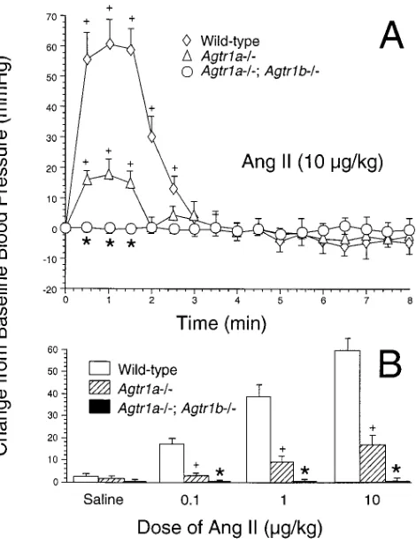
Blood pressure response to Ang II, losartan, CV 11974, PD 123319 and saralasin infusion
without effect on blood pressure in Agtr1a2/2; Agtr1b2/2 mice (n 5 5 for each, data not shown), indicating that non-AT1A/non-AT1B receptors exert no acute pressor or depres-sor effect on arterial pressure in these mutant mice lacking AT1A and AT1B (i.e., AT1) receptors.
Histological findings
Kidney. As shown in Fig. 2 and Fig. 5 A, kidneys from AT1
double nullizygotes at 0, 1, 3, 5, and 9 wk of age showed pro-gressive changes in the papilla and calyx that are similar to those described for Agt2/2 mice (4). AT1 double nullizygotes had a widening calyx space (calyx-to-papilla diameter ratio, 1.5760.21 in 1/1 vs. 1.7560.23 in Agtr1a2/2; Agtr1b2/2 mice at birth to 1.3860.25 in 1/1 vs. 5.6561.30 in Agtr1a2/2;
Agtr1b2/2 mice at 9 wk of age; P , 0.001) and hypoplastic pa-pilla (0.3260.04 mm in 1/1 vs. 0.3160.02 mm in Agtr1a2/2;
Agtr1b2/2 mice at birth to 0.8660.07 mm in 1/1 vs. 0.3060.09 mm in Agtr1a2/2; Agtr1b2/2 mice at 9 wk of age,
P , 0.001).
As shown in Fig. 3, Agtr1a2/2; Agtr1b2/2 mice had renal arterial lesions similar to those described for Agt2/2 mice; namely, prominent medial hyperplasia of the interlobular ar-teries and afferent arterioles. Morphometric examination of renal arteries in Agtr1a2/2; Agtr1b2/2 mice at 3 wk of age demonstrated a significant increase in both wall thickness (P , 0.001 vs. wild-type, P , 0.01 vs. Agtr1a2/2) and wall thickness ratio (P , 0.001 vs. wild-type).
As in Agt2/2 mice, delay in glomerular maturity was found in AT1 double nullizygotes at 1 wk of age, maturity in-dex averaging 1.8460.16 in 1/1 mice (n 5 3) and 1.4060.23 in
Agtr1a2/2; Agtr1b2/2 mice (n 5 3; P , 0.05). At 3 wk of age, however, the glomeruli of AT1 double nullizygotes were as mature as those in wild-type mice.
Heart. No gross anatomical deformity was recognized in Agtr1a2/2; Agtr1b2/2 mice (n 5 37) when they were exam-ined after weaning. Among the mice that died before weaning (n 5 28), however, two Agtr1a2/2; Agtr1b2/2 mice were found to have a large ventricular septal defect involving both membranous and muscular portions (Fig. 4), whereas none was found in mice with the other genotypes inspected before weaning (n 5 71).
The heart weight of animals randomly selected at birth was on average lowest in Agtr1a2/2; Agtr1b2/2 mice (8.860.4 mg, n 5 5) and highest in 1/1 (9.660.6 mg, n 5 6), although the difference did not reach statistical significance.
Adrenal glands. AT1 double nullizygotes had no gross or
microscopic abnormalities in adrenal glands (Fig. 5 B).
Histochemical analysis for b-galactosidase staining
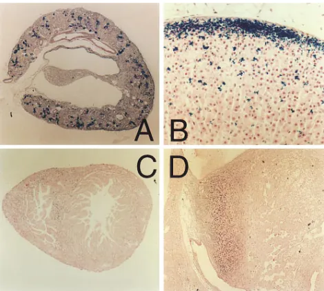
Expression of the reporter gene LacZ was systematically ex-amined in AT1 double nullizygotes by b-galactosidase stain-ing. In the kidney, b-galactosidase staining was most intensely observed in juxtaglomerular cells and hyperplastic renal arter-ies (Fig. 5 A). The proximal tubule, glomerular mesangial and endothelial cells, and occasionally interstitial cells of the
kid-Figure 1. (A) Effect of intravenous bolus infusion of Ang II (10 mg/kg Bwt) on mean arterial pressure in anesthetized mice pretreated with enalapril. Changes in mean arterial pressure from baseline assessed at 30 s intervals are depicted for wild-type (n 5 5), Agtr1a2/2 (n 5 4), and Agtr1a2/2; Agtr 1b2/2 (n 5 4) animals (*P , 0.001 vs. wild-type and Agtr1a2/2. 1P , 0.001 vs. baseline blood pressure). (B)
[image:4.612.62.297.58.365.2]changes in mean arterial pressure assessed 60 s after injection of sa-line, 0.1, 1, and 10 mg/kg body wt of Ang II in wild-type (n 5 5), Agtr1a2/2 (n 5 4), and Agtr1a2/2; Agtr 1b2/2 (n 5 4) mice pre-treated with enalapril. Data are expressed as means6SE (*P , 0.001 vs. wild-type and Agtr1a2/2. 1P , 0.001 vs. wild-type).
[image:4.612.317.555.490.664.2]ney cortex, also showed positive staining. However, no expres-sion was identified in the medulla or papilla of AT1 double nullizygotes of any age. In the adrenal glands, zona glomeru-losa cells showed strong intensity of b-galactosidase staining within the cytosol as previously reported (11). Additionally, positive staining was visible at the cell membrane in both zona fasciculata and zona reticularis cells (Fig. 5 B). In the heart,
b-galactosidase staining appeared evenly distributed through-out the tissue, except that the staining was intensified within the neonatal interventricular septum (Fig. 5 C). Positive stain-ing was not clearly demonstrable in interstitial fibroblasts. In the placenta from mice at 10.5 d of gestation, positive staining was observed in extravillous cytotrophoblasts, whereas b -galac-tosidase staining was negligible within the villi or endometrium (Fig. 5 D). The placenta at 19 d of gestation showed no posi-tive staining. b-galactosidase staining was not detectable in the lung, spleen, or peripheral resistance vessels.
Discussion
In the present study, our assessment of AT1 double nullizy-gotes focused on the several key functional and structural pa-rameters that had been found to be abnormal in Agt2/2 mice
[image:5.612.56.297.57.288.2](3–5). These are in utero and ex utero survival rates, body weight, systemic arterial pressure, heart weight, kidney mor-phology, particularly with regard to the maturity of the glo-merulus, growth of the papilla, and renal arterial wall dimension. The analyses revealed that, unlike that of any other angio-tensin receptor null mutants studied (10–15), the phenotypes of AT1 double nullizygotes duplicate those of Agt2/2 mice. The study has thus verified the notion that the AT1 receptor transduces the major biological actions of Ang II known today, and that no other receptors are required for these actions. Of all the abnormalities described above, a particularly puzzling one is the hypoplastic papilla, which becomes evident only a
[image:5.612.316.540.62.201.2]Figure 3. Morphometric analyses of renal arteries in wild-type (n 5 4), Agtr1a2/2 (n 5 4), Agtr1a2/2; Agtr1b2/2 (n 5 4), and Agt2/2 (n 5 3) mice at 3 wk of age. Each dot represents a single vessel. Hori-zontal bars represent mean values. Arteries from Agtr1a2/2; Agtr1b2/2 mice have higher values for wall thickness when com-pared with those of 1/1 or Agtr1a2/2 mice (*P , 0.001 versus wild-type, **P , 0.01 versus wild-type and Agtr1a2/2). Values for wall thickness averaged 12.3160.92, 19.6861.12, 25.7461.52, and 27.186 1.90 mm in 1/1, Agtr1a2/2, Agtr1a2/2; Agtr1b2/2, and Agt2/2 mice, respectively. (B) Arteries from Agtr1a2/2; Agtr1b2/2 mice reveal higher values for wall thickness ratio when compared with those of wild-type mice (*P , 0.001 vs. wild-type, **P , 0.001 vs. wild-type and Agtr1a2/2). Values for wall thickness ratio averaged 0.5360.01, 0.6160.02, 0.6160.01, and 0.6960.03 in 1/1, Agtr1a2/2, Agtr1a2/2; Agtr1b2/2, and Agt2/2 mice, respectively.
Figure 4. A large ven-tricular septal defect found in a AT1 double nullizygote that died at 2 d of age. Left and right lateral ventricle walls were cut open and removed with mi-croscissors. The large septal defect is viewed from the lateral aspect. LA, left atrium; LV, left ventricle; VSD, ventric-ular septal defect.
[image:5.612.315.550.380.591.2]few weeks after birth. We therefore vigorously searched for AT1 receptor gene expression in the renal medulla and papilla using a reporter gene system. Although the reporter gene could disclose the localization of the receptor within finite structures of several organs including the placenta and adrenal and renal cortex, no expression was identified in the medulla or papilla. Results of our previous study using in situ hybrid-ization with the antisense cRNA probe for the AT1 receptor (23) are consistent with the above results from the present study. The hypoplastic papilla seen in Agt2/2 and AT1 dou-ble nullizygotes may therefore reflect an indirect effect of the inhibition of AT1 function on the renal papilla. The results from our most recent study support this possibility (24).
In addition to these abnormalities, other organs, e.g., the adrenal gland, were found to be morphologically intact in AT1 double nullizygotes as well as in Agt2/2 mice (22). Taken to-gether, therefore, the results indicate that the markedly high endogenous Ang II present in AT1 double nullizygotes (19) does not exert discernible actions via non-AT1 receptors since AT1 double nullizygotes are phenotypically identical to Agt2/2
mice, which completely lack Ang’s actions. Moreover, infusion of Ang II, AT1 blockers, or an AT2 blocker was without effect on blood pressure in Agtr1a2/2; Agtr1b2/2 mice, indicating that AT2 receptor, which is believed to transduce the depres-sor effect of Ang II (12), does not exert an acute depresdepres-sor ef-fect in the absence of AT1 receptors. It is conceivable, how-ever, that an important role of the AT2 receptor may be demonstrable when its function is chronically inhibited.
Of interest is our additional finding that two out of 28 AT1 double nullizygotes inspected had a ventricular septum defect. This finding is in concert with the findings in humans that an abnormality in chromosome 3q on which the AT1 gene resides (25) accompanies a ventricular septum defect (26), although these findings may just be coincidental. Our recent demonstra-tion of AT1 gene expression in embryonic hearts of wild-type mice (23) further supports the important ontogenic role of the AT1 receptor in the formation of ventricular septum. In this regard, lack of similar cardiac defect in Agt2/2 mice (4) raised the possibility that activation of non-AT1 receptor due to en-hanced Ang II generation in AT1 double nullizygotes, but not in Agt2/2 mice, can account for the cardiac muscle defect seen only in AT1 double nullizygotes. If this is indeed the case, the AT2 receptor that is expressed in rodent embryonic hearts (27) is one such candidate, since recent studies suggest a role of this receptor in cell apoptosis (28, 29).
Acknowledgments
The authors thank Mr. Edward Price, Jr., Ms. Teresa Bills, Ms. Tracy Moore-Jarrett, and Ms. Ellen Donnert for their superb technical as-sistance. Dr. Taiji Matsusaka is the recipient of a fellowship grant from the National Kidney Foundation. Dr. Agnes Fogo is a recipient of an Established Investigator Award from the American Heart As-sociation.
This work was supported by National Institutes of Health grants DK-44757, DK-37868, and HL-58205.
References
1. Matsusaka, T., J. Hymes, and I. Ichikawa. 1996. Angiotensin in progres-sive renal diseases: theory and practice. J. Am. Soc. Nephrol. 7:2025–2043.
2. Timmermans, P.B., P.C. Wong, A.T. Chiu, W.F. Herblin, and R.D.
Smith. 1993. Angiotensin II receptors and angiotensin II receptor antagonists. Pharmacol. Rev. 45:205–251.
3. Kim, H.S., J.H. Krege, K.D. Kluckman, J.R. Hagaman, J.B. Hodgin, C.F. Best, J.C. Jennette, T.M. Coffman, N. Maeda, and O. Smithies. 1995. Genetic control of blood pressure and the angiotensinogen locus. Proc. Natl. Acad. Sci. USA. 92:2735–2739.
4. Niimura, F., P.A. Labosky, J. Kakuchi, S. Okubo, H. Yoshida, T. Oikawa, T. Ichiki, A.J. Naftilan, A. Fogo, T. Inagami, B.L.M. Hogan, and I. Ichikawa. 1995. Gene targeting reveals a requirement for angiotensin in the development and maintenance of kidney morphology and growth regulation. J. Clin. Invest. 96:2947–2954.
5. Tanimoto, K., F. Sugiyama, Y. Goto, J. Ishida, E. Takimoto, K. Yagami, A. Fukamizu, and K. Murakami. 1994. Angiotensinogen-deficient mice with hy-potension. J. Biol. Chem. 269:31334–31337.
6. Sasaki, K., Y. Yamano, S. Bardham, N. Iwai, J. Murray, M. Hasegawa, Y. Matsuda, and T. Inagami. 1991. Cloning and expression of a complementary DNA encoding a bovine adrenal angiotensin II type-1 receptor. Nature. 351: 230–232.
7. Murphy, T.J., R.W. Alexander, K.K. Griendling, M.S. Runge, and K.E. Bernstein. 1991. Isolation of a cDNA encoding the vascular type-1 angiotensin II receptor. Nature. 351:233–236.
8. Kambayashi, Y., S. Bardhan, K. Takahashi, S. Tsuzuki, H. Inui, T. Ha-makubo, and T. Inagami. 1993. Molecular cloning of a novel angiotensin II re-ceptor isoform involved in phosphotyrosine phosphatase inhibition. J. Biol. Chem. 268:24543–24546.
9. Mukoyama, M., M. Nakajima, M. Horiuchi, H. Sasamura, R.E. Pratt, and V.J. Dzau. 1993. Expression cloning of type 2 angiotensin II receptor reveals a unique class of seven-transmembrane receptors. J. Biol. Chem. 268:24539– 24542.
10. Matsusaka, T., H. Nishimura, H. Utsunomiya, J. Kakuchi, F. Niimura, T. Inagami, A. Fogo, and I. Ichikawa. 1996. Chimeric mice carrying ‘regional’ targeted deletion of the angiotensin type 1A receptor gene: Evidence against the role for local angiotensin in the in vivo feedback regulation of renin synthe-sis in juxtaglomerular cells. J. Clin. Invest. 98:1867–1877.
11. Chen, X., W. Li, H. Yoshida, S. Tsuchida, H. Nishimura, F. Takemoto, S. Okubo, A. Fogo, T. Matsusaka, and I. Ichikawa. 1997. Targeting deletion of angiotensin type 1B receptor gene in the mouse. Am. J. Physiol. 272:F299– F304.
12. Ichiki, T., P. Labosky, C. Shiota, S. Okuyama, Y. Imagawa, A. Fogo, F. Niimura, I. Ichikawa, B. Hogan, and T. Inagami. 1995. Effects on blood pres-sure and exploratory behaviour of mice lacking angiotensin II type-2 receptor. Nature. 377:748–750.
13. Hein, L., G.S. Barsh, R.E. Pratt, V.J. Dzau, and B.K. Kobilka. 1995. Be-havioural and cardiovascular effects of disrupting the angiotensin II type-2 re-ceptor in mice. Nature. 377:744–747.
14. Ito, M., M.I. Oliverio, P.J. Mannon, C.F. Best, N. Maeda, O. Smithies, and T.M. Coffman. 1995. Regulation of blood pressure by the type 1A angio-tensin II receptor gene. Proc. Natl. Acad. Sci. USA. 92:3521–3525.
15. Sugaya, T., S. Nishimura, K. Tanimoto, E. Tanimoto, T. Yamagishi, K. Imamura, S. Goto, K. Imaizumi, Y. Hisada, A. Otsuka, et al. 1995. Angiotensin II type 1a receptor–deficient mice with hypotension and hyperreninemia. J. Biol. Chem. 270:18719–18722.
16. Yoshida, H., J. Kakuchi, D.F. Guo, H. Furuta, N. Iwai, R. van der Meer-de Jong, T. Inagami, and I. Ichikawa. 1992. Analysis of the evolution of angio-tensin II type 1 receptor gene in mammals (mouse, rat, bovine and human). Biochem. Biophys. Res. Comm. 186:1042–1049.
17. MacTaggart, T.E., M. Ito, O. Smithies, and S.W. John. 1997. Mouse an-giotensin receptor genes Agtr1a and Agtr1b map to chromosomes 13 and 3. Mamm. Genome. 8:294–295.
18. Clauser, E., K.M. Curnow, E. Davies, S. Conchon, B. Teutsch, B. Vi-anello, C. Monnot, and P. Corvol. 1996. Angiotensin II receptors: protein and gene structures, expression and potential pathological involvements. Eur. J. En-docrinol. 134:403–411.
19. Nishimura, H., T. Matsusaka, A. Fogo, V. Kon, and I. Ichikawa. 1997. A novel in vivo mechanism for angiotensin type 1 receptor regulation. Kidney Int. 52:345–355.
20. Burson, J.M., G. Aguilera, K.W. Gross, and C.D. Sigmund. 1994. Differ-ential expression of angiotensin receptor 1A and 1B in mouse. Am. J. Physiol. 267:E260–E267.
21. Oliverio, M., C. Best, H. Kim, W. Arendshorst, O. Smithies, and T. Coffman. 1997. Angiotensin II responses in AT (1A) receptor-deficient mice; a role for AT (1B) receptors in blood pressure regulation. Am. J. Physiol. 272: F515–F520.
22. Okubo, S., F. Niimura, H. Nishimura, F. Takemoto, A. Fogo, T. Matsu-saka, and I. Ichikawa. 1997. Angiotensin-independent mechanism for aldoster-one synthesis during chronic extracellular fluid volume depletion. J. Clin. In-vest. 99:855–860.
23. Kakuchi, J., T. Ichiki, S. Kiyama, B.L. Hogan, A. Fogo, T. Inagami, and I. Ichikawa. 1995. Developmental expression of renal angiotensin II receptor genes in the mouse. Kidney Int. 47:140–147.
type 1 (AT1) and type 2 (AT2) receptors. J. Am. Soc. Nephrol. 8:405A. 25. Curnow, K., L. Pascoe, and P. White. 1992. Genetic analysis of the hu-man type-1 angiotensin II receptor. Mol. Endocrinol. 6:1113–1118.
26. Wilson, G., M. Dasouki, and M. Barr. 1985. Further delineation of the dup (3q) syndrome. Am. J. Med. Genet. 22:117–123.
27. Sechi, L.A., C.A. Griffin, E.F. Grady, J.E. Kalinyak, and M. Scham-belan. 1992. Characterization of angiotensin II receptor subtypes in rat heart.
Circ. Res. 71:1482–1489.
28. Yamada, T., M. Horiuchi, and V.J. Dzau. 1996. Angiotensin II type 2 re-ceptor mediates programmed cell death. Proc. Natl. Acad. Sci. USA. 93:156–160.