Expression of the Na-K-2Cl cotransporter by
macula densa and thick ascending limb cells of
rat and rabbit nephron.
N Obermüller, … , D H Ellison, S Bachmann
J Clin Invest.
1996;
98(3)
:635-640.
https://doi.org/10.1172/JCI118834
.
Sodium and chloride transport by the macula densa and thick ascending limb of Henle's
loop participates importantly in extracellular fluid volume homeostasis, urinary concentration
and dilution, control of glomerular filtration, and control of renal hemodynamics.
Transepithelial Na and Cl transport across the apical membrane of thick ascending limb
(TALH) cells is mediated predominantly by a loop diuretic sensitive Na-K-2Cl cotransport
pathway. The corresponding transport protein has recently been cloned. Functional studies
suggest that the cotransporter is expressed by macula densa cells as well as by TALH cells.
The current studies were designed to identify sites of Na-K-2Cl cotransporter expression
along distal nephron in rabbit and rat. Non-isotopic high-resolution in situ hybridization,
using an antisense probe for the apical form of the Na-K-2Cl cotransporter identified
expression throughout the TALH, from the junction between inner and outer medulla to the
transition to distal convoluted tubule. Expression by macula densa cells was confirmed by
colocalization using markers specific for macula densa cells. First, Na-K-2Cl cotransporter
mRNA was detected in macula densa cells that did not stain with anti-Tamm-Horsfall
protein antibodies. Second, Na-K-2Cl cotransporter mRNA was detected in macula densa
cells that show positive NADPH-diaphorase reaction, indicating high levels of constitutive
nitric oxide synthase activity. In rat, levels of Na-K-2Cl cotransporter mRNA expression
were similar in TALH and macula densa cells. In rabbit, expression […]
Research Article
Find the latest version:
http://jci.me/118834/pdf
J. Clin. Invest.
© The American Society for Clinical Investigation, Inc. 0021-9738/96/08/0635/06 $2.00
Volume 98, Number 3, August 1996, 635–640
Rapid Publication
Expression of the Na-K-2Cl Cotransporter by Macula Densa and Thick Ascending
Limb Cells of Rat and Rabbit Nephron
Nicholas Obermüller,* Shanti Kunchaparty,‡§ David H. Ellison,‡§ and Sebastian Bachmann*i
*Department of Anatomy, University of Heidelberg, D-69120 Heidelberg, Germany; ‡Section of Nephrology, Yale University School
of Medicine; and §VA Medical Center, West Haven, Connecticut 06516; and iDepartment of Anatomy, Charité, Humboldt University,
D-10115 Berlin, Germany
Abstract
Sodium and chloride transport by the macula densa and thick ascending limb of Henle’s loop participates impor-tantly in extracellular fluid volume homeostasis, urinary concentration and dilution, control of glomerular filtration, and control of renal hemodynamics. Transepithelial Na and Cl transport across the apical membrane of thick ascending limb (TALH) cells is mediated predominantly by a loop di-uretic sensitive Na-K-2Cl cotransport pathway. The corre-sponding transport protein has recently been cloned. Func-tional studies suggest that the cotransporter is expressed by macula densa cells as well as by TALH cells. The current studies were designed to identify sites of Na-K-2Cl cotrans-porter expression along distal nephron in rabbit and rat. Non-isotopic high-resolution in situ hybridization, using an antisense probe for the apical form of the Na-K-2Cl cotransporter identified expression throughout the TALH, from the junction between inner and outer medulla to the transition to distal convoluted tubule. Expression by macula densa cells was confirmed by colocalization using markers specific for macula densa cells. First, Na-K-2Cl cotrans-porter mRNA was detected in macula densa cells that did not stain with anti-Tamm-Horsfall protein antibodies. Sec-ond, Na-K-2Cl cotransporter mRNA was detected in mac-ula densa cells that show positive NADPH-diaphorase reac-tion, indicating high levels of constitutive nitric oxide synthase activity. In rat, levels of Na-K-2Cl cotransporter mRNA expression were similar in TALH and macula densa cells. In rabbit, expression levels were higher in macula densa cells than in surrounding TALH cells. The present data provide morphological support for a previously estab-lished functional concept that Na-K-2Cl cotransport at the TALH is accomplished by the expression of a well-defined cotransporter. At the macula densa, this transporter may
establish a crucial link between tubular salt load and glom-erular vascular regulation. (J. Clin. Invest. 1996. 98:635– 640.) Key words: tubulo-glomerular feedback• nitric oxide •
NADPH-diaphorase • Tamm-Horsfall protein • distal nephron
Introduction
A furosemide/bumetanide-sensitive sodium-potassium-chloride cotransport pathway plays a central role in transcellular reab-sorption of NaCl in the thick ascending limb (TALH)1 of the
mammalian loop of Henle. Transport is accomplished by a lu-minally located electroneutral cotransporter with a typical stoi-chiometry of 1Na:1K:2Cl which operates in conjunction with the Na pump, a K channel and a Cl channel (1, 2). The apical Na-K-2Cl cotransporter is functionally similar to a basolateral Na-K-2Cl cotransporter that participates in Cl secretion by several epithelia. Recently, cDNAs encoding both secretory (3) and absorptive (4, 5) Na-K-2Cl cotransporters have been cloned. Both secretory (identified as the bumetanide-sensitive cotransporter, BSC2, or Na-K-2Cl transporter, NKCC1) and absorptive (BSC1 or NKCC2) forms of the Na-K-2Cl porter are members of the electroneutral Na-(K)-Cl cotrans-porter gene family which also includes the thiazide-sensitive Na-Cl cotransporter (4). The predicted Na-K-2Cl protein has 12 putative transmembrane segments and a molecular mass of 121 kD, which is consistent with the size (150 kD) of the glyco-sylated transporter defined earlier by photoaffinity labeling (6). The transporter is expressed as an z 5.0-kb transcript (7).
The intrarenal distribution of Na-K-2Cl cotransporter ex-pression has recently been demonstrated in rat and mouse (4, 7, 8) using radiolabeled molecular probes and specific anti-body against recombinant fusion protein. Localization was as-signed to the TALH. Expression of the transporter was also shown during development to occur in mouse immature loops of Henle after induction at 14.5 d post conception (7). Due to limited structural resolution at the cellular level in these stud-ies, it was not possible to determine whether Na-K-2Cl cotransporter expression extends to the macula densa (MD). Immunoreactivity for the cotransporter was reported to be ab-sent from the MD, although lack of sensitivity or isoform-related specificity of the antibody have been discussed as pos-sible reasons for this result (8). Since physiological studies Address correspondence to Sebastian Bachmann, AG Anatomie der
HUB, Klinikum Charlottenburg, Haus 31, Spandauer Damm 130, D-14050 Berlin, Germany. Phone: 49 30 3651; FAX: 49 30 3035-2055. E-mail: [email protected]. Dr. Nicholas Obermüller’s present address is Department of Nephrology, Klinkum Mannheim, D-68167 Mannheim, Germany.
Received for publication 23 February 1996 and accepted in revised form 28 May 1996.
have postulated the existence of a furosemide-sensitive Na-K-Cl cotransporter to act as a sensor by which macula densa cells may detect changes in luminal [NaCl] (1, 2, 9) it was of particular interest to study the juxtaglomerular expression of the cotransporter at higher resolution. The present study de-scribes localization of the cotransporter in rat and rabbit TALH and MD using nonradioactive in situ hybridization cou-pled with other histochemical procedures to permit cellular identification of the NKCC2 mRNA-expressing cells.
Methods
Kidney preparation. Six adult male Sprague Dawley rats (300–500 g body weight) and two adult male New Zealand white rabbits (2.9 and 2.6 kg body weight) were allowed free access to standard laboratory chow and tap water. For histochemical evaluation of kidney tissue, animals were anesthetized with pentobarbital sodium (40 mg/kg body weight i.p.) and perfused retrogradely through the abdominal aorta with freshly prepared 3% paraformaldehyde in phosphate-buffered saline (PBS) at pH 7.4 for 5 min as described (10). For cryoprotection of the tissues, kidneys were subsequently rinsed with a sucrose-PBS solution adjusted to 800 mOsm/kg. Kidneys were then removed and cut into 3-mm-thick slices that were immediately frozen in liquid ni-trogen-cooled isopentane.
Molecular cloning and preparation of riboprobes. Riboprobe was prepared from a 383 bp partial cDNA fragment of the mouse bumet-anide-sensitive Na-K-2Cl cotransporter, NKCC2 (7). This cDNA fragment was generated by amplification from mouse kidney cDNA using the polymerase chain reaction as described previously (11). The fragment was subcloned into the EcoRV site of pBluescript KS1 and sequenced using the dideoxynucleotide method of Sanger (12). The nucleotide sequence was found to be identical to that recently re-ported for mouse NKCC2 (7) and spans the region between predicted amino acid residues 61–188. The sequence is 96% identical at the nu-cleotide level to the rat NKCC2 (4). RNA probes were synthesized and labeled by in vitro transcription using digoxigenin (DIG)-11-UTP (13). To generate an antisense probe, plasmid was linearized with SmaI and then transcribed using T3 polymerase. To obtain the sense probe (control), plasmid was linearized with HindIII and transcribed using T7 polymerase.
Histochemical protocols. Cryostat sections (5–7 mm thick) were thawed onto silanized glass slides for in situ hybridization alone or combined with immunohistochemistry; for NADPH-diaphorase reac-tion, chrome-alum-coated slides were used. For in situ hybridizareac-tion, slides were processed essentially as described (13). Concentrations of the DIG-labeled probes were between 5–10 ng/ml of hybridization mix-ture. Hybridization was performed at 408C for 16–18 h. Slides were then washed sequentially at 478C for 30 min in 13 SSC containing 50% form-amide, 0.53 SSC containing 50% formamide (two washes for 30 min), 0.13 SSC containing 50% formamide (two washes for 30 min). Sub-sequently slides were rinsed twice in 0.53 SSC at room temperature for 10 min followed by a rinse in 0.23 SSC for 10 min followed by two rinses in Buffer I (100 mM Tris-HCl, 150 mM NaCl, pH 7.5).
To identify TALH cells, rabbit anti-Tamm-Horsfall protein (THP) antibody was used (kindly provided by John Hoyer, Philadel-phia, PA; 14). For the simultaneous detection of NKCC2 message and THP expression, non-specific antibody binding sites were blocked by incubating sections with a mixture of 2% normal sheep se-rum, 0.5% bovine serum albumin, and 3% Triton X-100 in Buffer I. Blocking medium was then removed and a mixture of sheep anti-DIG-alkaline phosphatase conjugate and anti-THP antibody was di-luted in blocking medium (dilution of anti-DIG-conjugate was 1:500, dilution of anti-THP antibody was 30 mg/ml). Sections were incu-bated with this mixture for 2 h at room temperature in a moist cham-ber, followed by 16 h at 48C. Slides were then washed two times for 15 min in Buffer I. THP expression was detected using a biotin-strepta-vidin-Texas-red staining system as described (13). Expression of NKCC2 mRNA was detected using a nitroblue tetrazolium/5-bromo-4-chloro-3-indolyl-phosphate system (13). In situ hybridization with-out combined application of immunohistochemistry was carried with-out omitting the steps involving incubation with specific antibody and corresponding agents for fluorescence detection.
Macula densa cells were identified by NADPH-diaphorase staining as described (10). Since combined in situ hybridization and NADPH-diaphorase staining cannot be performed on the same section, label-ing was performed separately on consecutive cryostat sections.
[image:3.612.57.222.180.679.2]Controls. Antibody specificity for THP detection has been char-acterized extensively (14). Specificity of the immunohistochemical
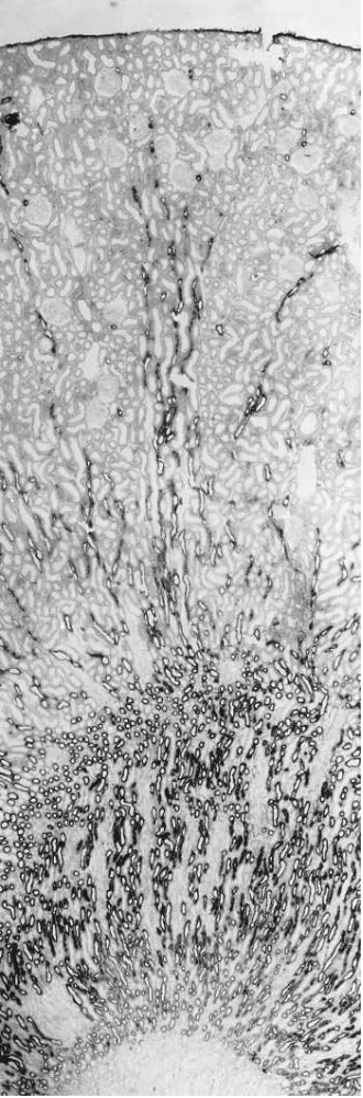
Figure 1.Expression of the furosemide/bumetanide sensitive Na-K-2Cl cotransporter (NKCC2) in rat kidney cortex: in situ hybridization with antisense DIG-labeled NKCC2 riboprobe. TALH segments are labeled throughout the outer medulla and cortex. Magnification
staining in the present study was tested by replacing the specific anti-body with non-immune serum; no signal was obtained under this con-dition. The specificity of the in situ hybridization signal was tested by parallel application of sense and antisense riboprobes on neighboring
sections. Throughout all experiments, incubation with the sense probes did not produce a detectable signal. Specificity of the NADPH-diaphorase reaction was verified by replacing NADPH with NADH, which resulted in a total absence of staining.
Results
Expression of NKCC in adult rabbit and rat kidney was exam-ined using nonradioactive in situ hybridization. For these ex-periments, DIG-labeled riboprobes were transcribed from a 383-bp fragment that is specific for the apical isoforms of the Na-K-2Cl cotransporter (NKCC2/BSC1) but does not distin-guish between three alternatively spliced isoforms of NKCC2 that are expressed in rabbit and mouse kidney (5, 7).
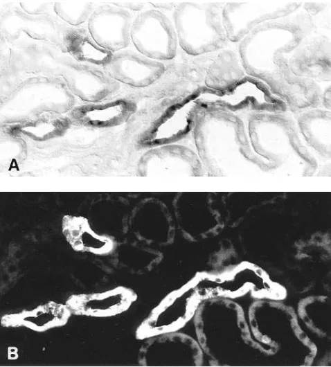
Both rat and rabbit kidney showed significant tubular la-beling with the NKCC2 hybridization probe in the cortex and outer medulla, while in the inner medulla or papillary surface epithelium, no staining was observed (Figs. 1 and 2). Vascular structures were unreactive throughout the kidney. In both spe-cies, NKCC2 mRNA was expressed exclusively in the distal straight tubule. In rat this finding was corroborated by simulta-neous in situ hybridization for NKCC2 mRNA and immuno-histochemistry with anti-Tamm-Horsfall protein antibody, a specific marker of TALH. Fig. 3 shows clearly that the two sig-nals overlap completely within tubules of medullary rays and the cortical labyrinth. Just as for Tamm-Horsfall protein
[image:4.612.316.556.59.357.2]im-Figure 2. Expression of NKCC2 at higher resolution in rat (A) and rabbit (B) kidney cortex. Only the TALH in the medullary rays are labeled. Neighboring proximal tubular segments and collecting ducts are negative. Contrast enhancement by differential interference con-trast optics in A. Magnification 3400.
[image:4.612.57.298.60.280.2]Figure 3. Combined histochemical labeling of rat kidney TALH with in situ hybridization by NKCC2 antisense probe (A) and by immuno-histochemistry using antibody to THP (B). There is complete overlap of the two signals. Magnification 3260.
[image:4.612.58.297.424.689.2]munoreactivity, the onset of NKCC2 mRNA labeling was along the medullary part of the TALH, at the border between outer and inner medulla (Fig. 1). The distal end of NKCC2 ex-pression was at the transition between the post-macula seg-ment of TALH and the distal convoluted tubule (DCT). In the rabbit, although not determined by double staining, the distri-bution of renal NKCC2 mRNA expression corresponded ex-actly to the labeling pattern observed in rat, i.e., the only la-beled structure was the TALH. While in rat, staining intensity of medullary and cortical TALH portions was roughly equal (Fig. 1), cortical TALH staining for NKCC2 in rabbit was somewhat weaker than in the medulla.
Figs. 4–7 show expression of NKCC2 in juxtaglomerular portions of the TALH. The macula densa clearly expresses NKCC2 mRNA in both rat and rabbit kidney. In rat signal in TALH cells is approximately equal to that in MD cells (Figs. 4, 5, and 6) whereas in rabbit, NKCC2 mRNA expression is stronger in MD cells than in adjacent TALH cells (Fig. 7). In some experiments the identity of MD cells was confirmed us-ing additional techniques. First, MD cells were identified as Horsfall protein unreactive cells surrounded by
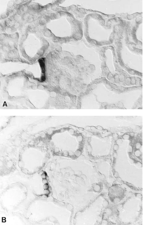
Tamm-Horsfall protein reactive TALH cells (14): on the same section, Tamm-Horsfall protein-unreactive MD cells clearly expressed NKCC2 mRNA (Fig. 4). Second, MD cells were identified by preparing consecutive sections for NKCC2 mRNA in situ hy-bridization and histochemical diaphorase staining, respec-tively. Earlier studies (10, 15) indicated that MD cells from several species stain positively for diaphorase; specifically, in both rabbit and rat kidney MD cells are the only epithelial cells of the entire nephron and collecting duct system that show significant nitric oxide synthase activity (10). Figs. 6 and 7 show a clear coincidence of NKCC2 mRNA expression and NADPH-diaphorase staining in all MD cells.
Discussion
[image:5.612.57.297.60.422.2]These experiments document NKCC2 mRNA expression along the distal nephron of both rat and rabbit kidney. The technique of combined in situ hybridization with immunohis-tochemistry and NADPH-diaphorase hisimmunohis-tochemistry permit-ted a precise delineation of the onset and distal extent of NKCC2 mRNA expression with particular respect to the MD
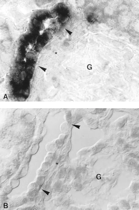
[image:5.612.315.554.60.413.2]Figure 5. Higher resolution of NKCC2 expression in rat macula densa (between arrowheads) by antisense in situ hybridization (A). B shows analogous topography with typical macula densa cells adjacent to the extraglomerular mesangium (asterisks in A and B). G, glomerulus. Magnification 3680.
Figure. 6. Labeling of the juxtaglomerular portion of TALH in rat kidney using in situ hybridization with NKCC2 antisense probe (A) and NADPH-diaphorase staining (B) on consecutive sections. Mac-ula densa cells (between arrowheads) are positively stained in A and
cells. By non-isotopic in situ hybridization, NKCC2 expression has been localized to the entire TALH of rat and rabbit kid-ney. To confirm the identity of cells that express NKCC2 mRNA, an anti-Tamm-Horsfall protein antibody was em-ployed. In rat, this antibody recognizes cells of the entire TALH except for the MD (14, 11). Thus in rat, double labeling with the in situ hybridization NKCC2 probe and anti-Tamm-Horsfall protein antibody revealed clear overlap. Onset of the signal was at the cortico-medullary border; the signal ended at the transition of the post-macula densa segment of TALH to the distal convoluted tubule. The pattern of NKCC2 expres-sion in rabbit appeared to be much the same as in rat, even though double staining with Tamm-Horsfall protein was not performed. Although previous studies have suggested that NKCC2 is expressed in additional nephron segments (5), NKCC2 in the present study was shown to be restricted to the TALH, at least within the methodological limitations of the in situ hybridization procedure. Low but physiologically signifi-cant expression of NKCC2 outside the TALH or expression of
structurally different furosemide/bumetanide-sensitive trans-port proteins is not excluded by the current results. Further, under the conditions of in situ hybridization used in the cur-rent experiments, it is unlikely that NKCC1 (BSC2), the secre-tory form of the furosemide/bumetanide-sensitive Na-K-2Cl cotransporter, was detected. There is only 62% nucleotide identity between the two forms of the furosemide/bumetanide-sensitive transporter within the region of the nucleotide probe used in the current experiments, and there are not smaller re-gions of significantly higher homology.
The difference between the robust expression by TALH cells and the complete absence of signal elsewhere suggest that NKCC2 expression is highly cell type-specific. Our results are corroborated by three previous in situ hybridization studies demonstrating Na-K-Cl-cotransporter expression in mouse (7) and rat kidney (4, 8) along the TALH. Forbush and colleagues have reported that NKCC2 is alternatively spliced, leading to expression of three unique isoforms in rabbit and mouse (5, 7). In mouse isoform F is expressed in the inner stripe of the outer medulla; isoform A is expressed predominantly in the outer stripe of the outer medulla; and isoform B is expressed most highly in the cortical TALH (7). The different isoforms differ only in a 96-bp sequence encoding a portion of the second pu-tative transmembrane segment. However, the probe used in the present study does not distinguish between the isoforms because it does not encompass any of the alternatively spliced region. Thus, no obvious differences in TALH subsegment-specific expression of NKCC2 were evident in the current ex-periments. Rather, labeling intensity appeared to be much the same in cortical and medullary TALH when considered per unit sectioned cell surface. In rabbit, however, cortical TALH labeling appeared considerably weaker than in the medulla. This difference may reflect an axial heterogeneity of Na trans-port previously noted by Knepper and Burg (16).
[image:6.612.58.298.60.437.2]The most prominent result of the present study is the cellu-lar identification of NKCC2 mRNA label in the juxtaglomeru-lar portion of rat and rabbit TALH. Igarashi et al. (7), who used radiolabeled probes, suggested that juxtaglomerular por-tions of TALH express high levels of NKCC2 isoform B in mouse kidney; yet owing to insufficient structural resolution of the autoradiographic label, the NKCC2 signal in their study could not be clearly assigned to the MD. To address the prob-lem of proper signal identification at the MD, we have combined high-resolution interference contrast microscopic non-isotopic in situ hybridization for NKCC2 with immunohistochemical detection of THP and histochemical detection of NADPH-dia-phorase reaction. The results indicate that NKCC2 mRNA is expressed in juxtaglomerular portions of TALH in rat and rab-bit that are clearly identified as MD cells. This identification is based on the characteristic cellular structure, the concomitant activity of NADPH-diaphorase and, in rat, on the absence of intracellular anti-THP staining. This result is in agreement with previous studies which have demonstrated by physiologi-cal measurements that NaCl movement across the luminal membrane of macula densa cells occurs, at least in part, through a Na-K-2Cl cotransporter (1, 9, 17). Apical Na-K-2Cl cotransport is potentially involved as a major factor linking single nephron GFR with tubular reabsorption (2). The results are not consistent with a recent report by Kaplan et al. (8) in which antibodies to NKCC2 were used to study its localization along the nephron. Those results showed that NKCC2 was present at the apical membrane of rat TALH cells but was
absent from the MD. More recent work by the same group, however, has shown that pre-treatment of renal tissue with microwaves unmasks NKCC2 immunoreactivity at the apical membrane of MD cells (M. Kaplan, personal communication), in agreement with the current results.
The intensity of NKCC2 mRNA expression is roughly equal in MD and surrounding TALH epithelium in rat; in rab-bit, however, NKCC2 expression is even significantly more in-tense in the MD compared to cortical TALH cells. Increased mRNA expression may reflect either enhanced transcription of NKCC2 mRNA or altered mRNA stability; increased ex-pression would be expected to lead to a higher synthetic rate for the cotransporter, perhaps contributing to the distinct ki-netic properties of the Na-K-2Cl cotransporter in MD, com-pared with surrounding TALH (1, 9, 18).
Coexpression of NO synthase activity reflected by NADPH-diaphorase staining (10), and NKCC2 expression within MD cells may represent two compounds of the tubulovascular sig-naling mechanism. NKCC2 expression may participate impor-tantly in luminal sensing of distal tubular [NaCl], and NO is currently considered a prominent modulator in MD-mediated information transfer at the juxtaglomerular apparatus (19, 20, 21). However, to date there is no direct evidence for a func-tional link between NKCC2-dependent salt transport and the release of NO from or within MD cells.
In conclusion, this study describes expression of NKCC2 mRNA along the entire TALH from the cortico-medullary boundary to its end at the transition to the DCT. The distribu-tion corresponds to the immunoreactivity pattern of Tamm-Horsfall protein with the exception of the macula densa, which is lacking THP but solidly expresses NKCC2 mRNA. Thus, morphological evidence is provided in rat and rabbit kidney for a previously established functional concept that Na-K-2Cl cotransport at the TALH is accomplished by the expression of a well-defined cotransporter. At the MD, this transporter may establish the link between tubular salt load and glomerular vascular regulation.
Acknowledgments
This work was supported by funds from the Deutsche Forschungsge-meinschaft (Ba 700/10-1) and the American Heart Association and the Department of Veterans Affairs, and was conducted during the tenure of an Established Investigatorship of the American Heart As-sociation (DHE). Nicholas Obermüller was a scholar from the Heidelberger Graduiertenkolleg für experimentelle Nieren- und Kreislaufforschung which was funded by the Deutsche Forschungsge-meinschaft.
References
1. Schlatter, E., M. Salomonsson, A.E.G. Persson, and R. Greger. 1989. Macula densa cells sense luminal NaCl concentration via furosemide sensitive
Na12Cl2K1 cotransport. Pflugers Arch. 414:286–290.
2. Schnermann, J., and J.P. Briggs. 1992. Function of the juxtaglomerular
apparatus. In The Kidney: Physiology and Pathophysiology. D.W. Seldin and
G. Giebisch, editors. Raven Press, Ltd., Orlando. 1249–1289.
3. Xu, J.-C., C. Lytle, T. Zhu, J.A. Payne, E. Benz, Jr., and B. Forbush, III. 1994. Molecular cloning and functional expression of the bumetanide-sensitive
Na-K-Cl cotransporter. Proc. Natl. Acad. Sci. USA. 92:2201–2205.
4. Gamba, G., A. Miyanoshita, M. Lombardi, J. Lytton, W.-S. Lee, M. He-diger, and S.C. Hebert. 1994. Molecular cloning, primary structure, and charac-terization of two members of the mammalian electroneutral
sodium-(potas-sium)-chloride cotransporter family expressed in kidney. J. Biol. Chem. 269:
17713–17722.
5. Payne, J.A., and B. Forbush, III. 1994. Alternatively spliced isoforms of the putative renal Na-K-Cl cotransporter are differentially distributed within
rabbit kidney. Proc. Natl. Acad. Sci. USA. 91:4544–4548.
6. Haas, M., and B. Forbush, III. 1987. Photolabeling of a 150-kDa (Na 1 K
1 Cl) cotransport protein from dog kidney with a bumetanide analogue. Am. J.
Physiol. 253:C243–C252.
7. Igarashi, P., G.B. Vanden Heuvel, J.A. Payne, and B. Forbush, III. 1995. Cloning, embryonic expression, and alternative splicing of a murine
kidney-spe-cific Na-K-Cl cotransporter. Am. J. Physiol. 269:F405–F418.
8. Kaplan, M.R., M.D. Plotkin, W.S. Lee, Z.C. Xu, J. Lytton, and S. Hebert. 1996. Apical localization of the Na-K-Cl-cotransporter, rBSC1, on rat thick
as-cending limbs. Kidney Int. 49:40–47.
9. Lapointe, J.-Y., A. Laamarti, A.M. Hurst, B.C. Fowler, and P.D. Bell. 1995. Activation of Na:2Cl:K cotransport by luminal chloride in macula densa
cells. Kidney Int. 47:752–757.
10. Bachmann, S., H.M. Bosse, and P. Mundel. 1995. Topography of nitric oxide synthesis by localizing constitutive NO synthases in mammalian kidney.
Am. J. Physiol. 268:F885–F898.
11. Obermüller, N., P.L. Bernstein, H. Velázquez, R. Reilly, D. Moser, D.H. Ellison, and S. Bachmann. 1995. Expression of the thiazide-sensitive Na-Cl
cotransporter in rat and human kidney. Am. J. Physiol. 269:F900–F910.
12. Sanger, F., S. Nicklen, and A.R. Coulson. 1977. DNA sequencing with
chain-terminating inhibitors. Proc. Natl. Acad. Sci. USA. 54:5463–5467.
13. Bachmann, S., M. Le Hir, and K.U. Eckardt. 1993. Co-localization of
erythropoietin mRNA and ecto-59-nucleotidase immunoreactivity in
peritubu-lar cells of rat renal cortex indicates that fibroblasts produce erythropoietin. J.
Histochem. Cytochem. 41:335–341.
14. Hoyer, J.R., S.P. Sisson, and R.L. Vernier. 1979. Tamm-Horsfall
glyco-protein: ultrastructural immunoperoxidase localization in rat kidney. Lab.
In-vest. 41:168–173.
15. Bachmann, S., and P. Mundel. 1994. Nitric oxide in the kidney:
synthe-sis, localization, and function. Am. J. Kid. Dis. 24:112–129.
16. Knepper, M.A., and M.B. Burg. 1983. Organization of nephron
func-tion. Am. J. Physiol. 244:F579–F589.
17. Bell, P.D., J.-Y. Lapointe, J. Cardinal, and Y.-S. Chang. 1991. Transport
pathways in macula densa cells. Kidney Int. 39 (Suppl. 32): S59–S64.
18. Lapointe, J.-Y., P.D. Bell, and J. Cardinal. 1990. Direct evidence for
api-cal Na1:2Cl2:K1 cotransport in macula densa cells. Am. J. Physiol. 258:F1466–
F1469.
19. Wilcox, C.S., W.J. Welch, F. Murad, S.S. Gross, G. Taylor, R. Levi, and H.H.H.W. Schmidt. 1992. Nitric oxide synthase in macula densa regulates
glom-erular capillary pressure. Proc. Natl. Acad. Sci. USA. 89:11993–11997.
20. Ito, S. Role of nitric oxide in glomerular arterioles and macula densa.
1994. News Physiol. Sci. 9:115–119.
21. Bosse, H.M., R. Böhm, S. Resch, and S. Bachmann. 1995. Parallel regu-lation of constitutive NO synthase and renin at JGA of rat kidney under various