0095-1137/96/$04.0010
Copyrightq1996, American Society for Microbiology
HSP60 Gene Sequences as Universal Targets for
Microbial Species Identification: Studies with
Coagulase-Negative Staphylococci
SWEE HAN GOH,
1,2,3† SHEILA POTTER,
1,2JULIAN O. WOOD,
1,2SEAN M. HEMMINGSEN,
4ROBERT P. REYNOLDS,
1,2ANDANTHONY W. CHOW
1,2,3,5*
Departments of Medicine
1and Microbiology and Immunology,
5Division of Infectious Diseases, University of
British Columbia, the Vancouver Hospital Health Sciences Centre,
2and the Canadian Bacterial
Diseases Network,
3Vancouver, British Columbia, and the Canadian National Research
Council Plant Biotechnology Institute, Saskatoon, Saskatchewan,
4Canada
Received 25 September 1995/Returned for modification 17 November 1995/Accepted 19 January 1996
A set of universal degenerate primers which amplified, by PCR, a 600-bp oligomer encoding a portion of the
60-kDa heat shock protein (HSP60) of both
Staphylococcus aureus
and
Staphylococcus epidermidis
were
devel-oped. However, when used as a DNA probe, the 600-bp PCR product generated from
S. epidermidis
failed to
cross-hybridize under high-stringency conditions with the genomic DNA of
S. aureus
and vice versa. To
investigate whether species-specific sequences might exist within the highly conserved HSP60 genes among
different staphylococci, digoxigenin-labelled HSP60 probes generated by the degenerate HSP60 primers were
prepared from the six most commonly isolated
Staphylococcus
species (
S. aureus
8325-4,
S. epidermidis
9759,
S.
haemolyticus
ATCC 29970,
S. schleiferi
ATCC 43808,
S. saprophyticus
KL122, and
S. lugdunensis
CRSN 850412).
These probes were used for dot blot hybridization with genomic DNA of 58 reference and clinical isolates of
Staphylococcus
and non-
Staphylococcus
species. These six
Staphylococcus
species HSP60 probes correctly
iden-tified the entire set of staphylococcal isolates. The species specificity of these HSP60 probes was further
demonstrated by dot blot hybridization with PCR-amplified DNA from mixed cultures of different
Staphylo-coccus
species and by the partial DNA sequences of these probes. In addition, sequence homology searches of
the NCBI BLAST databases with these partial HSP60 DNA sequences yielded the highest matching scores for
both
S. epidermidis
and
S. aureus
with the corresponding species-specified probes. Finally, the HSP60
degen-erate primers were shown to amplify an anticipated 600-bp PCR product from all 29
Staphylococcus
species and
from all but 2 of 30 other microbial species, including various gram-positive and gram-negative bacteria,
mycobacteria, and fungi. These preliminary data suggest the presence of species-specific sequence variation
within the highly conserved HSP60 genes of staphylococci. Further work is required to determine whether these
degenerate HSP60 primers may be exploited for species-specific microbic identification and phylogenetic
investigation of staphylococci and perhaps other microorganisms in general.
Coagulase-negative staphylococci have emerged as
predom-inant pathogens in hospital-acquired infections (3, 16, 17). In
light of this, it has become increasingly important to accurately
identify these isolates to the species level in reference
labora-tories in order to further define the epidemiology and clinical
significance of these microorganisms in the hospital setting.
Several commercial kits are now available for the identification
of coagulase-negative staphylococci to the species level (13,
21). Unfortunately, the overall accuracy of these systems,
which are based on phenotypic discrimination, has been low,
ranging from 50 to 70%. Genotypic methods of identification,
either directed at unique genes specific for a microbial species
or directed at unique sequences of ubiquitous genes such as
16S rRNA, may provide better results in terms of both
sensi-tivity and specificity (14, 16a, 23). Several nucleic acid-based
methods for species identification of staphylococci have been
reported and are primarily based on restriction analysis of the
16S rRNA genes (4, 7, 8, 10, 20, 27). Besides being
labor-intensive, a potential drawback with these procedures is
within-species microheterogeneity, presumably because there may be
multiple, evolutionarily diverged rRNA gene copies in some
microorganisms (23). A more ideal universal DNA target for
microbial identification to the species level would be one which
has well-conserved DNA sequences within a given species, but
with sufficient sequence variation to allow for species-specific
identification. Here, we present preliminary data that the
ubiq-uitous and highly conserved, single-copy 60-kDa heat shock
protein (HSP60) [also known as GroEL]) genes may be an
alternate DNA target for species-specific identification of
staphylococci and perhaps other microorganisms in general.
We have designed and synthesized a set of universal
degen-erate primers based on highly conserved regions within the
HSP60 genes from different microorganisms reported in the
published literature. These degenerate primers amplified by
PCR an anticipated 600-bp product from both Staphylococcus
aureus and Staphylococcus epidermidis. However, when used as
a DNA probe, the 600-bp PCR product generated from S.
epidermidis failed to cross-hybridize under high-stringency
con-ditions with the genomic DNA of S. aureus and vice versa (data
not shown). This suggested to us that there may be variable
sequences within the highly conserved HSP60 genes that may
* Corresponding author. Mailing address: Division of InfectiousDiseases, G. F. Strong Research Laboratory, Vancouver Hospital Health Sciences Centre, 2733 Heather St., Vancouver, British Colum-bia, Canada V5Z 3J5. Phone: (604) 875-4148. Fax: (604) 875-4013.
† Present address: Department of Pathology and Laboratory Med-icine, University of British Columbia, and the Provincial Laboratory, British Columbia Centre for Disease Control, Vancouver, British Co-lumbia, Canada V5Z 1L8.
818
on May 15, 2020 by guest
http://jcm.asm.org/
be useful for species-specific microbic identification among
different staphylococci. To test this hypothesis, DNA probes
were prepared from 600-bp PCR products generated by the
degenerate HSP60 primers from six reference staphylococcal
species (S. aureus 8325-4, S. epidermidis 9759, S. haemolyticus
ATCC 29970, S. lugdunensis CRSN 850412, S. saprophyticus
KL122, and S. schleiferi ATCC 43808). These were used in dot
blot hybridization studies to identify a set of 58 reference and
clinical isolates of Staphylococcus and non-Staphylococcus
spe-cies in a coded fashion. The results, which demonstrated 100%
accuracy in species identification of staphylococci compared
with that of the reference method of Kloos and Lambe (17),
strongly suggest that species-specific sequence variation exists
within the highly conserved HSP60 genes of staphylococci.
MATERIALS AND METHODS
Bacterial isolates.The bacterial isolates used in this study consisted of 35
reference strains of various Staphylococcus species obtained from the American Type Culture Collection and from W. Kloos, North Carolina State University, Raleigh, N.C., and 20 clinical isolates from our own collection identified in the Clinical Microbiology Laboratory of the Vancouver Hospital and Health Sci-ences Centre, Vancouver, British Columbia, Canada (Table 1). In addition, three non-staphylococcal reference isolates, Escherichia coli ATCC 25922, Pseudomo-nas aeruginosa ATCC 27853, and Bacillus subtilis ATCC 12432, were included as negative controls. All cultures were grown in brain heart infusion (BHI) broth and subcultured on BHI plates for examination of purity and colony character-istics. Clinical isolates that gave either false-positive or false-negative results when probed with the 600-bp HSP60 staphylococcal species probes were coded and sent to the reference laboratory, the Provincial Laboratory of the British Columbia Centre for Disease Control, Vancouver, for reidentification according to the method of Kloos and Lambe (17).
Isolation of genomic DNA.Genomic DNA of pure cultures after overnight
growth in BHI broth were prepared by the standard sodium dodecyl sulfate (SDS)-proteinase K-cetyl trimethyl ammonium bromide-phenol-chloroform method (2). For staphylococci, lysostaphin (from Sigma, or a recombinant prod-uct from Applied Microbiology Inc., New York, N.Y.) was substituted for ly-sozyme in facilitating cell lysis. DNA was resuspended in TE buffer (10 mM Tris, [pH 8.0], 1 mM EDTA), the concentration was determined by UV spectroscopy at A260, and the purity was estimated by the A260/A280ratio.
Input DNAs of mixed staphylococcal species for PCR experiments were pre-pared by pooling of 50ml of overnight cultures in BHI broth from different organisms in various combinations. Crude DNA was prepared with the Instagene purification matrix (Bio-Rad) according to the manufacturer’s instructions. Uni-noculated BHI broth was mock processed and used as a negative control. The final extract was in 500ml of purification matrix, and 20ml of the matrix extract was used as the target DNA for PCR.
PCR amplification.The PCR mixture contained (in final concentration) 50
mM KCl, 10 mM Tris (pH 8.3), 1.5 mM MgCl2, 200mM (each) deoxynucleoside triphosphate (dNTP), 50 ng of genomic DNA or 20ml of Instagene extract, 2 U of Taq DNA polymerase (GIBCO), and 0.5mg of each of the degenerate HSP60 primers, in a final volume made up to 100ml with distilled H2O. The sequences of the 59and 39HSP60 primers, designated H279 and H280, are 59-GAATTC GAIIIIGCIGGIGA(TC)GGIACIACIAC-39 and 59-CGCGGGATCC(TC)(T G)I(TC)(TG)ITCICC(AG)AAICCIGGIGC(TC)TT-39, respectively. Inosine (I) was used to reduce the degeneracy of the primers. The thermal cycling conditions were 3 min at 958C for 1 cycle, followed by 40 cycles of 1 min at 948C, 2 min at 378C, and 5 min at 728C. The last cycle was for 10 min at 728C. After PCR amplification, 10ml of each reaction mixture was analyzed on a 2.0% TAE (Tris-acetate-EDTA) agarose gel. The DNA fragments were visualized and pho-tographed under UV light after ethidium bromide staining.
Purification and digoxigenin labelling of 600-bp HSP60 PCR products.The
600-bp PCR products amplified from genomic DNA prepared from S. aureus (8325-4), S. epidermidis (9759), S. haemolyticus (ATCC 29970), S. lugdunensis (CRSN 850412), S. saprophyticus (KL 122), and S. schleiferi (ATCC 43808) were purified by electrophoresis on a 2.0% low-melting-point agarose gel and ex-tracted with eitherb-agarase I (New England Biolabs) or the QIA Quick Gel extraction kit (Qiagen Inc.) according to manufacturer’s instructions. Labelling of the 600-bp fragments for use as DNA probes was carried out with digoxigenin– 11-dUTP and the standard random primer method (24) according to Boehringer Mannheim protocols.
Dot blot hybridization.Genomic DNAs (300 ng, 0.4 M NaOH denatured) of
pure cultures or PCR-amplified DNAs from mixed cultures were dot blotted onto nylon membranes (Boehringer Mannheim). For mixed cultures, an over-night growth in BHI broth was first subjected to PCR amplification with the degenerate HSP60 primers as described earlier. PCR-amplified DNA was then purified with the QIA Quick-spin PCR purification kit (Qiagen Inc.). A 1:8,000 dilution of the purified PCR product in 50ml of distilled H2O was prepared, and
5ml of each of the diluted samples was spotted on the filters. After baking of the filters at 1208C for 30 min, the blots were neutralized with 0.5 M Tris (pH 7.5) and then dried before use. The filters were prehybridized at 428C in 50% form-amide–53SSC (13SSC is 0.15 M NaCl plus 0.015 M sodium citrate)–2% Boehringer Mannheim blocking reagent–0.1% N-lauryl sarcosine–0.02% SDS for at least 1 h. Hybridization with the same prehybridization buffer and digoxi-genin probes (40 to 50 ng/ml) was allowed to proceed overnight at 428C. After hybridization, the filters were washed sequentially with 23SSC–0.1% (wt/vol) SDS twice for 15 min at room temperature and then with 0.13SSC–0.1% SDS twice for 15 min at 688C. Detection of hybridization by chemiluminescence was performed as described in the Boehringer Mannheim protocols.
DNA sequencing.Direct DNA sequencing of the six Staphylococcus species
HSP60 probes was performed by the fluorescence-based dideoxy termination method with a cycle sequencing protocol and reagents supplied by Applied Biosystems, Inc. (26). The cycle sequencing reaction mixture contained 400 mM Tris-HCl; 10 mM MgCl2; 100 mM (NH4)2SO4(pH 9.0); 750mM dITP; 150mM (each) dATP, dTTP, and dCTP; 150mM (each) dye-labeled ddNTP; 4 U of AmpliTaq DNA polymerase; 7ml of template DNA; and 3.2 pM either primer H279 or primer H280, in a final volume made up with distilled H2O to 20ml. The thermal cycling conditions were 30 s at 968C, 15 s at 508C, and 4 min at 608C for 25 cycles. Sequenced products were purified with the QIA Quick-spin PCR purification kit, ethanol precipitated, and resuspended in 4ml of loading buffer (5:1 [vol/vol] deionized formamide–50 mM EDTA [pH 8.0]). Samples were heated at 908C for 2 min prior to loading on a 6% (wt/vol) polyacrylamide gel containing 7 M urea for electrophoresis and sequencing in an automated DNA sequencer (Applied Biosystems model 373A). Emission data from the fluores-cence-tagged reaction mixtures were collected and analyzed with the proprietary Macintosh-based software (version 1.2.0). The automatically assigned base calls were examined with the analysis software, and the sequence was edited manually when necessary. A nucleotide sequence homology search was performed through the National Center for Biotechnology Information (NCBI) BLAST Network Service according to the algorithm of Altschul et al. (1). A phylogenetic tree of the six HSP60 staphylococcal species probes was derived from multiple sequence alignments based on the Higgins-Sharp algorithm with the CLUSTAL4 package (15).
RESULTS
Dot blot hybridization of genomic DNA from pure cultures.
The results of dot blot hybridization of the 58 reference and
clinical isolates of Staphylococcus and non-Staphylococcus
spe-cies with the six Staphylococcus spespe-cies HSP60 probes are
summarized in Table 1. An example of the dot blot results
from the S. epidermidis HSP60 probe is shown in Fig. 1. None
of the control isolates, E. coli ATCC 25922, P. aeruginosa
ATCC 27853, and B. subtilis ATCC 12432, gave positive
hy-bridization signals with any of the six Staphylococcus species
HSP60 probes. The dot blot results from all 35 reference
iso-lates of Staphylococcus species with the six HSP60 probes were
in complete agreement regarding their species designation. In
contrast, 7 of 20 (35%) clinical staphylococcal isolates gave
either false-positive or false-negative results when hybridized
with the six Staphylococcus species HSP60 probes. These seven
clinical isolates included two strains identified as S.
haemolyti-cus by the clinical microbiology laboratory, which did not
hy-bridize with the S. haemolyticus probe but hyhy-bridized with the
S. epidermidis probe and the S. lugdunensis probe, respectively;
one strain identified as S. saprophyticus, which did not
hybrid-ize with the S. saprophyticus probe but hybridhybrid-ized with the S.
epidermidis probe; and four other strains identified as S.
homi-nis, S. sciuri, S. xylosus, and S. capitis, which hybridized with the
S. epidermidis probe, the S. aureus probe, the S. lugdunensis
probe, and the S. haemolyticus probe, respectively. These seven
clinical isolates were coded and sent to the provincial reference
laboratory in a blinded fashion for reidentification according to
the method of Kloos and Lambe (17). The results obtained
from the reference laboratory indicated that all seven isolates
had been previously misidentified in the clinical microbiology
laboratory and that their corrected species designation agreed
completely with the hybridization results obtained with the
Staphylococcus species HSP60 probes (Table 2). Thus, all three
apparently false-negative results from the HSP60 probes were
in fact true negative, and all seven apparently false-positive
on May 15, 2020 by guest
http://jcm.asm.org/
results were in fact true-positive. Taken together, the dot blot
results with these six Staphylococcus species HSP60 probes
were 100% accurate when tested with the set of 58 bacterial
isolates compared with those tested by the reference
identifi-cation method of Kloos and Lambe (17).
Dot blot hybridization with HSP60 products from mixed
cultures.
The species specificity of the HSP60 probes was
as-sessed in two sequential steps: by determination of whether the
degenerate primers can amplify species-specific staphylococcal
targets from mixed cultures by PCR and whether
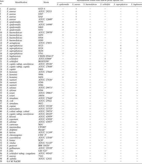
species-spe-TABLE 1. Order of genomic DNA used in dot blots and hybridization resultsIsolate
no. Identification Strain
Hybridization results with HSP60 probe specific for:
S. epidermidis S. aureus S. haemolyticus S. schleiferi S. saprophyticus S. lugdunensis
1 S. aureus 8325-4 2 1 2 2 2 2
2 S. aureus ATCC 29213 2 1 2 2 2 2
3 S. aureus 8387 2 1 2 2 2 2
4 S. aureus SA8 2 1 2 2 2 2
5 S. aureus ATCC 12600a 2 1 2 2 2 2
6 S. epidermidis 9759 1 2 2 2 2 2
7 S. epidermidis ATCC 14990a 1 2 2 2 2 2
8 S. epidermidis 8469 1 2 2 2 2 2
9 S. epidermidis 8331 1 2 2 2 2 2
10 S. haemolyticus ATCC 29970a 2 2 1 2 2 2
11 S. haemolyticus 8459 1 2 2 2 2 2
12 S. haemolyticus 8564 2 2 1 2 2 2
13 S. haemolyticus 8589 2 2 2 2 2 1
14 P. aeruginosa ATCC 27853 2 2 2 2 2 2
15 S. saprophyticus 8523 2 2 2 2 1 2
16 S. saprophyticus 8524 2 2 2 2 1 2
17 S. saprophyticus 8638 2 2 2 2 1 2
18 S. saprophyticus 9761 1 2 2 2 2 2
19 S. lugdunensis CRSN 850412a 2 2 2 2 2 1
20 S. schleiferi ATCC43808a 2 2 2 1 2 2
21 S. schleiferi BOSTONa 2 2 2 1 2 2
22 S. capitis subsp. ureolyticus ATCC 49326a 2 2 2 2 2 2
23 S. capitis subsp. capitis ATCC 27840a 2 2 2 2 2 2
24 S. capitis 8468 2 2 1 2 2 2
25 S. hominis ATCC 27844a 2 2 2 2 2 2
26 S. hominis 9998 2 2 2 2 2 2
27 S. hominis 8458 1 2 2 2 2 2
28 S. warneri ATCC 27836a 2 2 2 2 2 2
29 S. warneri 9290 2 2 2 2 2 2
30 S. warneri 8586 2 2 2 2 2 2
31 S. xylosus ATCC 29971a 2 2 2 2 2 2
32 S. xylosus 8584 2 2 2 2 2 1
33 S. sciuri ATCC 29061a 2 2 2 2 2 2
34 S. sciuri 10034 2 1 2 2 2 2
35 S. simulans ATCC 27848a 2 2 2 2 2 2
36 E. coli ATCC 25922 2 2 2 2 2 2
37 S. simulans 9852 2 2 2 2 2 2
38 S. caprae ATCC 35538a 2 2 2 2 2 2
39 S. auricularis ATCC 33753a 2 2 2 2 2 2
40 S. cohnii subsp. cohnii ATCC 29974a 2 2 2 2 2 2
41 S. capitis subsp. ureolyticus ATCC 49325a 2 2 2 2 2 2
42 S. kloosii ATCC 43959a 2 2 2 2 2 2
43 S. equorum ATCC 43958a 2 2 2 2 2 2
44 S. arlettae ATCC 43957a 2 2 2 2 2 2
45 S. carnosus MAa 2 2 2 2 2 2
46 S. intermedius CFDDa 2 2 2 2 2 2
47 S. delphini Heidya 2 2 2 2 2 2
48 S. hyicus ATCC 11249a 2 2 2 2 2 2
49 S. chromogenes CDC 2a 2 2 2 2 2 2
50 S. caseolyticus ATCC 13548a 2 2 2 2 2 2
51 S. lentus K20a 2 2 2 2 2 2
52 S. vitulus ATCC 51145a 2 2 2 2 2 2
53 S. pasteuri BM 10426a 2 2 2 2 2 2
54 S. gallinarum ATCC 35539a 2 2 2 2 2 2
55 S. felis GD 521a 2 2 2 2 2 2
56 S. schleiferi subsp. coagulans ATCC 49545a 2 2 2 1 2 2
57 S. aureus 7162 2 1 2 2 2 2
58 B. subtilis ATCC 12432 2 2 2 2 2 2
59 0.4 M NaOH 2 2 2 2 2 2
aIsolates kindly provided by W. Kloos, North Carolina St. University, Raleigh.
on May 15, 2020 by guest
http://jcm.asm.org/
[image:3.612.66.550.90.650.2]cific HSP60 probes can identify the correct target from mixed
PCR products by dot blot hybridization. Mixed PCR targets
containing various combinations of all but one of the six
dif-ferent Staphylococcus species (i.e., S. aureus, S. epidermidis, S.
haemolyticus, S. lugdunensis, S. saprophyticus, and S. schleiferi)
were prepared with the Bio-Rad Instagene purification matrix
and were amplified by PCR with our degenerate HSP60
prim-ers (Fig. 2a). PCR-amplified mixed-species DNAs were probed
with each of four Staphylococcus species HSP60 probes (i.e., S.
aureus, S. epidermidis, S. haemolyticus, and S. lugdunensis) (Fig.
2b). In all instances, when the four respective probes were
used, there was either absent or markedly reduced signal in the
hybridized mixed-species DNA targets lacking the
probe-spec-ified Staphylococcus species DNA, indicating the excellent
spe-cies specificity of these HSP60 probes (Fig. 2b). Although
some residual background signal can be observed in the blots
hybridized with the S. haemolyticus and S. aureus probes, the
signal-to-noise ratio was high with each probe compared with
those of the positive and negative controls (Fig. 2b, lanes 1 and
2).
Partial DNA sequences and phylogenetic tree of HSP60
Staphylococcus
species probes.
Results from the DNA
[image:4.612.60.296.72.216.2]se-quences of the six HSP60 Staphylococcus species probes are
only partially complete (data not shown and to be separately
reported). However, species-specific sequence variation within
the 600-bp probes was clearly evident in each instance.
Fur-thermore, nucleotide sequence homology searches of
data-bases available through the NCBI BLAST Network Service
with these partial HSP60 DNA sequences yielded the highest
matching scores for both S. epidermidis and S. aureus with the
corresponding species-specified probes, the only two
staphylo-coccal species for which HSP60 gene sequence data have been
published (Table 3). Finally, the partial HSP60 staphylococcal
species DNA sequences were also analyzed by multiple
align-ments, and a tentative phylogenetic tree demonstrating the
[image:4.612.316.551.77.292.2]FIG. 1. Determination of the specificity of the S. epidermidis 9759 HSP60 probe by dot blot hybridization to bacterial chromosomal DNA. Numbers refer to the numerical order and designation of bacterial isolates listed in Table 1. Positive hybridization reactions were observed for isolates 6 (S. epidermidis), 7 (S. epidermidis), 8 (S. epidermidis), 9 (S. epidermidis), 11 (misidentified as S. haemolyticus by the clinical laboratory and confirmed as S. epidermidis by the reference laboratory), 18 (misidentified as S. saprophyticus by the clinical labo-ratory and confirmed as S. epidermidis by the reference labolabo-ratory), and 27 (misidentified as S. hominis by the clinical laboratory and confirmed as S. epi-dermidis by the reference laboratory).
FIG. 2. Determination of the species specificity of Staphylococcus HSP60 probes by dot blot hybridization with PCR-amplified DNA from mixed cultures. (a) Mixed cultures consisting of the combination of different Staphylococcus species (represented by numbers shown at the top [1, present;2, absent]) were first prepared from overnight cultures and used as target DNA for PCR ampli-fication with the degenerate HSP60 primers. (b) PCR-amplified DNAs from mixed cultures (corresponding to the numbers at the top in panel a) were used for dot blot hybridization with each of four Staphylococcus species HSP60 probes. Absent or markedly reduced signals were observed in the blots lacking the probe-specified Staphylococcus species DNA.
TABLE 2. Species designation of seven clinical isolates with apparent false-negative and false-positive hybridization reactions with the six HSP60 staphylococcal species probes
Isolate
Staphylococcus species designation Positive hybridization
with HSP60 staphylococcal
probes Clinical
laboratory
Reference laboratory
9761 S. saprophyticus S. epidermidis S. epidermidis
8458 S. hominis S. epidermidis S. epidermidis
10034 S. sciuri S. aureus S. aureus
8468 S. capitis S. haemolyticus S. haemolyticus
8459 S. haemolyticus S. epidermidis S. epidermidis
8584 S. xylosus S. lugdunensis S. lugdunensis
8589 S. haemolyticus S. lugdunensis S. lugdunensis
TABLE 3. Results of homology searches of Staphylococcus species partial HSP60 DNA sequences from databases through the
NCBI BLAST Network Service
Staphylococcus species HSP60 sequence
High-scoring sequence identified from databases
S. epidermidis HSP60a S. aureus HSP60b
%
Similarity Probability
c %
Similarity Probability c
S. epidermidis 95 6.0 e2136 78 2.1 e278
S. aureus 80 4.2 e2102 93 3.2 e2135
S. haemolyticus 80 2.0 e2101 82 1.8 e293
S. lugdunensis 80 3.1 e285 82 4.8 e280
S. saprophyticus 82 2.4 e288 82 2.7 e276
S. schleiferi 77 7.4 e2100 79 1.4 e286
a
GenBank accession number U13618 (12). b
GenBank accession number D14711 (19). c
Probability of a random match.
on May 15, 2020 by guest
http://jcm.asm.org/
[image:4.612.316.556.588.703.2] [image:4.612.57.297.612.727.2]interspecies relationships among these six HSP60 probes was
derived (Fig. 3). The data revealed that S. aureus and S.
hae-molyticus are highly related (match ratio, 69.7%). Similarly, S.
lugdunensis and S. saprophyticus are highly related (match
ra-tio, 72.3%). Both of these clusters are related to S. epidermidis
(match ratios, 65.1 and 70.0%, respectively), while S. schleiferi
is the least related species (match ratio, 53.1%).
DISCUSSION
The finding that our six staphylococcal HSP60 probes
accu-rately identified all 35 reference isolates of various
Staphylo-coccus species, including S. aureus and five species representing
the most clinically important coagulase-negative staphylococci, is
most encouraging. The observation that 7 of 20 clinical isolates
of coagulase-negative staphylococci were misidentified by
com-mercially available identification systems further supports the
notion that existing laboratory methods based on phenotypic
characterization of coagulase-negative staphylococci are
unre-liable.
The hybridization results from the 58 bacterial isolates in
this study and the partial DNA sequence data from the six
Staphylococcus species HSP60 probes are highly suggestive
that HSP60 gene sequences can be useful targets for species
identification of S. aureus and at least some coagulase-negative
staphylococci. Whether this approach can be successfully
ap-plied to all Staphylococcus species and perhaps other
microor-ganisms will require extensive study. Nonetheless, the selection
of the HSP60 gene as a possible universal DNA target for
microbial species identification offers a number of theoretical
advantages. First, HSP60 genes are ubiquitous in both
pro-caryotes and eupro-caryotes and encode highly conserved
house-keeping proteins which are essential for the survival of these
cells (5, 9, 18, 22, 29). These characteristics may render them
less subject to random mutations or intraspecies variation.
Second, these highly conserved proteins have been examined
in a limited fashion for phylogenetic relationships among
eu-bacterial lineages (6, 11, 28). Evolutionary trees drawn from
the protein sequences of these molecules demonstrate
remark-able similarities to those derived from 16S rRNA genes.
More-over, in some instances, these phylogenetic studies on the basis
of HSP60 analysis have indicated specific relationships
be-tween certain eubacterial groups which appear not to be clearly
defined or are controversial in rRNA-based phylogenetic
eval-uation (28). Finally, the degenerate HSP60 primers that we
have developed were capable of amplifying an anticipated
600-bp HSP60 product from all 29 Staphylococcus species
listed in Table 1 and all but 2 of 30 other microbial species,
including the following gram-positive and gram-negative
bac-teria, mycobacbac-teria, and candida: Streptococcus agalactiae and
S. pneumoniae; Enterococcus faecalis; Listeria monocytogenes;
B. subtilis; E. coli; Salmonella typhi, S. typhimurium, and S.
gallinarum; Yersinia pseudotuberculosis; Vibrio cholerae; P.
aeruginosa; Haemophilus influenzae; Campylobacter jejuni, C.
coli, and C. laridis; Helicobacter pylori and H. mustelae;
Flex-ispira rappini; Neisseria gonorrhoeae; Legionella pneumophila;
Bartonella henselae, B. quintana, and B. bacilliformis; Borrelia
hermsii; Mycobacterium marinum and M. fortuitum; and
Can-dida albicans. The two exceptions were Borrelia burgdorferi and
Mycobacterium smegmatis MC155. It is likely that the HSP60
gene targets of these two species contained mismatches with
our degenerate primer set. Indeed, further optimization of our
degenerate primer set in subsequent studies did result in
suc-cessful amplification of the anticipated 600-bp HSP60 product
from B. burgdorferi (data not shown). In addition, Steingrube et
al. (25) were able to amplify the GroEL gene sequences of M.
smegmatis by utilizing a different set of HSP primers. Thus, for
the majority of microbial species, it would be possible to
gen-erate the 600-bp HSP60 sequences with our degengen-erate
prim-ers and to determine by direct sequencing the degree of
se-quence similarity or divergence within the HSP60 genes among
different microbial species. Such information will be invaluable
in ascertaining whether HSP60 gene sequences might be useful
for species-specific microbial identification or phylogenetic
in-vestigation of certain microorganisms. For this reason, we are
currently in the process of obtaining the DNA sequence data
of the entire set of the 600-bp HSP60 products generated with
our degenerate HSP60 primers from the genus Staphylococcus.
If these HSP60 genes are ultimately proven to contain
in-ternal sequences that are species specific, several approaches
can be utilized to exploit these universal targets for microbial
identification and identification to the species level both from
pure cultures and directly from clinical specimens. The latter
approach may be particularly useful for the rapid diagnosis of
infections due to slowly growing, fastidious, or nonculturable
microorganisms. Either species-specific HSP60 DNA probes
can be developed, or PCR-amplified HSP60 sequences
ob-tained from infected clinical specimens can be used for
nucle-otide homology searches from expanded databases, much in
the same way that 16S rRNA sequences have been utilized to
identify uncultured microbial pathogens (23).
In summary, our preliminary data from staphylococci do
suggest the presence of species-specific sequence variation
within the highly conserved HSP60 genes of this genus. Further
work is clearly warranted to determine whether these
degen-erate HSP60 primers may be exploited for species-specific
mi-crobic identification and phylogenetic investigation of all
staphylococci and perhaps other microorganisms in general.
ACKNOWLEDGMENTS
This work was supported in part by a grant from the Canadian Bacterial Diseases Network, National Centers of Excellence Program. We thank Wesley Kloos and C. George, North Carolina State Uni-versity, Raleigh, N.C., for the reference isolates of coagulase-negative staphylococci used in this study. We express our appreciation to Carol E. Shaw and Tazim Rahim from the reference laboratory, the Provin-cial Laboratory of the British Columbia Center for Disease Control, for reidentification of clinical staphylococcal isolates. We thank Lucy Tompkins and Stanley Falkow, Stanford University, Palo Alto, Calif., for helpful advice and provision of some of the microbial species used for PCR amplification with our HSP60 primers. We also thank Gabe Kalmar and Ewa Lis, Institute of Molecular Biology and Biochemistry, Simon Fraser University, Burnaby, British Columbia, for assisting in DNA sequencing and homology searches.
REFERENCES
1. Altschul, S. F., W. Gish, W. Miller, E. W. Myers, and D. J. Lipmann. 1990. Basic local alignment search tool. J. Mol. Biol. 215:403–410.
2. Ausubel, F. M., R. Brent, R. E. Kingston, D. D. Moore, J. G. Seidman, J. A. FIG. 3. Phylogenetic tree derived from multiple alignments of partial DNA
sequences of six Staphylococcus species HSP60 probes. Match percentages are shown at branch points.
on May 15, 2020 by guest
http://jcm.asm.org/
Smith, and K. Struhl. 1994. Current protocols in molecular biology, p. 2.4.1–2.4.2. John Wiley & Sons Inc., New York.
3. Banarjee, S. N., T. G. Emori, D. H. Culver, R. P. Gaynes, W. R. Jarvis, T.
Horan, J. R. Edwards, J. Tolson, T. Henderson, and W. J. Martone.1991.
The national nosocomial infections surveillance system: secular trends in nosocomial primary bloodstream infections in the United States 1980–1989. Am. J. Med. 91(Suppl. 3B):86S–89S.
4. Bialkowska-Hobrzanska, H., H. V. Harry, D. Jaskot, and O. Hammerberg. 1990. Typing of coagulase-negative staphylococci by Southern hybridization of chromosomal DNA fingerprints using a ribosomal RNA probe. Eur. J. Clin. Microbiol. Infect. Dis. 9:588–594.
5. Craig, E. A., J. S. Weissman, and A. L. Horwich. 1994. Heat shock proteins and molecular chaperones: mediators of protein conformation and turnover in the cell. Cell 78:365–372.
6. Dasch, G. A., W. M. Ching, P. Y. Kim, H. Pham, C. K. Stover, E. V. Oaks,
M. E. Dobson, and E. Weiss.1990. A structural and immunological
compar-ison of rickettsial HSP60 antigens with those of other species. Ann. N. Y. Acad. Sci. 590:352–369.
7. De Buyser, M. L., A. Morvan, S. Aubert, F. Dilasser, and N. El Solh. 1992. Evaluation of a ribosomal RNA gene probe for the identification of species and subspecies within the genus Staphylococcus. J. Gen. Microbiol. 138:889– 899.
8. De Buyser, M. L., A. Morvan, F. Grimont, and N. El Solh. 1989. Character-ization of Staphylococcus species by ribosomal RNA gene restriction pat-terns. J. Gen. Microbiol. 135:989–991.
9. Ellis, R. J., and S. M. van der Vies. 1991. Molecular chaperones. Annu. Rev. Biochem. 60:321–347.
10. El Solh, N., M. L. De Buyser, A. Morvan, F. Grimont, S. Salesse-Walcher, S.
Aubert, C. Monzon-Moreno, O. Chesneau, and J. Allignet.1990. Use of
Bacillus subtilis 16S rRNA genes as a probe to identify species, subspecies and types in the genus Staphylococcus, p. 585–593. In R. P. Novick (ed.), Molecular biology of the staphylococci. VCH Publishers, Inc. New York. 11. Felsenstein, J. 1988. Phylogenies from molecular sequences: inference and
reliability. Annu. Rev. Genet. 22:521–565.
12. Goh, S. H., J. Wood, S. Hemmingsen, and A. W. Chow. 1995. Analysis of cloned HSP60 and HSP10 genes from Staphylococcus epidermidis. GenBank Database, accession no. U13618.
13. Grant, C. E., D. L. Sewell, M. Pfaller, R. V. Bumgardner, and J. A. Williams. 1994. Evaluation of two commercial systems for identification of coagulase-negative staphylococci to species level. Diagn. Microbiol. Infect. Dis. 18:1–5. 14. Greisen, K., M. Loeffelholz, A. Purohit, and D. Leong. 1994. PCR primers and probes for the 16S rRNA gene of most species of pathogenic bacteria, including bacteria found in cerebrospinal fluid. J. Clin. Microbiol. 32:335– 351.
15. Higgins, D. G., and P. M. Sharp. 1988. CLUSTAL: a package for performing multiple sequence alignment on a microcomputer. Gene 73:237–244. 16. Jarvis, W. J., and W. S. Martinec. 1992. Predominant pathogens in hospital
infections. J. Antimicrob. Chemother. 29:19–24.
16a.Kirschner, P., B. Springer, U. Vogel, A. Meier, A. Wrede, M. Kiekenbeck,
F. C. Bange, and E. C. Bo¨ttger.1993. Genotypic identification of
mycobac-teria by nucleic acid sequence determination: report of a 2-year experience in a clinical laboratory. J. Clin. Microbiol. 31:2882–2889.
17. Kloos, W. E., and D. W. Lambe, Jr. 1991. Staphylococcus, p. 222–237. In A. Balows, W. J. Hausler, Jr., K. L. Herrmann, H. D. Isenberg, and H. J. Shadomy (ed.), Manual of clinical microbiology, 5th ed. American Society of Microbiology, Washington, D.C.
18. Martin, J., M. Mayhew, T. Langer, and F. U. Hartl. 1993. The reaction cycle of GroEL and GroES in chaperonin-assisted protein folding. Nature (Lon-don) 366:228–233.
19. Ohta, T., K. Honda, M. Kuroda, K. Saito, and H. Hayashi. 1993. Molecular characterization of the gene operon of heat shock proteins HSP 60 and HSP 10 in methicillin resistant Staphylococcus aureus. Biochem. Biophys. Res. Commun. 193:730–737.
20. Pennington, T. H., C. Harker, and F. Thomson-Carter. 1991. Identification of coagulase-negative staphylococci by using sodium dodecyl sulfate-polyac-rylamide gel electrophoresis and rRNA restriction patterns. J. Clin. Micro-biol. 29:390–392.
21. Perl, T. M., P. R. Rhomberg, M. J. Bale, P. C. Fuchs, R. N. Jones, F. P.
Koontz, and M. A. Pfaller.1994. Comparison of identification systems for
Staphylococcus epidermidis and other coagulase negative Staphylococcus spe-cies. Diagn. Microbiol. Infect. Dis. 18:151–155.
22. Qoronfleh, M. W., U. N. Streips, and B. J. Wilkinson. 1990. Basic features of the staphylococcal heat shock response. Antonie Leeuwenhoek 58:79–86. 23. Relman, D. A. 1993. The identification of uncultured microbial pathogens. J.
Infect. Dis. 168:1–8.
24. Sambrook, J., E. F. Fritsch, and T. Maniatis. 1989. Molecular cloning: a laboratory manual, 2nd ed. Cold Spring Harbor Laboratory Press, Cold Spring Harbor, N.Y.
25. Steingrube, V. A., J. L. Gibson, B. A. Brown, Y. Zhang, R. W. Wilson, M.
Rajagopalan, and R. J. Wallace, Jr.1995. PCR Amplification and restriction
endonuclease analysis of a 65-kilodalton heat shock protein gene sequence for taxonomic separation of rapidly growing mycobacteria. J. Clin. Microbiol.
33:149–153.
26. Tamary, H., S. Surrey, H. Kirschmann, L. Shalmon, R. Zaizov, E. Schwartz,
and E. F. Rappaport.1994. Systematic use of automated fluorescence-based
sequence analysis of amplified genomic DNA for rapid detection of point mutations. Am. J. Hematol. 46:127–133.
27. Thomson-Carter, F. M., P. E. Carter, and T. H. Pennington. 1989. Differ-entiation of staphylococcal species and strains by ribosomal RNA gene restriction patterns. J. Gen. Microbiol. 135:2093–2097.
28. Viale, A. M., A. K. Arakaki, F. C. Soncini, and R. G. Ferreyra. 1994. Evo-lutionary relationships among eubacterial groups as inferred from GroEl (chaperonin) sequence comparisons. Int. J. Syst. Bacteriol. 44:527–533. 29. Yura, T., H. Nagai, and H. Mori. 1993. Regulation of the heat shock
re-sponse in bacteria. Annu. Rev. Microbiol. 47:321–350.