J
OURNAL OFC
LINICALM
ICROBIOLOGY,
0095-1137/98/$04.00
1
0
Mar. 1998, p. 768–776
Vol. 36, No. 3
Copyright © 1998, American Society for Microbiology
Molecular Typing of Borrelia burgdorferi Sensu Lato by
Randomly Amplified Polymorphic DNA Fingerprinting Analysis
GUIQING WANG, ALJE P.
VANDAM,* LODEWIJK SPANJAARD,
ANDJACOB DANKERT
Department of Medical Microbiology, Academic Medical Center, University of Amsterdam,
Meibergdreef 15, 1105 AZ Amsterdam, The Netherlands
Received 15 August 1997/Returned for modification 4 November 1997/Accepted 2 December 1997
To study whether pathogenic clusters of Borrelia burgdorferi sensu lato strains occur, we typed 136 isolates,
cultured from specimens from patients (n
5
49) with various clinical entities and from ticks (n
5
83) or dogs
(n
5
4) from different geographic regions, by randomly amplified polymorphic DNA (RAPD) fingerprinting
with four arbitrary primers. The RAPD patterns were reproducible up to the 95% similarity level as shown in
duplicate experiments. In these experiments the purified DNAs prepared on different days, from different
colonies, and after various passages were used as templates. With an intergroup difference of 55%, the 136
strains could be divided into seven genetic clusters. Six clusters comprised and corresponded to the established
species B. burgdorferi sensu stricto (n
5
23), Borrelia garinii (n
5
39), Borrelia afzelii (n
5
59), Borrelia japonica
(n
5
1), Borrelia valaisiana (n
5
12), and genomic group DN127 (n
5
1). One strain from a patient with
erythema migrans (EM) did not belong to any of the species or genomic groups known up to now. The RAPD
types of B. burgdorferi sensu stricto, B. garinii, and B. afzelii isolates, which may give rise to human Lyme
borreliosis (LB), were associated with their geographic origins. A high degree of genetic diversity was observed
among the 39 B. garinii strains, and six subgroups could be recognized. One of these comprised eight isolates
from patients with disseminated LB only and no tick isolates. B. afzelii strains from patients with EM or
acrodermatitis chronica atrophicans were not clustered in particular branches. Our study showed that RAPD
analysis is a powerful tool for discriminating different Borrelia species as well as Borrelia isolates within species.
Lyme borreliosis (LB) is a tick-borne spirochetal disease
endemic to regions in temperate climates throughout the world
(7). The clinical spectrum of LB varies from cutaneous
ery-thema migrans (EM) to severe arthritis, acrodermatitis
chronica atrophicans (ACA), and cardiac and neurological
manifestations (37). Borrelia burgdorferi sensu lato (19), the
etiologic agent of LB, is genetically divergent. On the basis of
DNA-DNA reassociation and the MseI restriction enzyme
pat-terns of the 5S-23S rRNA intergenic spacer, B. burgdorferi
sensu lato can be divided into at least 10 different species or
genomic groups: B. burgdorferi sensu stricto, B. garinii, B. afzelii
(5, 11), B. japonica (20), B. valaisiana (43), B. lusitaniae (22), B.
andersonii (25), B. tanukii, B. turdi (15, 26), and group DN127
(33). In North America, only B. burgdorferi sensu stricto, B.
andersonii, and group DN127 have been identified (3, 5, 25),
whereas B. burgdorferi sensu stricto, B. garinii, B. afzelii, B.
valaisiana, and B. lusitaniae have been found in Europe (5, 11,
22, 43). B. japonica, B. tanukii, and B. turdi are limited to Japan
(15, 20).
Not all strains from these species or genomic groups are
pathogenic for humans. Up to now, only B. burgdorferi sensu
stricto, B. garinii, B. afzelii, and group DN127 strains have been
cultured from patients with LB (5, 32). Therefore, it may be
that B. japonica, B. valaisiana, B. lusitaniae, B. andersonii, B.
tanukii, and B. turdi are not pathogenic for humans. In
addi-tion, different species have been associated with distinct
clini-cal manifestations of LB (4, 5, 11, 41). Arthritis is associated
with B. burgdorferi sensu stricto infection and neuroborreliosis
is associated with B. burgdorferi sensu stricto and B. garinii
infection (4, 41), whereas B. afzelii has more frequently been
cultured from skin biopsy specimens from patients with EM
and ACA (10, 11, 41). However, it is not clear whether within
species certain pathogenic clusters of strains which are
respon-sible for the particular clinical syndromes of human LB occur
and whether genetically distinguishable clusters causing EM,
ACA, or neuroborreliosis can be found.
Randomly amplified polymorphic DNA (RAPD) analysis
(46) or arbitrarily primed PCR (AP-PCR) (44), both of which
use low-stringency PCR amplification with a single primer with
an arbitrary sequence to generate strain-specific arrays of
anonymous DNA fragments, has been used for the molecular
typing of various microorganisms (39). In two studies, this
technique was also used to type B. burgdorferi, although the
procedure, including the use of radiolabelled nucleotides and
analysis of the fragments on large sodium dodecyl sulfate-urea
gels, was complex. In the first study, 29 B. burgdorferi isolates
were divided by AP-PCR into three genospecific groups (45).
In the second study, reported recently, the genetic diversity of
65 B. burgdorferi sensu stricto strains from various geographic
locations in North America and Europe was also evaluated by
AP-PCR (14).
In the present study we simplified and optimized RAPD
analysis for the typing of Borrelia strains. Subsequently, we
analyzed 136 B. burgdorferi sensu lato strains by using this
method. The objectives of our study were to evaluate the
usefulness of RAPD analysis as a molecular typing method for
Borrelia strains, to identify pathogenic clusters of Borrelia
strains causing particular clinical syndromes in patients with
LB, and to assess the geographic diversity within different
Borrelia species.
MATERIALS AND METHODS
Bacterial strains and DNA preparation.The 136 B. burgdorferi sensu lato strains used in this study are listed in Table 1. Forty-nine strains were isolated from human clinical specimens (of which 20 and 12 were cultured from skin
* Corresponding author. Mailing address: Department of Medical
Microbiology, Academic Medical Center, Meibergdreef 15, 1105 AZ
Amsterdam, The Netherlands. Phone: 31 20 566 4863. Fax: 31 20 697
9271. E-mail: A.P.vanDam@amc.uva.nl.
768
on May 15, 2020 by guest
http://jcm.asm.org/
TABLE 1. B. burgdorferi sensu lato strains used in this study
Species and strain Biological sourcea Geographic location RAPD typeb RAPD clusterc RFLP patternd Reference(s) or
provider
B. burgdorferi sensu stricto (n
5
23)
M20
I. ricinus
The Netherlands
1
I
A
29
M26
I. ricinus
The Netherlands
1
I
A
29
M16
I. ricinus
The Netherlands
2
I
A
29
VS293
I. ricinus
Switzerland
3
I
A
30
M11
I. ricinus
The Netherlands
4
I
A
29
M24
I. ricinus
The Netherlands
4
I
A
29
M12
I. ricinus
The Netherlands
5
I
A
29
VS219
I. ricinus
Switzerland
6
I
A
30
M29
I. ricinus
The Netherlands
7
I
A
29
VS215
I. ricinus
Switzerland
8
I
A
30
M2
I. ricinus
The Netherlands
9
I
A
29
M48
I. ricinus
The Netherlands
10
I
A
29
VS130
I. ricinus
Switzerland
11
I
A
30
VS134
I. ricinus
Switzerland
11
I
A
30
A44S
Human skin (EM)
The Netherlands
11
I
A
41
A91-15
Dog
United States
12
I
A
S. Rijpkema
Hum3336
I. pacificus
United States
13
I
A
33
A92-1-2
Dog
United States
14
I
A
2
N40
I. scapularis
United States
15
I
A
13, 33
A91-10
Dog
United States
16
I
A
2
HB19
Human (blood)
United States
17
I
A
6
A91-9
Dog
United States
18
I
A
S. Rijpkema
B31
I. scapularis
United States
19
I
A
ATCC 35210
B. garinii (n
5
39)
NT29
I. persulcatus
Japan
20
IIa
C
33
Ip89
I. persulcatus
Russia
21
IIa
C
33
HT7
I. persulcatus
Japan
22
IIa
C
16
A91S
Human skin (NB)
The Netherlands
23
IIb
B
21, 41
A91C
Human (CSF)
The Netherlands
23
IIb
B
41
A76S
Human skin (CA)
The Netherlands
23
IIb
B
21, 41
A104S
Human skin (NB)
The Netherlands
23
IIb
B
This study
A94S
Human skin (MY)
The Netherlands
23
IIb
B
21, 41
A19S
Human skin (NB)
The Netherlands
24
IIb
B
21, 41
PBi
Human (CSF)
Germany
25
IIb
B
47
A01C
Human (CSF)
The Netherlands
26
IIb
B
41
HT55
I. persulcatus
Japan
27
IIc
B
16
JP5
I. persulcatus
China
28
IId
B
23
JP12
I. persulcatus
China
28
IId
B
Z. F. Zhang
JP2
I. persulcatus
China
28
IId
B
23
HT19
I. persulcatus
Japan
29
IId
B
16
M63
I. ricinus
The Netherlands
30
IIe
B
29
M45
I. ricinus
The Netherlands
31
IIf
B
29
PHei
Human (CSF)
Germany
32
IIf
B
47
A87S
Human skin (NB)
The Netherlands
33
IIf
B
21, 41
M3
I. ricinus
The Netherlands
34
IIf
B
29
TN
I. ricinus
Germany
35
IIf
B
47
M4
I. ricinus
The Netherlands
36
IIf
B
29
M64
I. ricinus
The Netherlands
36
IIf
B
29
M6
I. ricinus
The Netherlands
37
IIf
B
29
M44
I. ricinus
The Netherlands
38
IIf
B
29
A77S
Human skin (NB)
The Netherlands
39
IIf
B
21, 41
A77C
Human (CSF)
The Netherlands
39
IIf
B
41
VSBM
Human (CSF)
Switzerland
39
IIf
B
31
M41
I. ricinus
The Netherlands
40
IIf
B
29
VSBP
Human (CSF)
Switzerland
41
IIf
B
30, 31
20047
I. ricinus
France
42
IIf
B
33
VS102
I. ricinus
Switzerland
43
IIf
B
30
M8
I. ricinus
The Netherlands
44
IIf
B
29
T25
I. ricinus
Germany
45
IIf
B
47
M42
I. ricinus
The Netherlands
46
IIf
B
29
M59
I. ricinus
The Netherlands
47
IIf
B
29
PBr
Human (CSF)
Germany
48
IIf
B
47
VSDA
Human (CSF)
Switzerland
49
IIf
B
31
B. afzelii (n
5
59)
A51T
I. ricinus
The Netherlands
50
III
D
41
A48T
I. ricinus
The Netherlands
51
III
D
41
A49T
I. ricinus
The Netherlands
52
III
D
41
A57T
I. ricinus
The Netherlands
53
III
D
41
A50T
I. ricinus
The Netherlands
54
III
D
41
A53T
I. ricinus
The Netherlands
55
III
D
41
A105S
Human skin (EM)
The Netherlands
56
III
D
This study
A106S
Human skin (EM)
The Netherlands
57
III
D
This study
Continued on following page
769
on May 15, 2020 by guest
http://jcm.asm.org/
TABLE 1—Continued
Species and strain Biological sourcea Geographic location RAPD typeb RAPD clusterc RFLP patternd Reference(s)
or provider
A40S
Human skin (EM)
The Netherlands
58
III
D
41
A60T
I. ricinus
The Netherlands
59
III
D
41
A58T
I. ricinus
The Netherlands
60
III
D
41
A59T
I. ricinus
The Netherlands
61
III
D
41
A16S
Human skin (ACA)
The Netherlands
62
III
D
41
A95S
Human skin (ACA)
The Netherlands
62
III
D
This study
A116S
Human skin (EM)
The Netherlands
63
III
D
This study
A43S
Human skin (EM)
The Netherlands
64
III
D
41
A56T
I. ricinus
The Netherlands
65
III
D
41
PKo
Human skin (EM)
Germany
66
III
D
33, 47
A88S
Human skin (ACA)
The Netherlands
67
III
D
41
M10
I. ricinus
The Netherlands
68
III
D
29
A11S
Human skin (ACA)
The Netherlands
69
III
D
41
A86S
Human skin (ACA)
The Netherlands
69
III
D
41
A10S
Human skin (EM)
The Netherlands
70
III
D
41
A13S
Human skin (EM)
The Netherlands
71
III
D
41
A72T
I. ricinus
The Netherlands
72
III
D
41
A61T
I. ricinus
The Netherlands
72
III
D
41
A71T
I. ricinus
The Netherlands
72
III
D
41
A63T
I. ricinus
The Netherlands
72
III
D
41
A67T
I. ricinus
The Netherlands
73
III
D
41
A108S
Human skin (EM)
The Netherlands
74
III
D
This study
A64T
I. ricinus
The Netherlands
75
III
D
41
A68T
I. ricinus
The Netherlands
76
III
D
41
A65T
I. ricinus
The Netherlands
77
III
D
41
A20S
Human skin (EM)
The Netherlands
78
III
D
41
M55
I. ricinus
The Netherlands
79
III
D
29
A09S
Human skin (ACA)
The Netherlands
80
III
D
41
A02S
Human skin (EM)
The Netherlands
80
III
D
41
A84S
Human skin (EM)
The Netherlands
81
III
D
41
A03S
Human skin (EM)
The Netherlands
82
III
D
41
A21S
Human skin (EM)
The Netherlands
82
III
D
41
A69T
I. ricinus
The Netherlands
83
III
D
41
A70T
I. ricinus
The Netherlands
84
III
D
41
A62T
I. ricinus
The Netherlands
85
III
D
41
A110S
Human skin (ACA)
The Netherlands
86
III
D
This study
A114S
Human skin (ACA)
The Netherlands
86
III
D
This study
A117S
Human skin (EM)
The Netherlands
87
III
D
This study
A111aS
Human skin (ACA)
The Netherlands
88
III
D
This study
A111bS
Human skin (ACA)
The Netherlands
88
III
D
This study
A33S
Human skin (EM)
The Netherlands
89
III
D
41
A38S
Human skin (EM)
The Netherlands
90
III
D
41
VS461
I. ricinus
Switzerland
91
III
D
6, 33
A73T
I. ricinus
The Netherlands
92
III
D
41
A54T
I. ricinus
The Netherlands
93
III
D
41
A27S
Human skin (EM)
The Netherlands
94
III
D
41
A52T
I. ricinus
The Netherlands
95
III
D
41
A17S
Human skin (ACA)
The Netherlands
96
III
D
41
M7C
eI. persulcatus
China
97
III
D
23, 33
A66T
I. ricinus
The Netherlands
98
III
D
41
A26S
Human skin (ACA)
The Netherlands
99
III
D
41
B. japonica HO14 (n
5
1)
I. persulcatus
Japan
100
IV
E
20, 33
B. valaisiana (n
5
12)
UK
I. ricinus
England
101
V
F
33, 43
M53
I. ricinus
The Netherlands
102
V
F
29, 43
M57
I. ricinus
The Netherlands
103
V
F
29, 43
AR-2
I. ricinus
The Netherlands
104
V
F
33, 43
M50
I. ricinus
The Netherlands
105
V
F
29, 43
M7
I. ricinus
The Netherlands
106
V
F
29, 43
VS116
I. ricinus
Switzerland
107
V
F
30, 43
M19
I. ricinus
The Netherlands
107
V
F
29, 43
M38
I. ricinus
The Netherlands
107
V
F
29, 43
M47
I. ricinus
The Netherlands
107
V
F
29, 43
M49
I. ricinus
The Netherlands
108
V
F
29, 43
M52
I. ricinus
The Netherlands
109
V
F
29, 43
Group DN127 25015 (n
5
1)
I. scapularis
United States
110
VI
K
3, 33
Borrelia sp. strain A14S (n
5
1)
Human skin (EM)
The Netherlands
111
VII
R
41
aFor human skin isolates, the clinical syndromes of the patient are indicated in parentheses, as follows: NB, neuroborreliosis; CA, carditis; MY, myalgia. CSF,
cerebrospinal fluid.
bRAPD types were defined at the 95% similarity level.
cRAPD clusters were defined at the 45% similarity level between genospecies, and the subclusters of B. garinii were defined at the 60% similarity level. dAll RFLP patterns except pattern R for strain A14S were described by Postic et al. (33).
eIn earlier studies, this strain was designated M7.
770
on May 15, 2020 by guest
http://jcm.asm.org/
biopsy specimens from patients with EM and ACA, respectively; 7 were cultured from skin biopsy specimens from patients with disseminated LB [21]; 9 were cultured from cerebrospinal fluid specimens; and 1 was from a blood specimen). Of the remaining 87 strains, 83 were isolated from Ixodes ticks and 4 were isolated from dogs. The origins of the isolates covered North America, Europe, and Asia. Most of these strains have previously been typed in our laboratory by reactivity with monoclonal antibodies and rRNA gene restriction analysis (41). Strains not included in previous analyses were identified as belonging to the species listed in Table 1 by PCR-restriction fragment length polymorphism (RFLP) analysis of the 5S-23S intergenic spacer (33, 43). Strains of B. hermsii and B. anserina were used as controls.
All spirochetal strains were grown in modified Kelly’s medium (34) at 33°C. Three B. burgdorferi strains (strains A87S, A48T, and UK) were also incubated in solid culture medium to obtain pure colonies (12). DNA was extracted as described by Wilson (49), and the concentration of DNA was determined by spectrophotometry.
RAPD-PCR amplification.Initially, 16 arbitrary oligonucleotide primers, syn-thesized by Perkin-Elmer Applied Biosystems (Perkin-Elmer, Cheshire, United Kingdom) and available in our laboratory, were tested for their usefulness for the typing of Borrelia species. On the basis of the initial experiments, four primers, primers 1254 (59-CCGCA GCCAA-39), 1283 (59-GCGAT CCCCA-39), 1247 (59-AAGAG CCCGT-39) (1), and AP13 (59-TTGTC TAGTG GCAAG GCT-39), were selected and used for the typing of the Borrelia isolates collected.
RAPD-PCR was performed under a layer of mineral oil in a 25-ml reaction mixture containing 20 ng of purified DNA, 10 mM Tris-HCl (pH 8.8), 50 mM KCl, 4.0 mM MgCl2, 0.1 mg of bovine serum albumin per ml, a 200mM
con-centration of each deoxynucleotide triphosphate (Pharmacia Biotech), 1 U of AmpliTaq polymerase (Perkin-Elmer, Gouda, The Netherlands), and a 0.4mM concentration of a single primer (0.8mM for primer AP13). The PCR was carried out with a Biometra thermocycler (Westburg B.V., Leusden, The Netherlands) by using the following steps: initial denaturation at 94°C for 2 min, followed by 3 cycles of 94°C for 5 min, 36°C for 5 min, and 72°C for 5 min and then 30 cycles of 94°C for 1 min, 36°C for 1 min, and 72°C for 2 min and a final incubation at 72°C for 10 min. Duplicate experiments with either the same DNA template or DNA extracted on different days or from different colonies or passages were performed to assess reproducibility.
RAPD fingerprinting analysis.The amplified DNA fragments were separated on 1% (wt/vol) agarose gels (Boehringer, Mannheim, Germany) containing 10 mg of ethidium bromide per ml in the gel and in 13Tris-acetate-EDTA buffer. For each experimental run, bacteriophagelDNA digested with BstEII and Neisseria meningitidis ET80 DNA amplified with primer 1254 by the same pro-tocol were included and were used as a size marker for the amplified fragments and as a reference for the normalization of different gels, respectively. All pictures were digitized with an Iris video digitizer and were analyzed with com-puter software (GelCompar; Applied Math, Kortrijk, Belgium). The bands with a faint intensity which were not reproducible in duplicate experiments were excluded in the final analysis.
For each Borrelia strain, the DNA fingerprinting patterns obtained with the four primers were combined. These combined patterns were used for the simi-larity estimation and cluster analysis, in which the simisimi-larity among strains was estimated by means of the Dice comparison, and the clustering of strains was determined by the unweighted average pair group method (36) to facilitate the plotting of the dendrogram. Isolates with a level of similarity of more than 0.95 were assigned to the same RAPD type.
PCR amplification and sequencing of the 16S rRNA gene.PCR amplification and DNA sequencing of the 59end of the 16S rRNA gene were performed as described previously (43).
Nucleotide sequence accession numbers.The partial 16S rRNA gene se-quences which we determined in this study have been assigned the following GenBank accession numbers: AF010163 (strain A01C), AF010164 (strain A76S), AF010165 (strain A87S), and AF010166 (strain M63). The accession numbers of the B. burgdorferi sensu lato strains which we used for comparison are as follows: U03396 (B. burgdorferi B31), D67018 (B. garinii 20047), X85199 (B. garinii PBi), and U78151 (B. afzelii VS461).
RESULTS
Reproducibility.
In order to assess the reproducibility and
the day-to-day variation of RAPD fingerprinting, we
per-formed PCR reamplification with 54 of the 136 Borrelia strains.
In these duplicate experiments we used either the same DNA
template (n
5
36) or DNA extracted on different occasions
(n
5
18). The DNA fingerprints of these strains were very
reproducible. Occasionally, a discrepancy in the appearance of
faint bands was seen. Usually, the intensities of these faint
bands were below the limit for inclusion in the analysis. In
addition, second DNA samples from 10 strains were tested by
one of the investigators who had no knowledge of the origins
of these samples. In these analyses, DNA samples from the
same strain showed at least 95% similarity. No differences were
seen between the RAPD patterns for two Borrelia strains
(A38S and A87S) at low passage numbers (5 and 3) and high
passage numbers (25 and 27) (data not shown). The RAPD
patterns of four different colonies of strain A48T were
identi-cal to each other, as were the RAPD patterns of 10 different
colonies of strain UK (data not shown). Four colonies of strain
A87S were also highly similar to each other; however, two of
these colonies lacked one band after amplification with primer
1247, leading to a similarity level of 95.8% (Fig. 1).
RAPD fingerprinting among species.
In the combined gels
obtained after four individual amplifications with different
primers, the DNA fingerprints of Borrelia strains comprised 20
to 35 fragments with sizes ranging from 0.3 to 4.5 kb (Fig. 2).
A total of 111 RAPD types, designated types 1 to 111, were
identified for the 136 B. burgdorferi sensu lato strains (Table 1).
The discrimination index of the RAPD technique calculated by
application of the Simpson numerical index of diversity (18)
was 0.996. Phylogenetic analysis showed that the 136 B.
burg-dorferi sensu lato strains used in this study could be divided
into seven genetic clusters with an intergroup difference at
least of 55% (Fig. 3). The RAPD fingerprints for
representa-tive strains of these seven clusters are included in Fig. 2.
Clus-ters I, II, III, IV, and V corresponded to the well-known
spe-cies B. burgdorferi sensu stricto (n
5
23), B. garinii (n
5
39), B.
afzelii (n
5
59), B. japonica (n
5
1), and B. valaisiana (n
5
12),
respectively (5, 11, 20, 43). Cluster VI included one isolate
from Borrelia genomic group DN127. The seventh cluster, with
only one Borrelia isolate cultured from a sample from a patient
in The Netherlands with EM who developed LB, has not been
described previously.
All Borrelia strains from the three pathogenic species B.
burgdorferi sensu stricto, B. garinii, and B. afzelii presented a
1.84-kb predominant band after amplification with primer
1254. This band was absent from Borrelia strains from species
which were cultured only from nonhuman sources.
RAPD fingerprinting within species.
Within the clusters of
B. burgdorferi sensu stricto, B. garinii, and B. afzelii, a
correla-tion between the geographic origins of the strains and their
RAPD patterns was observed. Among the B. burgdorferi sensu
stricto isolates, the European and North American isolates
were in different clusters (Fig. 4). B. garinii strains from various
sources exhibited high degrees of genetic diversity. At the 60%
similarity level, the 39 B. garinii strains could be divided into six
FIG. 1. Reproducibility of RAPD fingerprinting. The DNA fingerprints from different passages and different colonies of B. garinii A87S were obtained by PCR amplification with primers 1254 (A), 1283 (B), 1247 (C), and AP13 (D). Lanes 1 and 6, passages 3 and 27, respectively; lanes 2 to 5, four different colonies from passage 9, respectively; lane M, BstEII-digested bacteriophage lambda DNA. The molecular sizes (in kilobases) are indicated on the left.
V
OL. 36, 1998
RAPD FINGERPRINTING OF BORRELIA BURGDORFERI
771
on May 15, 2020 by guest
http://jcm.asm.org/
subgroups. The eight B. garinii isolates from Ixodes persulcatus
from the far eastern area of Russia, Japan, and China clustered
into subgroups IIa, IIc, and IId, respectively, whereas the 30
European isolates from Ixodes ricinus as well as from patients
were clustered into subgroups IIb and IIf (Fig. 5). Subgroup
IIe contained only one strain, strain M63, which had previously
been designated as an independent genomic group because of
its unique rRNA restriction pattern (29). The majority (95%;
56 of 59) of the B. afzelii strains showed more than 70%
identity in their RAPD fingerprints. However, strain M7C,
originating from China (RAPD type 97), was rather different
from the other strains, which were isolated from The
Nether-lands and Germany. Two other isolates from The NetherNether-lands
(RAPD types 98 and 99) were also distinct from the other
strains (Fig. 6). Some of the B. afzelii tick isolates that had been
collected from the same sampling site showed a tendency to
cluster. Of the 12 tick isolates from Santpoort, in the dunes in
the western part of The Netherlands, 9 clustered together into
two groups (RAPD types 50 to 55 and 59 to 61, respectively),
but 3 strains were divergent. A similar result was obtained for
13 tick isolates from a forest in Drenthe, in the northern part
of The Netherlands: 11 isolates grouped into two subclusters
(RAPD types 72 to 77 and 83 to 85, respectively) and 2 were
unrelated (Fig. 6).
[image:5.612.124.474.67.485.2]RAPD types among isolates from patients.
Of the 49 Borrelia
strains from human specimens, 48 were typed by RAPD
anal-ysis as B. burgdorferi sensu stricto (n
5
2), B. garinii (n
5
16),
or B. afzelii (n
5
30). These results were consistent with the
classification of these strains as designated in Table 1. Only
one strain, strain A14S, could not be classified by RAPD
anal-ysis into any of the six LB-related species or genomic groups
included in this study. This strain also had a quite remarkable
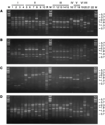
FIG. 2. RAPD fingerprints of the representative B. burgdorferi sensu lato strains from different species or genomic groups (RAPD clusters I to VII) obtained by using four primers as indicated in the legend to Fig. 1. Lanes 1 to 4, B. burgdorferi sensu stricto strains B31, A91-9, VS215, and A44S, respectively; lanes 5 to 10, B. garinii subgroup IIa to IIf strains NT29, PBi, HT55, HT19, M63, and 20047, respectively; lanes 11 to 15, B. afzelii VS461, A38S, A95S, A67T, and A71T, respectively; lane 16, B. japonica HO14; lanes 17 and 18, B. valaisiana VS116 and M19, respectively; lane 19, Borrelia group DN127 strain 25015; lane 20, Borrelia sp. strain A14S; lanes 21 and 22, B. hermsii and B. anserina, respectively; lane R, reference for normalization of different gels; lane M, BstEII-digested bacteriophage lambda DNA. The molecular sizes (in kilobases) are indicated on the right.
772
WANG ET AL.
J. C
LIN. M
ICROBIOL.
on May 15, 2020 by guest
http://jcm.asm.org/
PCR-RFLP pattern: instead of a 246- to 255-bp fragment, a
fragment of 225 bp was amplified. Digestion of this fragment
with MseI resulted in a unique pattern, which was designated
pattern R (Table 1). This pattern was not consistent with any
pattern produced by the 10 LB-related Borrelia species or
genomic groups identified up to now (unpublished data). The
16 B. garinii strains, which were all isolated from patients with
extracutaneous syndromes, were found in subgroups IIb and
IIf. Isolates from human skin biopsy specimens and
cerebro-spinal fluid were evenly distributed among both subgroups.
Interestingly, subgroup IIb included only eight isolates from
patients with disseminated LB and showed little heterogeneity.
Isolates from both humans and ticks were found in subgroup
IIf, with no particular clustering for the isolates from humans.
For B. afzelii, isolates from patients (n
5
30) did not differ from
those from ticks (n
5
29). Furthermore, no significant
subclus-ter was found among isolates recovered from patients with EM
(n
5
18) and ACA (n
5
12).
Further characterization of B. garinii subgroup IIb.
Since B.
garinii subgroup IIb only contained isolates from humans,
fur-ther characterization of this subgroup was performed by
se-quence analysis of the 16S rRNA gene. Partial 16S rRNA gene
analysis of three isolates in this subgroup (isolates PBi, A01C,
and A76S) showed the two conserved nucleotide substitutions
at positions 469 and 635 (B. burgdorferi B31 numbering) (17) in
comparison to the B. garinii consensus sequences (24) and to
sequences from B. burgdorferi sensu stricto and B. afzelii,
con-firming that strains in this subgroup are genetically distinct
from other B. garinii strains.
DISCUSSION
[image:6.612.307.549.71.246.2]RAPD analysis has been used with increasing frequency as a
method for the molecular typing and genetic characterization
[image:6.612.53.286.72.411.2]FIG. 3. Simplified dendrogram of B. burgdorferi sensu lato strains. The 136 LB-associated Borrelia strains used in this study could be divided into seven clusters with an intergroup difference of 55%. Six of them (clusters I to VI) corresponded to the indicated established species or genomic groups. One strain (branch VII) could not be classified into one of the described LB-related Borrelia species. Isolates from patients are in boldface. The numbers in parentheses indicate the corresponding RAPD type of each strain.
FIG. 4. Dendrogram of B. burgdorferi sensu stricto strains (n523). The Borrelia isolates derived from North America and Europe clustered into separate branches.
FIG. 5. Dendrogram of B. garinii strains (n539). The asterisks indicate strains from the cerebrospinal fluid of patients. See the legend to Fig. 3 for more information.
V
OL. 36, 1998
RAPD FINGERPRINTING OF BORRELIA BURGDORFERI
773
on May 15, 2020 by guest
http://jcm.asm.org/
[image:6.612.321.537.434.693.2]of various microorganisms (38, 39). However, considerable
attention is directed to its reliability and reproducibility, since
many studies have indicated that various factors can affect the
results of RAPD fingerprinting (8, 28, 38, 40). In our
experi-ments, optimal RAPD fingerprinting was found with the four
primers that we selected and purified DNA. With these four
different primers, the reproducibility of the RAPD fingerprints
for a random subset of 54 of the 136 isolates was up to at least
a 95% level of similarity. The number of passages did not affect
the results of RAPD analysis. However, PCR amplification of
different colonies of one strain resulted in a difference of one
band, amplified by one of the primers. This band was absent
from two colonies and was present in two other colonies.
Whether this difference is caused by the presence of a mixed
population of two related strains or by colonial variation within
one strain must be elucidated. Since these RAPD fingerprints
from different colonies still showed more than 95% similarity
to each other and belonged to the same RAPD type according
to our definition, we concluded that RAPD analysis is an
appropriate method for the typing of uncloned Borrelia strains.
By RAPD fingerprinting 135 of the 136 B. burgdorferi sensu
lato strains used in this study could be grouped into six
differ-ent species or genomic groups. This result was consistdiffer-ent with
our classification of these strains on the basis of rRNA gene
restriction analysis (41) or PCR-RFLP. One strain, strain
A14S, could not be classified into one of the six species or
genomic groups included in this study and also had a highly
divergent PCR-RFLP pattern. This strain has pathogenic
po-tential, since it was originally isolated from a patient with EM.
In our study, strains belonging to the same species but
orig-inating from different geographical regions clustered into
sep-arate branches by RAPD analysis. Regarding B. burgdorferi
sensu stricto, North American and European strains fell into
separate subgroups. With the exception of one B. garinii strain,
B. garinii strains from Europe clustered into two major
sub-branches. All eight B. garinii strains from far east Asia were
clearly different from the European strains included in these
two major subbranches. Among the B. afzelii strains, the
het-erogeneity was limited. Only 2 of the 56 Dutch strains and the
one Chinese strain tested were more divergent than the
ma-jority of the Dutch strains. Since only one Chinese strain was
studied, more strains from Asia should be typed by RAPD
fingerprinting before a conclusion can be drawn as to whether
B. afzelii strains from European and Asian sources differ.
Re-gional differences among North American B. burgdorferi sensu
stricto strains were also found with the use of pulsed-field gel
electrophoresis and sequence analysis (27). Another recent
study in which pulsed-field gel electrophoresis as well as the
AP-PCR technique was used showed that B. burgdorferi sensu
stricto strains could be subdivided into a number of subgroups,
generally consisting of only North American or only European
strains (14); however, a few North American strains, including
strain B31, clustered with the European strains and vice versa.
In contrast, in our study B31 clustered only with North
Amer-ican strains. Since Foretz et al. (14) studied different European
and North American isolates, this may partly account for
dif-ferences between the studies.
B. afzelii strains grown from ticks collected in a limited area
were often quite similar to each other, although we also found
some exceptions. This is in accordance with the assumption
that the migration rate of the vectors of B. afzelii, presumably
being ticks and rodents, is rather low. It would be interesting to
study a large collection of B. garinii strains in the same way,
since birds are thought to be involved in the transmission of
these spirochete species (9), and much more diversity of the
strains within one area may occur because of this route of
transmission.
Among the B. garinii strains, one cluster of eight strains
(subgroup IIb) consisted only of isolates from humans. The
strains were closely related to each other and were markedly
different from the other B. garinii strains tested. The three
subgroup IIb strains for which 16S rRNA gene sequencing was
performed differed from the other B. garinii strains. All eight
isolates in subgroup IIb originated from patients with
dissem-inated disease. In another study, we showed that these strains
all belonged to OspA type 4, as defined by Wilske et al. (47),
and that they were resistant to the activity of normal human
serum, in contrast to other B. garinii strains (42). Interestingly,
these type 4 B. garinii strains have only been recovered from
human specimens until now (48), and therefore, they may have
an increased pathogenic potential.
[image:7.612.53.279.72.426.2]The RAPD patterns of B. afzelii strains from ticks and
hu-mans were randomly distributed. In addition, strains from
pa-tients with ACA were not different from strains from papa-tients
with EM. This is in accordance with a recent study in which
pulsed-field gel electrophoresis analysis of strains from
pa-tients with EM or ACA did not reveal differences between
these groups (10). Therefore, those B. afzelii strains causing
FIG. 6. Dendrogram of B. afzelii strains from different geographic and bio-logical sources (n559). Tick isolates from two different regions in The Neth-erlands are indicated. Isolates from patients with ACA are underlined. Sa (Sant-poort) and Dr (Drenthe) are two geographic regions in the western and northern parts of The Netherlands, respectively. See the legend to Fig. 3 for more infor-mation.
774
WANG ET AL.
J. C
LIN. M
ICROBIOL.
on May 15, 2020 by guest
http://jcm.asm.org/
ACA have minor differences in their genomes in comparison
with other B. afzelii strains. Alternatively, the persistence of
these spirochetes, resulting in ACA, may mainly be determined
by host factors.
In conclusion, RAPD analysis is a reliable technique for
identifying the different Borrelia species, as well as for
discrim-inating between strains within Borrelia species. Further use of
this technique may lead to more information about the
path-ways of the geographic spread of the spirochetes. In addition,
if more pathogenic subgroups exist, these may also be
identi-fied by this technique, which can be easily and rapidly
per-formed after the isolation of a Borrelia strain.
ACKNOWLEDGMENTS
We thank S. J. Cutler (London, England), R. de Boer (Amsterdam,
The Netherlands), M. Fukunaga (Fukuyama, Japan), O. Pe´ter (Sion,
Switzerland), S. G. T. Rijpkema (Bilthoven, The Netherlands), B.
Wilske (Munich, Germany), and Z. F. Zhang (Beijing, People’s
Re-public of China) for supplying Borrelia strains. We also thank A. Oei
for technical assistance, A. van der Ende and I. Schuurman for help in
the development of RAPD analysis, and W. van Est for photography.
REFERENCES
1. Akopyanz, N., N. O. Bukanov, T. U. Westblom, S. Kresovich, and D. E. Berg. 1992. A diversity among clinical isolates of Helicobacter pylori detected by PCR-based RAPD fingerprinting. Nucleic Acids Res. 20:5137–5142. 2. Appel, M. J. G., S. Allan, R. H. Jacobson, T. L. Lauderdale, Y. F. Chang, S. J.
Shin, and B. A. Summers.1992. Experimental Lyme disease in dogs pro-duces arthritis and persistent infection. J. Infect. Dis. 167:651–654. 3. Assous, M. V., D. Postic, G. Paul, P. Nevot, and G. Baranton. 1994.
Indi-vidualisation of two new genomic groups among American Borrelia burgdor-feri sensu lato strains. FEMS Microbiol. Lett. 121:93–98.
4. Balmelli, T., and J.-C. Piffaretti. 1995. Association between different clinical manifestations of Lyme disease and different species of Borrelia burgdorferi sensu lato. Res. Microbiol. 146:329–340.
5. Baranton, G., D. Postic, I. Saint Girons, P. Boerlin, J.-C. Piffaretti, M.
Assous, and P. A. D. Grimont.1992. Delineation of Borrelia burgdorferi sensu stricto, Borrelia garinii sp. nov., and group VS461 associated with Lyme borreliosis. Int. J. Syst. Bacteriol. 42:378–383.
6. Barbour, A. G., S. L. Tessier, and W. J. Todd. 1983. Lyme disease spirochetes and Ixodid tick spirochetes share a common surface antigenic determinant defined by a monoclonal antibody. Infect. Immun. 41:795–804.
7. Bennett, B. E. 1995. Tick and Lyme disease. Adv. Parasitol. 36:344–405. 8. Benter, T., S. Papadopoulos, M. Manns, and H. Poliwoda. 1995.
Optimiza-tion and reproducibility of random amplified polymorphic DNA in human. Anal. Biochem. 230:92–100.
9. Bunikis, J., B. Olse´n, V. Fingerle, J. Bonnedahl, B. Wilske, and S.
Berg-stro¨m.1996. Molecular polymorphism of the Lyme disease agent Borrelia garinii in northern Europe is influenced by a novel enzootic Borrelia focus in the North Atlantic. J. Clin. Microbiol. 34:364–368.
10. Busch, U., C. Hizo-Teufel, R. Bo¨hmer, V. Fingerle, D. Ro¨ßler, B. Wilske, and
V. Preac-Mursic.1996. Borrelia burgdorferi sensu lato strains isolated from cutaneous Lyme borreliosis biopsies differentiated by pulsed-field gel elec-trophoresis. Scand. J. Infect. Dis. 28:583–589.
11. Canica, M. M., F. Nato, L. du Merle, J. C. Maizie, G. Baranton, and D.
Postic.1993. Monoclonal antibodies for identification of Borrelia afzelii sp. nov. associated with late cutaneous manifestation of Lyme borreliosis. Scand. J. Infect. Dis. 25:441–448.
12. Dever, L. L., J. H. Jorgensen, and A. G. Barbour. 1992. In vitro antimicrobial susceptibility testing of Borrelia burgdorferi: a microdilution MIC method and time-kill studies. J. Clin. Microbiol. 30:2692–2697.
13. Fikrig, E., S. W. Barthold, F. S. Kantor, and R. A. Flavell. 1990. Protection of mice against the Lyme disease agent by immunizing with recombinant OspA. Science 250:553–556.
14. Foretz, M., D. Postic, and G. Baranton. 1997. Phylogenetic analysis of Bor-relia burgdorferi sensu stricto by arbitrarily primed PCR and pulsed-field gel electrophoresis. Int. J. Syst. Bacteriol. 47:11–18.
15. Fukunaga, M., A. Hamase, K. Okada, and M. Nakao. 1996. Borrelia tanukii sp. nov. and Borrelia turdae sp. nov. found from Ixodes ticks in Japan: rapid species identification by 16S rRNA gene-targeted PCR analysis. Microbiol. Immunol. 40:877–881.
16. Fukunaga, M., and Y. Koreki. 1996. A phylogenetic analysis of Borrelia burgdorferi sensu lato isolates associated with Lyme disease in Japan by flagellin gene sequence determination. Int. J. Syst. Bacteriol. 46:416–421. 17. Gazumyan, A., J. J. Schwartz, D. Liveris, and I. Schwartz. 1994. Sequence
analysis of the ribosomal RNA operon of the Lyme disease spirochete, Borrelia burgdorferi. Gene 146:57–65.
18. Hunter, P. R., and M. A. Gaston. 1988. Numerical index of the discrimina-tory ability of typing systems: an application of Simpson’s index of diversity. J. Clin. Microbiol. 26:2465–2466.
19. Johnson, R. C., G. P. Schmid, F. W. Hyde, A. G. Steigerwalt, and D. J.
Brenner.1984. Borrelia burgdorferi sp. nov.: etiological agent of Lyme dis-ease. Int. J. Syst. Bacteriol. 34:496–497.
20. Karabata, H., T. Masuzawa, and Y. Yanagihara. 1993. Genomic analysis of Borrelia japonica sp. nov. isolated from Ixodes ovatus in Japan. Microbiol. Immunol. 37:843–848.
21. Kuiper, H., A. P. van Dam, L. Spanjaard, B. M. de Jongh, A.
Widjojoku-sumo, A. C. P. Ramselaar, I. Cairo, K. Vos, and J. Dankert.1994. Isolation of Borrelia burgdorferi from skin biopsy specimens taken from healthy-look-ing skin of patients with Lyme borreliosis. J. Clin. Microbiol. 32:715–720. 22. Le Fleche, A., D. Postic, K. Girardet, O. Peter, and G. Baranton. 1997.
Characterization of Borrelia lusitaniae sp. nov. by 16S ribosomal DNA se-quence analysis. Int. J. Syst. Bacteriol. 47:921–925.
23. Liang, J. G., and Z. F. Zhang. 1996. Analysis of rRNA gene restriction fragment length polymorphism of Borrelia burgdorferi sensu lato isolated in China. Chin. J. Microbiol. Immunol. 16:359–362.
24. Marconi, R. T., and C. F. Caron. 1992. Development of polymerase chain reaction sets for diagnosis of Lyme disease and for species-specific identifi-cation of Lyme disease isolates by 16S rRNA signature nucleotide analysis. J. Clin. Microbiol. 30:2830–2834.
25. Marconi, R. T., D. Liveris, and I. Schwartz. 1995. Identification of novel insertion elements, restriction fragment length polymorphism patterns, and discontinuous 23S rRNA in Lyme disease spirochetes: phylogenetic analyses of rRNA genes and their intergenic spacers in Borrelia japonica sp. nov. and genomic group 21038 (Borrelia andersonii sp. nov.) isolates. J. Clin. Micro-biol. 33:2427–2434.
26. Masuzawa, T., T. Komikado, A. Iwaki, H. Suzuki, K. Kaneda, and Y.
Yanagi-hara.1996. Characterization of Borrelia sp. isolated from Ixodes tanuki, I. turdus, and I. columnae in Japan by restriction fragment length polymor-phism of rrf (5S)-rrl (23S) intergenic spacer amplicons. FEMS Microbiol. Lett. 142:77–83.
27. Mathiesen, D. A., J. H. Oliver, Jr., C. P. Kolbert, E. D. Tullson, B. J. B.
Johnson, G. L. Campbell, P. D. Mitchell, K. D. Reed, S. R. Telford III, J. F. Anderson, R. S. Lane, and D. H. Persing.1997. Genetic heterogeneity of Borrelia burgdorferi in the United States. J. Infect. Dis. 175:98–107. 28. Meunier, J. R., and P. A. D. Grimont. 1993. Factors affecting reproducibility
of random amplified polymorphic DNA fingerprinting. Res. Microbiol. 144: 373–379.
29. Nohlmans, L. M. K. E., R. de Boer, A. E. J. M. van den Bogaard, and C. P. A.
Boven.1995. Genotypic and phenotypic analysis of Borrelia burgdorferi iso-lates from The Netherlands. J. Clin. Microbiol. 33:119–125.
30. Pe´ter, O., and A. G. Bretz. 1992. Polymorphism of outer surface proteins of Borrelia burgdorferi as a tool for classification. Zentralbl. Bakteriol. Para-sitenkd. Infektkrankh. Hyg. Abt. 1 Orig. Reihe A 277:28–33.
31. Pe´ter, O., A. G. Bretz, R. Zenha¨usern, H. Roten, and E. Roulet. 1993. Isolement de Borrelia burgdorferi du liquide ce´phalorachidien de trois enfants avec une atteinte neurologique. Schweiz. Med. Wochenschr. 123:14–19. 32. Picken, R. N., Y. Cheng, F. Strle, and M. M. Picken. 1996. Patients isolates
of Borrelia burgdorferi sensu lato with genotypic and phenotypic similarities to strains 25015. J. Infect. Dis. 174:1112–1115.
33. Postic, D., M. V. Assous, P. A. D. Grimont, and G. Baranton. 1994. Diversity of Borrelia burgdorferi sensu lato evidenced by restriction fragment length polymorphism of rrf (5S)-rrl (23S) intergenic spacer amplicons. Int. J. Syst. Bacteriol. 44:743–752.
34. Preac-Mursic, V., B. Wilske, and G. Schierz. 1986. European Borrelia burg-dorferi isolated from humans and ticks: culture conditions and antibiotic susceptibility. Zentralbl. Bakteriol. Parasitenkd. Infektkrankh. Hyg. Abt. 1 Orig. Reihe A 263:112–118.
35. Rijpkema, S. G. T., M. J. C. H. Molkenboer, L. M. Schouls, F. Jongejan, and
J. F. P. Schellekens.1995. Simultaneous detection and genotyping of three genomic groups of Borrelia burgdorferi sensu lato in Dutch Ixodes ricinus ticks by characterization of the amplified intergenic spacer region between 5S and 23S rRNA genes. J. Clin. Microbiol. 33:3091–3095.
36. Sneath, P. H. A., and R. R. Sokal. 1973. Numerical taxonomy: the principle and practices of numerical classification. W. H. Freeman & Co., San Fran-cisco, Calif.
37. Steere, A. C. 1989. Lyme disease. N. Engl. J. Med. 321:586–596. 38. Tyler, K. D., G. Wang, S. D. Tyler, and W. M. Johnson. 1997. Factors
affecting reliability and reproducibility of amplification-based fingerprinting of representative bacterial pathogens. J. Clin. Microbiol. 35:339–346. 39. van Belkum, A. 1994. DNA fingerprinting of medically important
microor-ganisms by PCR. Clin. Microbiol. Rev. 7:174–184.
40. van Belkum, A., J. Kluytmans, W. van Leeuwen, R. Bax, W. Quint, E. Peters,
A. Fluit, C. Vandenbboucke-Grauls, A. van der Brule, H. Koeleman, W. Melchers, J. Meis, A. Elaichouni, M. Vaneechoutte, F. Moonens, N. Maes, M. Struelens, F. Tenover, and H. Verbrugh.1995. Multicenter evaluation of arbitrarily primed PCR for typing of Staphylococcus aureus strains. J. Clin. Microbiol. 33:1537–1547.
41. Van Dam, A. P., H. Kuiper, K. Vos, A. Widjojokusumo, B. M. de Jongh, L.
V
OL. 36, 1998
RAPD FINGERPRINTING OF BORRELIA BURGDORFERI
775
on May 15, 2020 by guest
http://jcm.asm.org/
Spanjaard, A. C. P. Ramselaar, M. D. Kramer, and J. Dankert.1993. Dif-ferent genospecies of Borrelia burgdorferi are associated with distinct clinical manifestations of Lyme borreliosis. Clin. Infect. Dis. 17:708–717. 42. van Dam, A. P., A. Oei, R. Jaspars, C. Fijen, B. Wilske, L. Spanjaard, and
J. Dankert. 1997. Complement-mediated serum sensitivity among spiro-chetes that cause Lyme disease. Infect. Immun. 65:1228–1236.
43. Wang, G., A. P. van Dam, A. Le Fleche, D. Postic, O. Peter, G. Baranton, R.
de Boer, L. Spanjaard, and J. Dankert.1997. Genetic and phenotypic anal-ysis of Borrelia valaisiana sp. nov. (Borrelia genomic groups VS116 and M19). Int. J. Syst. Bacteriol. 47:926–932.
44. Welsh, J., and M. McClelland. 1990. Fingerprinting genomes using PCR with arbitrary primers. Nucleic Acids Res. 18:7213–7218.
45. Welsh, J., C. Pertzman, D. Postic, I. Saint Girons, G. Baranton, and M.
McClelland.1992. Genomic fingerprinting by arbitrarily primed polymerase chain reaction resolves Borrelia burgdorferi into three distinct phyletic groups.
Int. J. Syst. Bacteriol. 42:370–377.
46. Williams, J. G. K., A. R. Kubelik, K. J. Livak, J. Antoni Rafalski, and S. V.
Tingey.1990. DNA polymorphisms amplified by arbitrary primers are useful as genetic markers. Nucleic Acids Res. 18:6531–6535.
47. Wilske, B., V. Preac-Mursic, U. B. Gobel, B. Graf, S. Jauris, E. Soutschek, E.
Schwab, and G. Zumstein.1993. An OspA serotyping system for Borrelia burgdorferi based on reactivity with monoclonal antibodies and OspA se-quence analysis. J. Clin. Microbiol. 31:340–350.
48. Wilske, B., U. Busch, H. Eiffert, V. Fingerle, H.-W. Pfister, D. Rossler, and
V. Preac-Mursic.1996. Diversity of OspA and OspC among cerebrospinal fluid isolates of Borrelia burgdorferi sensu lato from patients with neurobor-reliosis in Germany. Med. Microbiol. Immunol. 184:195–201.
49. Wilson, K. 1988. Preparation of genomic DNA from bacteria, p. 2.4.1–2.4.5. In F. M. Ausubel (ed.), Current protocols in molecular biology, vol 1. John Wiley & Sons, Inc., New York, N.Y.


![Synthesis and Polymerization of N,N' [Bis(4,4' Hydroxypropyl Methacrylate Phenylester) Pyromellitimide] Thermoset Polymer by Microwave Irradiation](data:image/gif;base64,R0lGODlhAQABAIAAAP///wAAACH5BAEAAAAALAAAAAABAAEAAAICRAEAOw==)