R E S E A R C H A R T I C L E
Open Access
Associations between disc space narrowing,
anterior osteophytes and disability in
chronic mechanical low back pain: a cross
sectional study
Romain Shanil Perera
1*, Poruwalage Harsha Dissanayake
2, Upul Senarath
3, Lalith Sirimevan Wijayaratne
4,
Aranjan Lional Karunanayake
5and Vajira Harshadeva Weerabaddana Dissanayake
6Abstract
Background:Radiographic features of lumbar disc degeneration (LDD) are common findings in patients with chronic mechanical low back pain; however, its role in disability and intensity of pain is debatable. This study aims to investigate the associations of the x-ray features of LDD and lumbar spondylolisthesis with severity of disability and intensity of pain. Methods:A cross-sectional study was conducted on 439 patients with chronic mechanical low back pain who attended the rheumatology clinic, National Hospital of Sri Lanka, Colombo, from May 2012 to May 2014. Severity of disability was measured using Modified Oswestry Disability Index and intensity of pain was assessed using numeric rating scale (0–100). X-ray features of LDD (disc space narrowing, anterior osteophytes and overall LDD) and spondylolisthesis were assessed in lateral recumbent lumbar x-rays (L1/L2 to L5/S1) and graded by a consultant radiologist blinded to clinical data. Generalised linear model with linear response was used to assess the associations of x-ray features of LDD with severity of disability and intensity of pain adjusting for age, gender, body mass index and pain radiating into legs. Results:Mean age was 48.99 ± 11.21 and 323 (73.58%) were females. 87 (19.82%) were obese. Mean severity of disability was 30.95 ± 13.67 and mean intensity of pain was 45.50 ± 20.37. 69 (15.72%), 26 (5.92%) and 85 (19.36%) patients had grade 2 disc space narrowing, anterior osteophytes and overall LDD, respectively. 51 (11.62%) patients had lumbar spondylolisthesis. Grade of disc space narrowing and overall LDD were not associated with severity of disability or intensity of pain. The presence of lumbar spondylolisthesis was associated with severity of disability. Female gender and pain radiating into legs were associated with severity of disability and intensity of pain. Advancing age was associated with x-ray features of LDD and lumbar spondylolisthesis.
Conclusions:Lumbar spondylolisthesis is associated with severity of disability in patients with chronic mechanical low back pain. Associations of x-ray features of LDD with severity of disability and intensity of pain are inconclusive. Female gender and pain radiating into legs are significant confounders.
Keywords:Disability, Disc space narrowing, Anterior osteophytes, Low back pain, Lumbar disc degeneration
* Correspondence:[email protected]
1Department of Allied Health Sciences, Faculty of Medicine, University of
Colombo, 25, Kynsey Road, Colombo 8, Sri Lanka
Full list of author information is available at the end of the article
Background
Disability due to chronic low back pain is one of the leading health care problems in most regions of the world including South Asia [1]. It affects all aspects of life including physical, mental, and social well-being [2]. Disabling chronic low back pain is reported to be a major issue in occupational health in Sri Lanka [3, 4]. Most chronic low back pains are related to mechanical causes including injuries of the musculoskeletal struc-tures of the spine and pathologies associated with lum-bar disc degeneration (LDD) [5, 6]. LDD is a common finding in the aging spine and symptoms of chronic mechanical low back pain are not always correlated with the radiological features of LDD. Patients with chronic low back pain receive routine spinal imaging (lumbar x-ray, computed tomography, or magnetic resonance imaging [MRI]) and MRI of lumbar spine has become the popular choice for routine imaging as it gives a direct visualisation of the disc without exposure to the radiation. However MRI is not a cost effective method in routine spinal imaging in developing countries and clinicians in developing countries like Sri Lanka regu-larly use x-ray lumbar spine as a feasible option for assessing features related to LDD [7].
There are mixed evidence for the association of LDD with chronic mechanical low back pain and disability. Although, routine x-ray of lumbar spine does not affect the outcome of the treatment of uncomplicated acute and subacute low back pain [8], x-ray features related to LDD may benefit the clinical diagnosis and management of chronic low back pain and disability when combined with other factors such as proper history taking, severity of symptoms, surgical risks and costs [8]. Disc space nar-rowing and anterior osteophytes are the main x-ray fea-tures of LDD [9] and are proven to be highly correlated with the morphological stages of LDD [10]. Disc space narrowing is associated with lumbar spinal stenosis, disc herniation and spondylolisthesis which are also related to the pain and disability [11]. Disc space narrowing is associated with the presence of chronic low back pain [9, 12] and intensity of pain [13]. This association becomes stronger with increasing severity of disc space narrowing [12, 13]. Mostly these associations are reported in population based studies and their study samples were limited to middle aged and elderly indivi-duals [9, 12, 13]. There are a limited number of studies which have investigated the association of disc space narrowing with disability [9]. Although anterior osteo-phyte is the most frequently observed degenerative feature of the aging lumbar spine, it has variably corre-lated results on its association with intensity of pain [9, 13]. With regard to disability, we could not find enough evidence to prove its association with anterior osteo-phytes [9, 14]. Both disc space narrowing and anterior
osteophytes have been used to determine the grade of overall LDD [15] and high variability exists among the associations between the overall LDD and intensity of pain/disability [14, 16, 17].
Severity of disability/intensity of pain and x-ray features of LDD are further influenced by the effects of age, gender, body mass index (BMI) and the presence of pain radiating into legs. Advancing age increases the susceptibility for severe disability [18]. In most studies females have reported increased intensity of pain and severe disability [19, 20]. In addition obese patients have a higher risk for recurrent disabling low back pain [21]. Furthermore pain radiating into legs is associated with symptomatic disc herniation contributing to severe pain and disability [22]. Age, gender, BMI and the presence of pain radiating into legs may be helpful in predicting the severity of x-ray fea-tures of LDD. Advancing age increases the susceptibility for severe degeneration [23, 24]. In addition, there is evidence that males have more degenerative changes compared to females [9], but there are other studies that have given contradicting results [25]. Certain studies have reported that higher BMI has an add-on effect on LDD [26, 27]. However the evidence for associations of gender, BMI and the presence of pain radiating into legs with grade of x-ray features of LDD are inconsistent and need further investigation.
lumbar spondylolisthesis with severity of disability and intensity of pain in patients with chronic mechanical low back pain adjusting for age, gender, BMI and pain radia-ting into legs. In addition we assessed the associations of x-ray features of LDD with age, gender, BMI and pain radiating into legs.
Methods
Study design, setting and participants
A descriptive cross-sectional study was conducted on consecutive patients with chronic mechanical low back pain who attended the rheumatology clinic, National Hospital of Sri Lanka, Colombo, from May 2012 to May 2014. Both male and female patients of Sri Lankan origin with chronic mechanical low back pain aged 20 to 69 years were recruited to the study. Both patients with and without x-ray evidence of LDD and spondylolis-thesis were included. Low back pain was defined as pain, muscle tension, or stiffness localized below the costal margin and above the inferior gluteal folds, with or with-out pain radiating into the leg [19]. Back pain during day time worsening in the latter part of the day due to movements was considered to be due to a mechanical cause [28]. Chronicity was defined as pain on most days of the week for at least three months [2]. Patients with back pain due to inflammatory causes (seronegative spondyloarthropathies, diffuse idiopathic skeletal hype-rostosis, rheumatoid arthritis), visceral origin (urinary tract infections, inflammatory pelvic disease), systemic infections affecting spine (spinal tuberculosis), metabolic bone diseases (osteoporosis and osteomalacia), fractures in the vertebral column, past surgeries in the spine, and spinal tumours were excluded. Pregnant females and pa-tients who refuse to participate in the study were also excluded. The study was carried out in accordance with the Declaration of Helsinki and with the approval of the Ethics Review Committee of the Faculty of Medicine, University of Colombo. Patients who fulfilled the inclu-sion and excluinclu-sion criteria were recruited to the study after obtaining written informed consent.
Clinical evaluation
Demographic (age and gender) and clinical data (inten-sity of pain, severity of disability, presence of pain radia-ting into legs, and BMI) were recorded using a pretested interviewer administered questionnaire and clinical examination. The intensity of pain was measured using a 101 (0 to 100) point numeric rating scale. Patients were asked to score the average intensity of pain experienced during the past 7 days out of 100 [29–31]. Disability was assessed using the Modified Oswestry Disability Index (MODI). MODI is a low back pain specific disability questionnaire with ten items which assess pain and its impact on the activities of daily living including personal
care, lifting, walking, sitting, standing, sleeping, travel-ling, social work, home and work duties. Each item has six responses where higher values represent greater dis-ability. Sum of responses was calculated and presented as a percentage [32, 33]. Pain radiating into legs was positive if the pain radiated below the knee of either one or both legs. Height (cm) and weight (kg) of the patients were recorded with light clothing and without shoes to the nearest 0.1 cm and 0.1 kg, respectively, and BMI was calculated (kg/m2) [34]. International cut off values were used for categorisation of BMI [35].
Radiographic evaluation
was trained on radiographic evaluation according to the Lane atlas. On random evaluation of 25% of the ra-diographs were reported by the second medical officer who was blinded to the first reader’s interpretations.
Statistical analysis
Descriptive statistics were calculated to summarise the sample characteristics. Both univariable and multiva-riable analyses were carried out. For the univamultiva-riable analysis, severity of disability and intensity of pain were defined as continuous outcome/dependent variables and independent samples t-test was used when there were two categories and Analysis of Variance (ANOVA) was used when there were more than two categories.
Multivariable analysis was performed using different regression models considering the nature of the out-come/dependent variables. Multivariable generalised linear model with linear response was used when the severity of disability and intensity of pain were used as the continuous outcome variables. X-ray features of LDD (disc space narrowing, anterior osteophytes and overall LDD) and presence of lumbar spondylolisthesis were defined as main independent variables/predictor variables and were treated as categorical variables. Sepa-rate linear regression models were created for each fea-ture. In each multivariable generalised linear model with linear response, the magnitude of the association was presented asβcoefficients with 95% confidence intervals (CI). Multivariable ordinal logistic regression was used when the severity of x-ray features of LDD (disc space narrowing, anterior osteophytes and overall LDD) were used as the ordinal outcome variables (0, 1 and 2). Mul-tivariable logistic regression analysis was used when the
presence of lumbar spondylolisthesis (yes/no) was used as a binary outcome variable. Magnitude of the associa-tions was presented as adjusted odds ratios (aOR) with 95% CI in logistic regression models. Age, gender, BMI and presence of pain radiating into legs were defined as confounder variables in all regression models. Age and BMI were treated as continuous variables and gender (male/female) and presence of pain radiating into legs (yes/no) were treated as categorical variables.
Assumptions of ANOVA, independent samples t-test and regression models were verified. P value < 0.05 was used as the level of significance. Statistical analysis was carried out using SPSS version 17.
Results
Characteristics of the participants
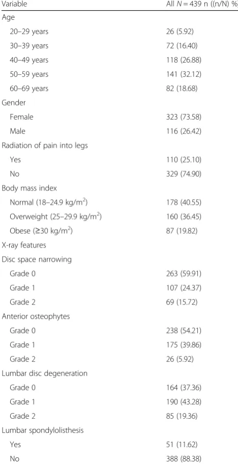
Table 1 summarises the characteristics of the study par-ticipants. Among 689 patients with chronic mechanical low back pain, 439 patients were recruited according to eligibility criteria. Thirteen patients had missing data for the variable BMI. Mean age ± SD was 48.99 ± 11.21 and 323 (73.58%) were females. BMI ± SD was 26.39 ± 4.65 and 87 (19.82%) were obese. Mean severity of disability was 30.95 ± 13.67 and mean intensity of pain was 45.50 ± 20.37. In addition, 110 (25.10%) patients had pain radiating into legs. With regard to interobserver repro-ducibility, intra-class correlation coefficient (ICC) of two readers for disc space narrowing was 0.88 (0.82-0.91) and ICC for anterior osteophytes was 0.81 (0.75 –0.85). Among patients, 176 (40.09%) had disc space narrowing and 201 (45.78%) had anterior osteophytes with 69 (15.72%) and 26 (5.92%) having grade 2 disc space nar-rowing and grade 2 anterior osteophytes, respectively.
Fig. 1Assesment of the x-ray features of lumbar disc degeneration - lateral x-ray of lumbar spine. Arrows -a–no disc space narrowing/anterior osteophyte (grade 0 lumbar disc degeneration),b–mild disc space narrowing and small anterior osteophyte, (grade 1 lumbar disc degeneration)
[image:4.595.56.540.87.288.2]LDD was present in 275 (62.64%) and 85 (19.36%) had grade 2 LDD. Lumbar spondylolisthesis was present in 51 (11.62%) patients.
Associations of x-ray features of lumbar disc degeneration, spondylolisthesis with severity of disability
There were no significant differences in severity of disability with the severity of disc space narrowing, an-terior osteophytes and LDD according to ANOVA and generalised linear models with linear response (Table 2 and 3). Patients with the presence of lumbar spondylo-listhesis had significantly severe disability in contrast to the patients without lumbar spondylolisthesis in both
univariable and multivariable analysis (Table 2 and 3). Female gender and presence of pain radiating into legs were significantly associated with the severity of disability in all the multivariable generalised linear models (Table 3).
Associations of x-ray features of lumbar disc degeneration, spondylolisthesis with intensity of pain
Disc space narrowing and LDD were not associated with intensity of pain in either univariable or multivariable regression analyses (Table 2 and 3). However patients with grade 1 anterior osteophytes had significantly higher intensity of pain compared to the patients with grade 0 anterior osteophytes. The presence of lumbar spondylo-listhesis was not associated with the intensity of pain. Female gender and pain radiating into legs were associated with the intensity of pain in all multivariable generalised linear models (Table 3). In addition increasing age was associated with two linear regression models involving anterior osteophytes and LDD (Table 3).
Associations of age, gender, BMI and presence of pain radiating into legs with x-ray features of lumbar disc degeneration and spondylolisthesis
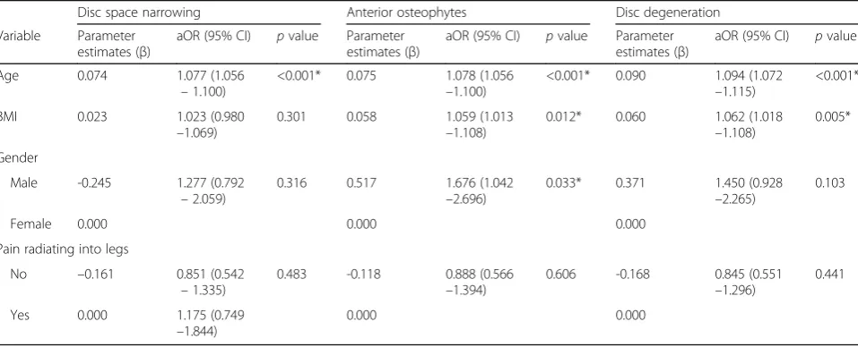
The presence of grade 2 disc space narrowing was reported from 20– 29 years age group, but presence of grade 2 anterior osteophytes was reported from 40 – 49 years age group. Furthermore the presence of lumbar spon-dylolisthesis was reported from 30 – 39 years age group. Advancing age was strongly associated with the severity of disc space narrowing, anterior osteophytes, LDD (Table 4) and presence of lumbar spondylolis-thesis (aOR 1.15; 95% CI: 1.1–1.21) after adjusting for gender, BMI and pain radiating into legs. Male gender was associated with the severity of anterior osteophytes, but was not associated with disc space narrowing, over-all LDD and lumbar spondylolisthesis. Furthermore, BMI was significantly associated with grades of anterior osteophytes and LDD.
Discussion
[image:5.595.55.289.102.558.2]In this study we assessed the associations of x-ray fea-tures of LDD and lumbar spondylolisthesis with severity of disability and intensity of pain in patients with chronic mechanical low back pain adjusting for age, gen-der, BMI and pain radiating into legs. In addition we assessed the associations of x-ray features of LDD with age, gender, BMI and presence of pain radiating into legs. We found that, the associations of x-ray features of LDD with severity of disability or intensity of pain (except anterior osteophytes) were inconclusive. The presence of lumbar spondylolisthesis was associated with increased severity of disability. However the Female gender and presence of pain radiating into legs were
Table 1Summary of sample characteristics
Variable AllN= 439 n ((n/N) %)
Age
20–29 years 26 (5.92)
30–39 years 72 (16.40)
40–49 years 118 (26.88)
50–59 years 141 (32.12)
60–69 years 82 (18.68)
Gender
Female 323 (73.58)
Male 116 (26.42)
Radiation of pain into legs
Yes 110 (25.10)
No 329 (74.90)
Body mass index
Normal (18–24.9 kg/m2) 178 (40.55)
Overweight (25–29.9 kg/m2) 160 (36.45)
Obese (≥30 kg/m2) 87 (19.82)
X-ray features
Disc space narrowing
Grade 0 263 (59.91)
Grade 1 107 (24.37)
Grade 2 69 (15.72)
Anterior osteophytes
Grade 0 238 (54.21)
Grade 1 175 (39.86)
Grade 2 26 (5.92)
Lumbar disc degeneration
Grade 0 164 (37.36)
Grade 1 190 (43.28)
Grade 2 85 (19.36)
Lumbar spondylolisthesis
Yes 51 (11.62)
associated with increased severity of disability and inten-sity of pain. Furthermore, x-ray features of LDD and lumbar spondylolisthesis were strongly associated with advancing age.
Lumbar intervertebral discs are fibrocartilage pads bet-ween adjacent lumbar vertebral bodies which distribute compressive loading evenly on to the vertebral bodies. Intervertebral discs contribute to spinal stability along with the apophyseal joints and supported by surroun-ding muscles and ligaments [38]. With LDD the normal architecture of the disc is disrupted leading to abnormal biomechanical force distribution which may cause severe and disabling low back pain. With degeneration, the height of the disc can be reduced due to inward or out-ward herniation of the disc material and is visible as disc space narrowing in x-ray lumbar spine. This results in abnormal load distribution to the surrounding structures and lead to segmental instability and spondylolisthesis. Formation of osteophytes is a compensatory mechanism to distribute increasing axial forces of spine on a larger articulating surface to prevent spinal instability [11]. Al-though x-ray features of LDD are not correlated with the outcome of the treatment, they can give important details for managing chronic mechanical low back pain especially in the presence of severe symptoms [39].
Disc space narrowing is used as a surrogate variable for LDD and many studies found positive association with the presence of chronic low back pain in popula-tion based studies [9, 15, 24]. However, studies done in clinical settings did not find significant association be-tween disc space narrowing and intensity of pain [17].
Similarly in our study disc space narrowing was not associated with intensity of pain. There are limited cross sectional clinical studies which have assessed the asso-ciation of LDD with disability. A study on 172 consecu-tive patients with chronic low back pain in United Kingdom did not find significant association between LDD (based on x-ray findings) and disability [15]. Authors of the previous study did not assess the association of features of LDD separately as disc space narrowing and anterior osteophytes, but rather assessed the overall LDD. According to our univariable and multivariable analyses disc space narrowing was not associated with disability, but gender and presence of pain radiating into legs had significant association with disability.
[image:6.595.57.536.109.336.2]Comparatively, the association between anterior osteo-phytes and chronic low back pain is largely considered as not significant, unless there is a higher grade of an-terior osteophytes [9, 24]. As mentioned previously we could not find cross sectional clinical studies which have assessed the associations between anterior phytes and disability. Higher grades of anterior osteo-phytes are frequently seen in elderly individuals (above 65 years). Our sample was restricted to patients below 70 years and there were only 26 patients with grade 2 anterior osteophytes. In our results, grade of anterior osteophytes was not associated with the severity of disability. However the patients with grade 1 anterior osteophytes had higher intensity of pain in contrast to patients with grade 0 anterior osteophytes. The overall association between the grades of anterior osteophytes and intensity of pain was inconsistent as there was no
Table 2Means of severity of disability/intensity of pain according to the severity of x-ray features of lumbar disc degeneration and lumbar spondylolisthesis–univariable analysis
Variable Mean disability ± SD pvalue Mean intensity of pain ± SD pvalue
Disc space narrowing 0.115 0.504
Grade 0 30.48 ± 12.92 44.63 ± 19.58
Grade 1 30.10 ± 13.84 47.31 ± 21.69
Grade 2 34.07 ± 15.81 46.01 ± 21.33
Anterior osteophytes 0.076 0.200
Grade 0 30.05 ± 13.61 44.10 ± 20.20
Grade 1 32.65 ± 14.15 47.64 ± 20.73
Grade 2 27.79 ± 9.17 43.87 ± 18.93
Lumbar disc degeneration 0.207 0.534
Grade 0 29.75 ± 12.85 44.13 ± 19.05
Grade 1 31.07 ± 13.79 46.11 ± 21.32
Grade 2 32.98 ± 14.79 46.79 ± 20.77
Lumbar spondylolisthesis 0.001* 0.289
Yes 36.65 ± 13.58 48.34 ± 19.97
No 30.20 ± 13.52 45.13 ± 20.42
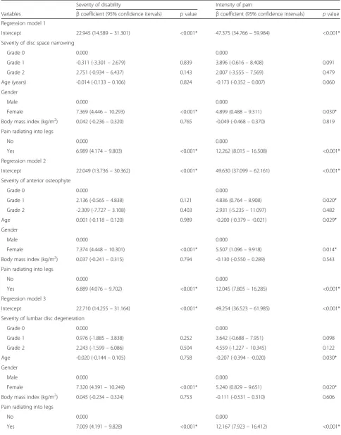
Table 3Associations of x-ray features of lumbar disc degeneration and spondylolisthesis with severity of disability and intensity of pain–multivariable generalised linear models with linear response
Severity of disability Intensity of pain
Variables βcoefficient (95% confidence itervals) p value βcoefficient (95% confidence intervals) pvalue
Regression model 1
Intercept 22.945 (14.589–31.301) <0.001* 47.375 (34.766–59.984) <0.001*
Severity of disc space narrowing
Grade 0 0.000 0.000
Grade 1 -0.311 (-3.301–2.679) 0.839 3.896 (-0.616–8.408) 0.091
Grade 2 2.751 (-0.934–6.437) 0.143 2.007 (-3.555–7.569) 0.479
Age (years) -0.014 (-0.133–0.106) 0.824 -0.173 (-0.352–0.007) 0.060
Gender
Male 0.000 0.000
Female 7.369 (4.446–10.293) <0.001* 4.899 (0.488–9.311) 0.030*
Body mass index (kg/m2) 0.042 (-0.236–0.320) 0.765 -0.049 (-0.468–0.370) 0.819
Pain radiating into legs
No 0.000 0.000
Yes 6.989 (4.174–9.803) <0.001* 12.262 (8.015–16.508) <0.001*
Regression model 2
Intercept 22.049 (13.736–30.362) <0.001* 49.630 (37.099–62.161) <0.001*
Severity of anterior osteophyte
Grade 0 0.000 0.000
Grade 1 2.136 (-0.565–4.838) 0.121 4.836 (0.764–8.908) 0.020*
Grade 2 -2.309 (-7.727–3.108) 0.403 2.931 (-5.235–11.097) 0.482
Age 0.001 (-0.118–0.120) 0.989 -0.200 (-0.379–-0.021) 0.029*
Gender
Male 0.000 0.000
Female 7.374 (4.448–10.301) <0.001* 5.507 (1.096–9.918) 0.014*
Body mass index (kg/m2) 0.037 (-0.241–0.315) 0.794 -0.130 (-0.550–0.289) 0.543
Pain radiating into legs
No 0.000 0.000
Yes 6.889 (4.076–9.702) <0.001* 12.045 (7.805–16.285) <0.001*
Regression model 3
Intercept 22.710 (14.255–31.164) <0.001* 49.254 (36.523–61.985) <0.001*
Severity of lumbar disc degeneration
Grade 0 0.000 0.000
Grade 1 0.976 (-1.885–3.838) 0.252 3.642 (-0.688–7.951) 0.098
Grade 2 2.243 (-1.599–6.086) 0.504 4.559 (-1.227–10.345) 0.122
Age -0.020 (-0.144–0.105) 0.758 -0.207 (-0.394 - -0.020) 0.030*
Gender
Male 0.000 0.000
Female 7.320 (4.391–10.249) <0.001* 5.240 (0.829–9.651) 0.020*
Body mass index (kg/m2) 0.045 (-0.234–0.324) 0.753 -0.111 (-0.531–0.310) 0.606
Pain radiating into legs
No 0.000 0.000
significant association between intensity of pain and grade 2 anterior osteophytes in contrast to grade 0 an-terior osteophytes.
In most studies, overall LDD poorly correlated with clinical symptoms including severity of disability and in-tensity of pain [14, 40]. In our results we could not find a significant association between LDD and severity of disability or LDD and intensity of pain, which agree with the findings of the previous evidence [15]. Most radio-graphic scoring systems including the Lane atlas have
[image:8.595.57.539.112.296.2]used disc space narrowing or anterior osteophytes or both features to determine the overall LDD. Accordingly, either higher grades of disc space narrowing or higher grades of anterior osteophytes could determine higher grades of LDD. Although grade 1 anterior osteophytes was associated with intensity of pain (in contrast to pa-tients with grade 0 anterior osteophytes), we could not find a significant association between overall LDD and intensity of pain. The strength of the association might have become further attenuated when both features (disc
Table 3Associations of x-ray features of lumbar disc degeneration and spondylolisthesis with severity of disability and intensity of pain–multivariable generalised linear models with linear response(Continued)
Regression model 4
Intercept 23.304 (15.086–31.523) <0.001* 48.248 (35.749–60.748) <0.001*
Presence of lumbar spondylolisthesis
No 0.000 0.000
Yes 5.670 (1.684–9.656) 0.005* 3.549 (-2.514–9.612) 0.251
Age -0.040 (-0.156–0.076) 0.501 -0.168 (-0.345–0.009) 0.063
Gender
Male 0.000 0.000
Female 7.011 (4.114–9.908) <0.001* 4.824 (0.418–9.231) 0.032*
Body mass index (kg/m2) 0.078 (-0.197–0.353) 0.578 -0.055 (-0.473–0.363) 0.795
Pain radiating into legs
No 0.000 0.000
Yes 6.820 (4.019–9.621) <0.001* 12.128 (7.868–16.388) <0.001*
Main predictor (independent) variables were analysed as follows: Regression model 1: severity of disc space narrowing (grade 0, 1 and 2) Regression model 2: severity of anterior osteophytes (grade 0, 1 and 2) Regression model 3: severity of lumbar disc degeneration (grade 0, 1 and 2) Regression model 4: presence of lumbar spondylolisthesis (yes/no) * -pvalue < 0.05
Table 4Associations of age, gender and BMI with x-ray features of lumbar disc degeneration–multivariable ordinal logistic
regression model
Disc space narrowing Anterior osteophytes Disc degeneration
Variable Parameter estimates (β)
aOR (95% CI) pvalue Parameter estimates (β)
aOR (95% CI) pvalue Parameter estimates (β)
aOR (95% CI) pvalue
Age 0.074 1.077 (1.056
–1.100)
<0.001* 0.075 1.078 (1.056 –1.100)
<0.001* 0.090 1.094 (1.072 –1.115)
<0.001*
BMI 0.023 1.023 (0.980
–1.069)
0.301 0.058 1.059 (1.013
–1.108)
0.012* 0.060 1.062 (1.018
–1.108)
0.005*
Gender
Male -0.245 1.277 (0.792
–2.059)
0.316 0.517 1.676 (1.042
–2.696)
0.033* 0.371 1.450 (0.928
–2.265)
0.103
Female 0.000 0.000 0.000
Pain radiating into legs
No –0.161 0.851 (0.542
–1.335)
0.483 -0.118 0.888 (0.566
–1.394)
0.606 -0.168 0.845 (0.551
–1.296)
0.441
Yes 0.000 1.175 (0.749
–1.844)
0.000 0.000
* -pvalue <0.05
[image:8.595.59.538.522.716.2]space narrowing and anterior osteophytes) were consid-ered in overall LDD.
Degenerative lumbar spondylolisthesis is related to LDD and degenerative changes of the apophyseal joints [11]. In our results presence of lumbar spondylolisthesis was associated with the increasing grade of disc space narrowing and overall LDD, and was more frequent at the L4–L5 level. The presence of lumbar spondylolis-thesis was associated with increased severity of disability in our study, but it was not associated with the intensity of pain. Narrowing of the disc space is associated with advanced LDD, annular tears and disc herniation, but these features do not always correlate well with the intensity of pain. Furthermore it can adversely affect the biomechanical stability of the lumbar spine which will increase the strain on apophyseal joints and surrounding structures where the combined effects can reduce the flexibility and stability of the spine leading to severe disability [11].
X-ray features of LDD are age related [9, 24] and our study results are compatible with the previous evidence. Interestingly, disc space narrowing was seen from an early age (20 –39 years), but anterior osteophytes was seen from the middle age group (40–49 years) onwards. Although previous studies have found significantly higher degenerative features in males [9], we found posi-tive association only with anterior osteophytes. Further-more, increasing BMI was associated with increasing grade of anterior osteophytes and LDD which was com-patible with previous findings [26, 27].
There is evidence that females are more susceptible to higher intensity of pain and disability and our results were compatible with the existing evidence. Finding reasons for this is beyond the objectives of our study, however, certain studies have suggested that females have higher sensitization to pain, higher chance of reporting of pain and differences in response to anal-gesics [41–43]. The presence of pain radiating into legs was strongly associated with severity of disability and intensity of pain. Pain radiating into legs is associated with symptomatic disc herniation, annular tears and nerve impingement which can cause severe disability and pain [22]. These two variables have strong con-founding effect on the associations between x-ray fea-tures of LDD/lumbar spondylolisthesis and severity of disability/intensity of pain.
As there are less certain radiographic recommenda-tions for uncomplicated chronic mechanical low back pain [44], regular radiographic assessment (x-ray lumbar spine) are taken into account during the decision making on different treatment options. In management of chronic mechanical low back pain weight training is a viable option in patients with mild LDD, but presence of moderate to severe features of LDD make this option
unjustifiable. Furthermore, patients with lumbar spondy-lolisthesis may require specific flexion strengthening ex-ercises during the management to reduce the pain and disability [45]. The presence of lumbar spondylolisthesis, female gender and pain radiating into legs increased the severity of disability in our patients and these features might provide helpful information when assessing the severity of disability and management decision on type of treatment to administer.
There are a few limitations in the study. Our study is cross-sectional and was conducted in a specific group of patients with chronic mechanical low back pain at a single centre. We have not assessed the other associated factors with disability and pain such as depression, anxiety and fear avoidance. In addition we have not assessed the dynamic stability of the lumbar spine which could have contributed to the severity of disability and intensity of pain. X-ray lumbar spine cannot visualise the intervertebral disc directly. There may be increased risk of type 1 error due to multiple comparisons and it may affect the significance of the findings.
Conclusions
This study shows that the predictive ability of x-ray features of LDD for severity of disability and intensity of pain is weak among the patients with chronic mecha-nical low back pain. However the presence of lumbar spondylolisthesis is associated with severe disability. Female gender and the presence of pain radiating into legs are associated with increased severity of disability and intensity of pain, hence acting as strong con-founders. Advancing age is associated with x-ray features of advanced LDD including spondylolisthesis. The pre-sence of lumbar spondylolisthesis, gender and pain radiating into legs are good predictive factors of severe disability and higher intensity of pain which may facili-tate the decision making process in management of chronic mechanical low back pain.
Abbreviations
aOR:Adjusted odds ratios; BMI: Body mass index; CI: Confidence intervals; ICC: Intra-class correlation coefficient; LDD: Lumbar intervertebral disc degeneration; MODI: Modified oswestry disability index; SD: Standard deviation
Acknowledgements
A special thanks to staff of the Rheumatology Clinic, NHSL and all the patients participated in the study.
Funding
This work was funded by the University Grants Commission, Sri Lanka (UGC/ ICD/2/RG2011/02/08) and the University of Colombo, Sri Lanka (AP/3/2012/ PG/03).
Availability of data and materials
Authors’contributions
RSP participated in the conception and design, acquisition of data, performed the statistical analysis and interpretation, and drafted the manuscript. PHD participated in the conception and design and helped to revise the manuscript. US performed the statistical analysis and interpretation and helped to draft and revise the manuscript. LSW participated in the conception and design, acquisition of data and helped to revise the manuscript. ALK participated in the conception and design, helped to perform the statistical analysis and interpretation and revised the manuscript. VHWD participated in the conception and design, helped to perform the statistical analysis and interpretation and revised the manuscript. All authors reviewed and approved the final version of the manuscript.
Competing interests
The authors declare that they have no competing interest.
Consent for publication
Not applicable.
Ethics approval and consent to participate
The study was carried out in accordance with the Declaration of Helsinki and with the approval of the Ethics Review Committee of the Faculty of Medicine, University of Colombo. Patients were recruited after taking informed written consent.
Publisher’s Note
Springer Nature remains neutral with regard to jurisdictional claims in published maps and institutional affiliations.
Author details
1Department of Allied Health Sciences, Faculty of Medicine, University of
Colombo, 25, Kynsey Road, Colombo 8, Sri Lanka.2Department of Anatomy, Faculty of Medical Sciences, University of Sri Jayewardenepura, Gangodawila, Nugegoda, Sri Lanka.3Department of Community Medicine, Faculty of Medicine, University of Colombo, 25, Kynsey Road, Colombo 8, Sri Lanka. 4
National Hospital of Sri Lanka, Colombo 10, Sri Lanka.5Department of Anatomy, Faculty of Medicine, University of Kelaniya, Annasihena Road, Ragama, Sri Lanka.6Department of Anatomy, Faculty of Medicine, University of Colombo, 25, Kynsey Road, Colombo 8, Sri Lanka.
Received: 1 April 2016 Accepted: 9 May 2017
References
1. Vos T, Flaxman AD, Naghavi M, Lozano R, Michaud C, Ezzati M, et al. Years lived with disability (YLDs) for 1160 sequelae of 289 diseases and injuries 1990-2010: a systematic analysis for the global burden of disease study 2010. Lancet. 2012;380(9859):2163–96. doi:10.1016/s0140-6736(12)61729-2. 2. North RB, Shipley J, Wang H, Mekhail N. A review of economic factors
related to the delivery of health care for chronic low back pain. Neuromodulation. 2014;17 Suppl 2:69–76. doi:10.1111/ner.12057. 3. Warnakulasuriya SS, Peiris-John RJ, Coggon D, Ntani G, Sathiakumar N,
Wickremasinghe AR. Musculoskeletal pain in four occupational populations in Sri Lanka. Occup Med (Lond). 2012;62(4):269–72. doi:10.1093/occmed/kqs057. 4. Lombardo SR, Vijitha De Silva P, Lipscomb HJ, Ostbye T. Musculoskeletal
symptoms among female garment factory workers in Sri Lanka. Int J Occup Environ Health. 2012;18(3):210–9. doi:10.1179/1077352512z.00000000029. 5. Boos N, Weissbach S, Rohrbach H, Weiler C, Spratt KF, Nerlich AG.
Classification of age-related changes in lumbar intervertebral discs: 2002 Volvo award in basic science. Spine. 2002;27(23):2631–44. doi:10.1097/01. brs.0000035304.27153.5b.
6. Deyo RA, Weinstein JN. Low back pain. N Engl J Med. 2001;344(5):363–70. doi:10.1056/NEJM200102013440508.
7. Muhogora WE, Ahmed NA, Almosabihi A, Alsuwaidi JS, Beganovic A, Ciraj-Bjelac O, et al. Patient doses in radiographic examinations in 12 countries in Asia, Africa, and eastern Europe: initial results from IAEA projects. AJR Am J Roentgenol. 2008;190(6):1453–61. doi:10.2214/ajr.07.3039.
8. Chou R, Qaseem A, Snow V, Casey D, Cross Jr JT, Shekelle P, et al. Diagnosis and treatment of low back pain: a joint clinical practice guideline from the American college of physicians and the American pain society. Ann Intern Med. 2007;147(7):478–91.
9. de Schepper EI, Damen J, van Meurs JB, Ginai AZ, Popham M, Hofman A, et al. The association between lumbar disc degeneration and low back pain: the influence of age, gender, and individual radiographic features. Spine. 2010;35(5):531–6. doi:10.1097/BRS.0b013e3181aa5b33.
10. Benneker LM, Heini PF, Anderson SE, Alini M, Ito K. Correlation of radiographic and MRI parameters to morphological and biochemical assessment of intervertebral disc degeneration. Eur Spine J. 2005;14(1): 27–35. doi:10.1007/s00586-004-0759-4.
11. Modic MT, Ross JS. Lumbar degenerative disk disease. Radiology. 2007;245(1):43–61. doi:10.1148/radiol.2451051706.
12. Pye SR, Reid DM, Smith R, Adams JE, Nelson K, Silman AJ, et al. Radiographic features of lumbar disc degeneration and self-reported back pain. J Rheumatol. 2004;31(4):753–8.
13. Cho NH, Jung YO, Lim SH, Chung CK, Kim HA. The prevalence and risk factors of low back pain in rural community residents of Korea. Spine. 2012;37(24):2001–10. doi:10.1097/BRS.0b013e31825d1fa8.
14. Peterson CK, Bolton JE, Wood AR. A cross-sectional study correlating lumbar spine degeneration with disability and pain. Spine. 2000;25(2):218–23. 15. Kettler A, Wilke HJ. Review of existing grading systems for cervical or
lumbar disc and facet joint degeneration. Eur Spine J. 2006;15(6):705– 18. doi:10.1007/s00586-005-0954-y.
16. Muraki S, Akune T, Oka H, Ishimoto Y, Nagata K, Yoshida M, et al. Incidence and risk factors for radiographic lumbar spondylosis and lower back pain in Japanese men and women: the ROAD study. Osteoarthritis Cartilage. 2012; 20(7):712–8. doi:10.1016/j.joca.2012.03.009.
17. Hicks GE, Morone N, Weiner DK. Degenerative lumbar disc and facet disease in older adults: prevalence and clinical correlates. Spine. 2009;34(12):1301–6. doi:10.1097/BRS.0b013e3181a18263.
18. Scheele J, Enthoven WT, Bierma-Zeinstra SM, Peul WC, van Tulder MW, Bohnen AM, et al. Characteristics of older patients with back pain in general practice: BACE cohort study. Eur J Pain. 2014;18(2):279–87. doi:10.1002/j. 1532-2149.2013.00363.x.
19. Manek NJ, MacGregor AJ. Epidemiology of back disorders: prevalence, risk factors, and prognosis. Curr Opin Rheumatol. 2005;17(2):134–40. 20. Gerdle B, Bjork J, Henriksson C, Bengtsson A. Prevalence of current and
chronic pain and their influences upon work and healthcare-seeking: a population study. J Rheumatol. 2004;31(7):1399–406.
21. Vincent HK, Omli MR, Day T, Hodges M, Vincent KR, George SZ. Fear of movement, quality of life, and self-reported disability in obese patients with chronic lumbar pain. Pain Med. 2011;12(1):154–64. doi:10.1111/j.1526-4637. 2010.01011.x.
22. Lin CW, Verwoerd AJ, Maher CG, Verhagen AP, Pinto RZ, Luijsterburg PA, et al. How is radiating leg pain defined in randomized controlled trials of conservative treatments in primary care? a systematic review. Eur J Pain. 2014;18(4):455–64. doi:10.1002/j.1532-2149.2013.00384.x.
23. Kalichman L, Guermazi A, Li L, Hunter DJ. Association between age, sex, BMI and CT-evaluated spinal degeneration features. J Back Musculoskelet Rehabil. 2009;22(4):189–95. doi:10.3233/bmr-2009-0232.
24. Goode AP, Marshall SW, Renner JB, Carey TS, Kraus VB, Irwin DE, et al. Lumbar spine radiographic features and demographic, clinical, and radiographic knee, hip, and hand osteoarthritis. Arthritis Care Res (Hoboken). 2012;64(10):1536–44. doi:10.1002/acr.21720.
25. Siemionow K, An H, Masuda K, Andersson G, Cs-Szabo G. The effects of age, sex, ethnicity, and spinal level on the rate of intervertebral disc degeneration: a review of 1712 intervertebral discs. Spine. 2011;36(17):1333–9. doi:10.1097/BRS. 0b013e3181f2a177.
26. Samartzis D, Karppinen J, Chan D, Luk KD, Cheung KM. The association of lumbar intervertebral disc degeneration on magnetic resonance imaging with body mass index in overweight and obese adults: a population-based study. Arthritis Rheum. 2012;64(5):1488–96. doi:10. 1002/art.33462.
27. Hassett G, Hart DJ, Manek NJ, Doyle DV, Spector TD. Risk factors for progression of lumbar spine disc degeneration: the Chingford study. Arthritis Rheum. 2003;48(11):3112–7. doi:10.1002/art.11321.
28. Walker BF, Williamson OD. Mechanical or inflammatory low back pain. What are the potential signs and symptoms? Man Ther. 2009;14(3):314–20. doi:10. 1016/j.math.2008.04.003.
30. Bijur PE, Latimer CT, Gallagher EJ. Validation of a verbally administered numerical rating scale of acute pain for use in the emergency department. Acad Emerg Med. 2003;10(4):390–2.
31. Jensen MP, Karoly P, Braver S. The measurement of clinical pain intensity: a comparison of six methods. Pain. 1986;27(1):117–26.
32. Fritz JM, Irrgang JJ. A comparison of a modified oswestry Low back pain disability questionnaire and the Quebec back pain disability scale. Phys Ther. 2001;81(2):776–88.
33. Fairbank JC, Pynsent PB. The oswestry disability index. Spine. 2000;25(22): 2940–52. discussion 52.
34. Arambepola C, Ekanayake R, Fernando D. Gender differentials of abdominal obesity among the adults in the district of Colombo, Sri Lanka. Prev Med. 2007;44(2):129–34. doi:10.1016/j.ypmed.2006.11.004.
35. Katulanda P, Jayawardena MA, Sheriff MH, Constantine GR, Matthews DR. Prevalence of overweight and obesity in Sri Lankan adults. Obes Rev. 2010; 11(11):751–6. doi:10.1111/j.1467-789×.2010.00746.x.
36. Whitley AS, Sloane C, Hoadley G, Moore AD. Clark’s Positioning in Radiography 12Ed. Boca Raton: CRC Press; 2005.
37. Lane NE, Nevitt MC, Genant HK, Hochberg MC. Reliability of new indices of radiographic osteoarthritis of the hand and hip and lumbar disc degeneration. J Rheumatol. 1993;20(11):1911–8.
38. Adams MA. Biomechanics of back pain. Acupunct Med. 2004;22(4):178–88. 39. Vining RD, Potocki E, McLean I, Seidman M, Morgenthal AP, Boysen J, et al.
Prevalence of radiographic findings in individuals with chronic low back pain screened for a randomized controlled trial: secondary analysis and clinical implications. J Manipulative Physiol Ther. 2014;37(9):678–87. doi:10. 1016/j.jmpt.2014.10.003.
40. Muraki S, Oka H, Akune T, Mabuchi A, En-Yo Y, Yoshida M, et al. Prevalence of radiographic lumbar spondylosis and its association with low back pain in elderly subjects of population-based cohorts: the ROAD study. Ann Rheum Dis. 2009;68(9):1401–6. doi:10.1136/ard.2007.087296.
41. Fillingim RB, Doleys DM, Edwards RR, Lowery D. Clinical characteristics of chronic back pain as a function of gender and oral opioid use. Spine. 2003;28(2):143–50. doi:10.1097/01.BRS.0000041582.00879.D3.
42. Stewart Williams J, Ng N, Peltzer K, Yawson A, Biritwum R, Maximova T, et al. Risk factors and disability associated with Low back pain in older adults in Low- and middle-income countries. Results from the WHO study on global AGEing and adult health (SAGE). PLoS One. 2015;10(6):e0127880. doi:10. 1371/journal.pone.0127880.
43. Bartley EJ, Fillingim RB. Sex differences in pain: a brief review of clinical and experimental findings. Br J Anaesth. 2013;111(1):52–8. doi:10.1093/bja/aet127. 44. Chou R, Fu R, Carrino JA, Deyo RA. Imaging strategies for low-back pain:
systematic review and meta-analysis. Lancet. 2009;373(9662):463–72. doi:10. 1016/S0140-6736(09)60172-0.
45. Kalichman L, Hunter DJ. Diagnosis and conservative management of degenerative lumbar spondylolisthesis. Eur Spine J. 2008;17(3):327–35. doi: 10.1007/s00586-007-0543-3.
• We accept pre-submission inquiries
• Our selector tool helps you to find the most relevant journal
• We provide round the clock customer support
• Convenient online submission
• Thorough peer review
• Inclusion in PubMed and all major indexing services
• Maximum visibility for your research
Submit your manuscript at www.biomedcentral.com/submit