Open Access
Research article
Simultaneous gene transfer of bone morphogenetic protein (BMP)
-2 and BMP-7 by
in vivo
electroporation induces rapid bone
formation and BMP-4 expression
Mariko Kawai*
1, Kazuhisa Bessho
2, Hiroki Maruyama
3, Jun-ichi Miyazaki
4and Toshio Yamamoto
1Address: 1Department of Oral Morphology, Graduate School of Medicine, Dentistry and Pharmaceutical Sciences, Okayama University, Okayama
700-8525, Japan, 2Department of Oral and Maxillofacial Surgery, Graduate School of Medicine, Kyoto University, Kyoto 606-8507, Japan, 3Division of Nephrology and Rheumatology, Niigata University Graduate School of Medical and Dental Sciences, Niigata 951-8120, Japan and 4Division of Stem Cell Regulation Research, Osaka University Medical School, Osaka 565-0871, Japan
Email: Mariko Kawai* - [email protected]; Kazuhisa Bessho - [email protected]; Hiroki Maruyama - [email protected]; Jun-ichi Miyazaki - [email protected]@med.niigata-u.ac.jp; Toshio Yamamoto - [email protected]
* Corresponding author
Abstract
Background: Transcutaneous in vivo electroporation is expected to be an effective gene-transfer method for promoting bone regeneration using the BMP-2 plasmid vector. To promote enhanced osteoinduction using this method, we simultaneously transferred cDNAs for BMP-2 and BMP-7, as inserts in the non-viral vector pCAGGS.
Methods: First, an in vitro study was carried out to confirm the expression of BMP-2 and BMP-7 following the double-gene transfer. Next, the individual BMP-2 and BMP-7 plasmids or both together were injected into rat calf muscles, and transcutaneous electroporation was applied 8 times at 100 V, 50 msec.
Results: In the culture system, the simultaneous transfer of the BMP-2 and BMP-7 genes led to a much higher ALP activity in C2C12 cells than did the transfer of either gene alone. In vivo, ten days after the treatment, soft X-ray analysis showed that muscles that received both pCAGGS-BMP-2 and pCAGGS-BMP-7 had better-defined opacities than those receiving a single gene. Histological examination showed advanced ossification in calf muscles that received the double-gene transfer. BMP-4 mRNA was also expressed, and RT-PCR showed that its level increased for 3 days in a time-dependent manner in the double-gene transfer group. Immunohistochemistry confirmed that BMP-4-expressing cells resided in the matrix between muscle fibers.
Conclusion: The simultaneous transfer of BMP-2 and BMP-7 genes using in vivo electroporation induces more rapid bone formation than the transfer of either gene alone, and the increased expression of endogenous BMP-4 suggests that the rapid ossification is related to the induction of BMP-4.
Published: 03 August 2006
BMC Musculoskeletal Disorders 2006, 7:62 doi:10.1186/1471-2474-7-62
Received: 11 May 2006 Accepted: 03 August 2006
This article is available from: http://www.biomedcentral.com/1471-2474/7/62
© 2006 Kawai et al; licensee BioMed Central Ltd.
Background
Non-viral gene delivery systems are potentially useful in gene therapies for tissue regeneration or repair [1,2]. In particular, electroporation is attractive, because it is an easy and inexpensive method that requires only a plasmid and a device for performing electroporation [3,4]. In addi-tion, the method does not require viral vectors, expensive proteins, or carrier matrices. Previously, we constructed a human BMP-2 gene expression vector (pCAGGS-BMP-2) and showed that transferring the BMP-2 gene into rat skel-etal muscles by in vivo transcutaneous electroporation induced ectopic bone formation [5]. However, there was no significant relationship between the dose of pCAGGS-BMP-2 plasmid vector used and the volume, quality, or time course of the ectopic bone formation. In this model, the surface area the electrodes can cover restricts the vol-ume of the injected plasmid to 50 μl. Furthermore, any interaction between the plasmid dose and the electrical parameters can affect the efficiency of the gene transfer [6]. For clinical applications, it is important to optimize the method to enhance bone formation at the level of the intrinsic osteoinductive activity.
Comparative analyses of the osteogenic activity of various human BMP adenoviral vectors have indicated that each BMP has a different potential to induce bone formation [7,8]. In culture, protein purified from the supernatant of adenoviral vector-infected epithelial cells expressing both BMP-2 and BMP-7 accelerates the differentiation of pre-osteoblastic or premyogenic cells into pre-osteoblastic cells [9]. In addition, culture supernatant from CHO cells that were transiently transfected with equal amounts of BMP-2 and BMP-7 expression vectors induces maximal alkaline phosphatase (ALP) activity in a mouse stromal cell culture system [10]. These researchers concluded that the com-bined transfer of the BMP-2 and BMP-7 genes to epithelial cells such as CHO or 293 cells produces the heterodimer BMP-2/7, and that it is the BMP-2/7 in the supernatant that enhances the differentiation of the preosteoblastic or myoblastic cells into osteogenic cells, leading to osteoin-duction [9,10]. However, there are few reports of the effect on bone formation of the simultaneous and direct gene transfer of two or more BMPs. The present study was undertaken to determine whether the combined gene transfer of BMP-2 and BMP-7 into skeletal muscles in rats using in vivo electroporation could induce ectopic bone formation more rapidly than the transfer of only one of these genes. In addition, the endogenous BMP-4 mRNA expression levels and BMP-4-expressing cells were exam-ined, since several reports show that exogenous BMPs ele-vate the levels of other BMPs or BMP-4 mRNA [11-14]. Prior to the in vivo study mentioned above, the effect of the combined direct gene transfer of BMP-2 and BMP-7 into myoblastic cells was also assessed using an in vitro
gene-transfer system.
Methods
Plasmid vector
Human BMP-7 cDNA was obtained by PCR with pUC BMP-7 as the template and the following primers: human BMP-7 forward primer, 5'-GAG AGA GAG AAGCTT GGA TCC ATG GTG GCC GGG ACC CGC (ATG, initial codon); human BMP-7 backward primer, 5'-AGA GAG AG
AAGCTT CTA GTG GCA GCC ACA GGC CCG GAC CA
(CTA, stop codon). Both primers had SwaI recognition sites (italicized). The PCR protocol consisted of 25 cycles of 15 sec at 98°C, 2 sec at 65°C, and 30 sec at 74°C, with KOD DNA polymerase (ToYoBo, Osaka, Japan). The PCR product was blunt-ended and ligated into the Eco RI-digested and blunt-ended cloning site of the pCAGGS expression vector, which contains the CAG (cytomegalo-virus immediate-early enhancer/chicken β-actin hybrid) promoter [15], to yield pCAGGS-BMP-7. The 1296-bp insert sequence was confirmed by DNA sequencing. PCAGGS-BMP-2 was described previously [5]. Plasmids were grown in Escherichia coli DH5α. Plasmid vectors were prepared using a Qiagen EndoFree plasmid Giga kit (Qia-gen GmbH, Hilden, Germany), as described previously [4].
ALP activity in C2C12 cells
We investigated the activity of ALP, which is a marker for osteoblastic differentiation, in C2C12 cells, a mouse myoblastic cell line, by directly and simultaneously trans-fecting them with equal doses of pCAGGS-BMP-2 and pCAGGS-BMP-7. The cells were cultured at 1 × 10⁵ cells
Animals
Nine-week-old male Wistar rats were purchased from Kurea (Osaka, Japan) and maintained under specific path-ogen-free conditions in our animal facility. All procedures were approved by the Animal Research Control Commit-tee of Okayama University (approval No.oku-2005110).
Intramuscular DNA injection and transcutaneous electroporation
Rats (N = 60) were anesthetized by intraperitoneal injec-tion of pentobarbital sodium (5.0 mg/100 g of body weight). The fur on the target area of the leg was removed with clippers. As shown in Fig 1, plate electrodes (Nepa Gene, Chiba, Japan) consisting of pairs of stainless steel plates with a fixed length of 5 mm were attached to the skin at the target site after being coated with keratin cream
(Fukuda Denshi, Tokyo, Japan). The accuracy of the elec-tric current applied was confirmed by measuring the resistance (usually below 800 Ω) between the electrodes, which surrounded the middle of the gastrocnemius mus-cle. Next, 50 μl of plasmid DNA (total 25 μg): 12.5 μg pCAGGS and 12.5 μg pCAGGS-BMP-2, 12.5 μg pCAGGS and 12.5 μg pCAGGS-BMP-7, 12.5 μg pCAGGS-BMP-2 and 12.5 μg pCAGGS-BMP-7, or 25 μg pCAGGS was injected with a 30-gauge needle into the center of the mus-cle between the electrodes. Electroporation was started immediately after the injection by applying 8 electrical pulses (100 V, 50 msec): four square pulses followed by 4 more of the opposite polarity at 1-sec intervals using an electroporator (CUY21EDIT; Nepa Gene).
Transcutaneous in vivo electroporation for skeletal muscle
Figure 1
Reverse transcription (RT)-PCR
Rats (each group N = 5) that had received injections of pCAGGS and BMP-2, pCAGGS and pCAGGS-BMP-7, pCAGGS-BMP-2 and pCAGGS-pCAGGS-BMP-7, or pCAGGS alone were killed under general anesthesia and the target muscles were resected (0.15 g) at 1, 3, 5, 7, and 9 days after gene transfer. Total RNA was isolated from the targeted muscle using Isogen (Nippon Gene, Tokyo, Japan). mRNAs for human BMP-2, human BMP-7, rat BMP-4, and glyceraldehyde-3-phosphate dehydrogenase (G3PDH) were detected by RT-PCR using the following primers: human BMP-2 backward primer, TCTCTGTT-TCAGGCCGAACA-3'; human BMP-2 forward primer, 5'-TCTGACTGACCGCGTTACTC-3'; human BMP-7 back-ward primer, 5'-CAAGCCCAAAATGGAGAGGA-3'; human BMP-7 forward primer, 5'-TCTGACTGACCGCGT-TACTC-3'; rat BMP-4 backward primer, TTCTCCAGAT-GTTCTTCGTG-3'; rat BMP-4 forward primer, 5'-ACTGCCGCAGCTTCTCTGAG-3'; G3PDH backward primer, 5'-TCCACCACCCTGTTGCTGTA-3'; G3PDH for-ward primer, 5'-ACTGGCGTCTTCACCACCAT-3'. The human BMP-2 and BMP-7 forward primers were designed to hybridize with the sequence immediately downstream of the transcriptional start site of the CAG promoter, so the PCR products were not contaminated by plasmid or genomic DNA. RNA (1 μg) was incubated at 42°C for 1 hr in a total volume of 20 μl and then at 94°C for 3 min, fol-lowed by 40 cycles of 94°C for 1 min, 55°C for 1 min, and 72°C for 1 min. The PCR products were analyzed by 2% agarose gel electrophoresis to detect the 285-bp human BMP-2 mRNA, 279-bp human BMP-7 mRNA, 546-bp rat BMP-4 mRNA, and 682-bp G3PDH mRNA.
Radiographic analysis
Ten days after the gene transfer, the rats (each group N = 6) were sacrificed with an overdose of sodium pentobar-bital. The injected regions of the calf muscles were excised and analyzed with a soft X-ray to detect calcified areas (SRO-M50, Saffron Inc., Tokyo, Japan).
Histological and immunohistochemical analyses
Specimens (each group N = 1) obtained 1, 3, 5, 7 and 10 days after electroporation were fixed with 0.05 M phos-phate-buffered 4% paraformaldehyde (pH 7.4), embed-ded in paraffin without decalcification, cut into 4-μ m-thick sections, and then stained with hematoxylin and eosin (HE), and von Kossa stain. For immunohistochem-ical detection of BMP-4, serial sections were rehydrated and immersed in 0.5% periodic acid solution for 10 min to eliminate the non-specific reaction by endogenous per-oxidase, then incubated in PBS containing 10% bovine serum albumin for 15 min, and washed with PBS. They were then incubated with an anti-BMP-4 monoclonal antibody (Novocastra laboratories, Newcastle, United Kingdom) diluted 1:400. Antibody incubation was
car-ried out for 12 h at 4°C, and the sections were rinsed sev-eral times before incubating them with peroxidase-labelled secondary antibody (Sigma, St. Louis, USA) diluted 1:200 for 1 h at room temperature. The incubation was terminated by washing the sections with PBS. The sec-tions were then immersed in a medium that consisted of 3, 3'– diaminobenzidine tetrahydrochloride (20 mg), 30% H2O2, (10 μl), and 0.05 M Tris-HCl buffer (pH 7.6) (100 ml) for 10 min at room temperature.
Statistical analysis
Results are presented as the mean ± standard error of the mean (SEM). The statistical analysis of differences in the ALP activity among the groups was performed by analysis of variance (ANOVA), followed by Fisher's comparison test.
Results
ALP activity in C2C12 cells
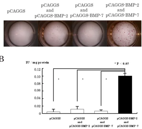
Ten days after the gene transfer by lipofection, the C2C12 cells that had been directly and simultaneously trans-fected with equal doses of pCAGGS-BMP-2 and pCAGGC-BMP-7 showed stronger ALP activity by histochemistry than cells transfected with the BMP-2 gene alone (Fig. 2A). In contrast, almost no positive cells were seen in the C2C12 cells transfected with pCAGGS-BMP-7 or pCAGGS alone. Quantitative analysis of the ALP activity revealed that the activity in the BMP-2 and pCAGGS-BMP-7-treated group was more than 5 times greater than in the single-gene transfer or control groups (p < 0.05) (Fig. 2B).
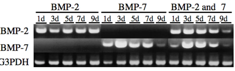
Human BMP-2 and BMP-7 mRNA expression in muscles electroporated with 2 and pCAGGS-BMP-7
To evaluate whether the human BMP-2 and -7 genes could be simultaneously and directly transferred by in vivo elec-troporation, we analyzed the BMPs' mRNA expression in the electroporated muscles by RT-PCR. The presence of human BMP-2 or BMP-7 mRNAs transcribed from pCAGGS-BMP-2 or pCAGGS-BMP-7 in the electroporated muscles was detected 1,3,5,7 and 9 days after treatment (Fig. 3). Both human BMP-2 and BMP-7 mRNAs were co-expressed in muscles after the simultaneous transfer of pCAGGS-BMP-2 and pCAGGS-BMP-7 using transcutane-ous in vivo electroporation up to 9 days. The G3PDH mRNA control was detected in all groups. These data revealed that multiple BMP genes could be successfully transferred by in vivo electroporation.
Simultaneous and direct electroporation-mediated in vivo
Simultaneous gene transfer with BMP-2 and BMP-7 in vitro
Figure 2
induces more rapid ossification than the transfer of either gene alone
The induction of bone formation was investigated by radi-ographic and histological observations 10 days after the gene transfer.
Radiographic findings
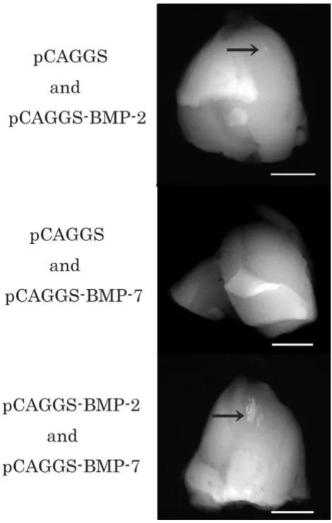
Radiographs revealed opacities that had well-defined mar-gins in the target muscles of groups receiving electropora-tion with pCAGGS and BMP-2, and pCAGGS-BMP-2 and pCAGGS-BMP-7. The opaque areas in the muscles treated simultaneously with the 2 and BMP-7 genes were larger and clearer than those in the other group (Fig. 4). In contrast, we could not detect well-defined opacities in muscles that received transferred pCAGGS-BMP-7 and pCAGGs or pCAGGS alone.
Histological findings
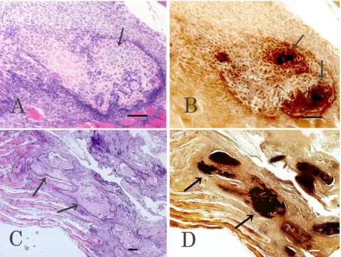
We examined sections from muscles harvested 1, 3, 5, 7, or 10 days after the electroporation histologically, by HE and von Kossa staining. By 10 days after the treatment, both cartilaginous tissues and bone had formed in speci-mens treated with pCAGGS and pCAGGS-BMP-2 or pCAGGS-BMP-2 and pCAGGS-BMP-7. However, in spec-imens from rats that received pCAGGS-BMP-2, the carti-laginous regions, rather than ossified regions, were predominant and widely distributed. Partially calcified areas were also observed by von Kossa stain (Fig. 5A,B). In contrast, bone was frequently observed among muscle fib-ers in the pCAGGS-BMP-2 and pCAGGS-BMP-7-treated specimens (Fig. 5C), and the bone contained osteoblasts, osteocytes, and osteoclasts. In addition, calcification was further advanced in these BMP-2 and pCAGGS-BMP-7-treated specimens than in the pCAGGS-BMP-2-treated specimens, as revealed by von Kossa stain (Fig. 5D). Neither cartilaginous tissue nor bone was observed
in the specimens treated with pCAGGS or pCAGGS and pCAGGS-BMP-7, 10 days after treatment.
Expression of rat endogenous BMP-4 mRNA
In the group treated with the combined 2 and BMP-7 genes, rat BMP-4 mRNA was expressed, and its level increased up to 3 days after gene transfer in a time-dependent manner. Its expression gradually decreased thereafter, and disappeared by 9 days after the gene trans-fer (Fig. 6). Rat BMP-4 mRNA expression was weak in the BMP-2 and in the BMP-7 single-gene transfer groups.
Immunohistochemical reactivity of BMP-4
The distribution of BMP-4-positive cells showed that, compared with specimens treated with single-gene trans-fer, there were higher numbers of positive cells in speci-mens treated with both the BMP-2 and BMP-7 genes. The BMP-4-positive cells were located in the matrix between the muscle fibers, which was greatly expanded in these specimens (Fig 7).
Discussion
In the present study, we evaluated the effect of the direct and simultaneous gene transfer of human BMP-2 and BMP-7 gene expression vectors (pCAGGS-BMP-2 and pCAGGS-BMP-7) on osteoinduction using in vivo electro-poration.
In vitro studies showed previously that BMP purified from the conditioned medium of cultured CHO cells that were transfected with equal amounts of BMP-2 and BMP-7 expression vectors increases the ALP activity of mouse stromal cells far better than BMP purified from the medium of cells transfected with one of these vectors alone. These results suggest that a BMP-2/7 heterodimer forms under these conditions and causes the high ALP
[image:6.612.62.551.86.235.2]RT-PCR evaluation of BMP-2 and BMP-7 gene expression after simultaneous gene transfer into skeletal muscles
Figure 3
activity, indicating that the BMP-2/7 heterodimer has a strong bone-inducing activity [9,10,16]. Furthermore, mixing the conditioned media from cells secreting either BMP-2 or BMP-7 alone does not increase the ALP activity synergistically [10,16]. These systems had three steps: the first was the combined gene transfer of BMP-2 and BMP-7 into epithelial cells to produce BMP-2/7. Next the BMP2/ 7 protein was purified from the supernatant of these cells. Finally, the purified protein was added to the preosteob-lastic or myobpreosteob-lastic cells. Here, we assessed the effect of the direct and simultaneous gene transfer of BMP-2 and
BMP-7 into skeletal muscles. Before our in vivo study, we carried out an in vitro experiment using the mouse myob-lastic C2C12 cell line to determine whether cells directly and simultaneously transfected with the 2 and BMP-7 genes would express these genes, as we hoped they would in vivo. In the present study, the C2C12 cells showed higher ALP activity in the group receiving the BMP-2 and BMP-7 genes than in the control group or the groups transfected with a single BMP expression plasmid. These results agreed with the previous studies [9,10]. Based on our in vitro study, we examined whether in vivo
electroporation with equal doses of pCAGGS-BMP-2 and pCAGGS-BMP-7 is effective for bone formation. Ten days after treatment, direct and simultaneous transfer of the BMP-2 and BMP-7 genes induced higher ALP activity (data not shown), and more intense calcification than did the transfer of either gene alone. In contrast, the calcified areas observed 10 days after the gene transfer with pCAGGS-BMP-2 alone were mostly cartilaginous tissue with very little bone. The muscles transfected with pCAGGS-BMP-7 did not form cartilaginous tissue, bone, or calcified areas during the observation period. There-fore, this study strongly suggests that the direct and simul-taneous gene transfer of equal doses of pCAGGS-BMP-2 and pCAGGS-BMP-7 into skeletal muscles by in vivo elec-troporation was successful and resulted in greater osteoin-ductive activity than the direct transfer of either gene alone. This effect may be owing to the formation of the BMP-2/7 heterodimer as suggested for the in vitro studies [9,10,16]. However, to fully understand our results, we may need to consider the more complicated and dynamic events underlying osteoinduction in vivo, because the extracellular matrix components that bind to BMPs [17] and/or the different affinities of the BMPs for their recep-tors [18-21] must be involved in the osteoinductive activ-ity. Further study is needed to clarify the mechanism of the osteoinduction reported here. For example, a single vector that can express BMP-2 and BMP-7 simultaneously might be useful for assessing the influence of the BMP-2/ 7 heterodimer on osteoinductive activity in vivo.
Endogenous BMP-4 mRNA was highly expressed in the skeletal muscle of rats receiving the pCAGGS-BMP-2 and pCAGGS-BMP-7 genes, but its expression was much weaker in the skeletal muscles of rats receiving only one of the genes. It is reported that 7 down-regulates BMP-4 mRNA through the stages of proliferation, matrix forma-tion, and mineralization in long-term primary cultures of fetal rat calvarial (FRC) cells [14]. In C2C12 cells, BMP-7 up-regulates the BMP-4 mRNA [22]. On the other hand, BMP-2 inhibits BMP-4 mRNA expression initially, but stimulates it during the mineralization phase [11,13]. In the costochondral growth-plate chondrocytes, BMP-2 induces the expression of BMP-4 mRNA in a cell
[image:7.612.62.297.85.455.2]matura-Soft X – ray analysis 10 days after in vivo electroporation
Figure 4
tion-dependent manner [12]. However, this is the first report that the simultaneous gene transfer by in vivo elec-troporation of BMP-2 and BMP-7 increases the level of BMP-4 mRNA.
Under conditions of normal osteoblastic cell differentia-tion, rat BMP-4 mRNA is expressed in the early phase with peak expression levels during the proliferation and matrix formation phases, but it declines afterwards in FRC cells [10,23]. In vivo, a transient elevation in the BMP-4 mRNA levels is reported in osteoprogenitor cells of the perios-teum in the early stages of fracture repair [24-27]. Moreo-ver, BMP-4 mRNA is detected in areas where
fibroblast-like cells and preosteoblasts migrate, in the early stage of distraction osteogenesis [28]. These results seem to sug-gest that BMP-4 plays an important role in the early phase of osteoinduction. Our histological results at 3 and 5 days, during the early phase of the BMP-4 mRNA increase, revealed abundant migratory spindle-shaped cells, i.e., fibroblast-like cells. These cells were in the same location as those that stained positive for BMP-4 by immunohisto-chemistry. Therefore, the expression of BMP-4 mRNA and its increase in the early phase might be involved with the effective bone formation seen when the 2 and BMP-7 genes were simultaneously transferred into rat skeletal muscles by transcutaneous in vivo electroporation.
[image:8.612.59.556.86.459.2]Histological analyses 10 days after in vivo electroporation
Figure 5
In this study, we used an animal model to study ectopic bone formation in the skeletal muscles to examine the efficiency of simultaneous and direct BMP-2 and BMP-7 gene transfer using in vivo electroporation. For clinical applications, it may be possible to reconstruct bone and the accompanying muscle in jaw areas that have been damaged, for example, by trauma or tumour treatment. Moreover, BMP-2 and BMP-7 gene transfer into the perio-stea to augment the alveolar ridge or fracture repair may enhance periosteal cell differentiation efficiently, because it is well known that periostea have multi-potential stem cells [29-31]. If successful, such clinical applications
would be very significant and wide-ranging, and could include fracture repair, orthognathic surgery, alveolar ridge augmentation, and dental implants.
Conclusion
We propose that the direct and simultaneous gene transfer of BMP-2 and BMP-7 by in vivo electroporation is more effective than the transfer of BMP-2 alone, which we explored in an earlier paper using the same conditions, such as the dose of plasmid and the size of the electrodes [5]. Since the major drawbacks of using adenovirus vec-tors, i.e., the need for titration, the potential toxicity, and
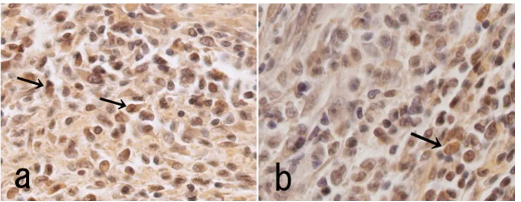
Immunohistochemical analysis of BMP-4 in skeletal muscle
Figure 7
Immunohistochemical analysis of BMP-4 in skeletal muscle. Immunohistochemical detection of BMP-4-positive cells 5 days after the combined transfer of the BMP-2 and BMP-7 genes (a), or the BMP-2 gene alone (b). The positive cells (arrows) were numerous in the matrix between the muscle fibres in the muscle transfected with the combined BMP genes compared with muscle transfected with BMP-2 alone.
[image:9.612.54.561.465.661.2]Rat BMP-4 expression after gene transfer with BMP-2, BMP-7, or BMP-2 and BMP-7
Figure 6
Rat BMP-4 expression after gene transfer with BMP-2, BMP-7, or BMP-2 and BMP-7. Rat BMP-4 mRNA expression
the need to suppress the host immune response, are not necessary using this system, gene therapy via in vivo elec-troporation is safer and simpler than via viral vectors. As a methodological consideration, electroporation is quite suitable for the direct and simultaneous transfer of two or more BMP genes for bone regeneration therapy.
Competing interests
The author(s) declare that they have no competing inter-ests.
Authors' contributions
MK performed all experiments, analyzed the data, and prepared the manuscript. KB and TY contributed to the in vivo electroporation experiments and provided useful dis-cussions about the results. HM and JM provided signifi-cant help to MK in constructing the plasmid vector.
Acknowledgements
This work was supported in part by a Grant-in-Aid for scientific research from Okayama University and a Grant-in-Aid for young investigators (B) 17791301 from the Japanese Ministry of Education, Culture, Sports, Science and Technology.
References
1. Ferber D, Marshall E: Gene therapy: safer and virus-free? Science
2001, 294:1638-1642.
2. Nishikawa M, Huang L: Nonviral vectors in the new millennium: delivery barriers in gene transfer. Hum Gene Ther 2001,
12:861-870.
3. Aihara H, Miyazaki J: Gene transfer into muscle by electropora-tion in vivo. Nature Biotechnol 1998, 16:867-870.
4. Maruyama H, Sugawa M, Moriguchi Y, Imazaki I, Ishikawa Y, Ataka K, Hasegawa S, Ito Y, Higuchi N, Kazama J, Gejyo F, Miyazaki J: Contin-uous erythropoietin delivery by muscle-targeted gene trans-fer using in vivo electroporation. Hum Gene Ther 2000,
11:429-437.
5. Kawai M, Bessho K, Kaihara S, Sonobe J, Oda K, Iizuka T, Maruyama H: Ectopic bone formation by bone morphogenetic protein-2 gene transfer to skeletal muscle using transcutaneous in vivo electroporation. Hum Gene Ther 2003, 14:1547-1556. 6. Wolf H, Rols MP, Boldt E, Meumann E, Teissie J: Control by pulse
parameters of electric field-mediated gene transfer in mam-malian cells. Biophys J 1994, 66:524-531.
7. Cehng H, Jiang W, Phillips FM, Haydon RC, Peng Y, Zhou L, Luu HH, An N, Breyer B, Vanicharn P, Szatkowski JP, Park JY, He TC: Osteo-genic activity of the fourteen types of human bone morpho-genetic proteins (BMPs). J Bone Joint Surg Am 2003, 85:1544-1552. 8. Li JZ, Li H, Sasaki T, Holman D, Beres B, Dumont RJ, Pittman DD, Hankins GR, Helm GA: Osteogenic potential of five different recombinant human bone morphogenetic protein adenovi-ral vectors in the rat. Gene Ther 2003, 10:1735-1743.
9. Zhu W, Rawlins BA, Boachie-Adjei O, Myers ER, Arimizu J, Choi E, Lieberman JR, Crystal RG, Hidaka C: Combined bone morphoge-netic protein-2 and -7 gene transfer enhances osteoblastic differentiation and spine fusion in a rodent model. J Bone Miner Res 2004, 19:2021-2032.
10. Israel DI, Nove J, Kerns KM, Kaufman RJ, Rosen V, Cox KA, Wozney
JM: Heterodimeric bone morphogenetic proteins show
enhanced activities in vitro and in vivo. Growth Factors 1996,
13:291-300.
11. Chen D, Harris MA, Rossini G, Dunstan CR, Dallas SL, Feng JQ, Mundy GR, Harris SE: Bone morphogenetic protein 2 (BMP-2) enhances BMP-3, BMP-4, and bone cell differentiation marker gene expression during the induction of mineralized bone matrix formation in cultures of fetal rat calvarial oste-oblasts. Calcif Tissue Intl 1997, 60:283-290.
12. Erickson DM, Harris SE, Dean DD, Harris MA, Wozney JM, Boyan BD, Schwartz Z: Recombinant bone morphogenetic protein (BMP)-2 regulates costochondral growth plate chondrocytes and induces expression of BMP-2 and BMP-4 in a cell matu-ration-dependent manner. J Orthop Res 1997, 15:371-380. 13. Hino J, Matsuo H, Kangawa K: Bone morphogenetic protein-3b
(BMP-3b) gene expression is correlated with differentiation in rat calvarial osteoblasts. Biochem Biophys Res Commun 1999,
256:419-424.
14. Yeh LC, Unda R, Lee JC: Osteogenic protein-1 differentially reg-ulates the mRNA expression of bone morphogenetic and their receptors in primary cultures of osteoblasts. J Cell Physiol
2000, 185:87-97.
15. Niwa H, Yamamura K, Miyazaki J: Efficient selection for high-expression transfectants with a novel eukaryotic vector. Gene 1991, 108:193-199.
16. Hazama M, Aono A, Ueno N: Efficient expression of het-erodimer of bone morphogenetic protein. Biochem Biophys Res Commun 1995, 209:859-866.
17. Reddy AH, Cunningham NS: Initiation and promotion of bone differentiation by bone morphogenetic proteins. J Bone Miner Res 1993, 8:499-502.
18. Dijke TP, Yamashita H, Sampath TK, Reddy AH, Estevez M, Riddle DL, Ichijyo H, Heldin CH, Miyazono K: Identification of type I receptors for osteogenic protein-1 and bone morphogenetic protein-4. J Biol Chem 1994, 269:16985-16988.
19. Rosenzweig BL, Lmamura T, Okadome T, Cox GN, Yamashita H, Dijke TP, Helden CH, Miyazono K: Cloning and characterization of a human type II receptor for bone morphogenetic pro-teins. Proc Natl Acad Sci U S A 1995, 92:7632-7636.
20. Nishitoh H, Ichijo H, Kimura M, Matsumoto T, Makishima F, Yamaguchi A, Yamashita H, Enomoto S, Miyazono K: Identification of type I and Type II serine/threonine kinase receptors for
growth/differentiation factor-5. J Biol Chem 1996,
271:21345-21352.
21. Hoffmann A, Gross G: BMP signaling pathways in cartilage and bone formation. Crit Rev Eukaryot Gene Expr Rev 2001, 11:23-45. 22. Yeh LC, Tsai AD, Lee JC: Osteogenic protein-1 (BMP-7) induces
osteoblastic cell differentiation of the pluripotent mesenchy-mal cell line C2C12. J Cell Biochem 2002, 87:292-304.
23. Harris SE, Sabatini M, Harris MA, Feng JQ, Wozney JM, Mundy GR:
Expression of bone morphogenetic protein messenger RNA in prolonged cultures of fetal rat calvarial cells. J Bone Miner Res 1994, 9:389-394.
24. Nakase T, Nomura S, Yoshikawa H, Hashimoto J, Kitamura Y, Oikawa S, Yakaoka K: Transient and localized expression of bone mor-phogenetic protein 4 messenger RNA during fracture heal-ing. J Bone Miner Res 1994, 9:651-659.
25. Onishi T, Ishidou Y, Nagamine T, Yone K, Imamura T, Kato M, Sam-path TK, Ten DP, Sakou T: Distinct and overlapping patterns of localization of bone morphogenetic protein (BMP) family members and a BMP type II receptor during fracture healing in rats. Bone 1998, 22:605-612.
26. Yaoita H, Orimo H, Shirai Y, Shimada T: Expression of bone mor-phogenetic proteins and rat distal-less homolog genes fol-lowing rat femoral fracture. J Bone Miner Res 2000, 18:63-70. 27. Yoshimura Y, Nomura S, Kawasaki S, Tsutsumimoto T, Shimizu T,
Takaoka K: Colocalization of noggin and bone morphogenetic protein-4 during fracture healing. J Bone Miner Res 2001,
16:876-884.
28. Berven LG, Simpson H, Triffitt JT: Expression of BMP-4 mRNA during distraction osteogenesis in rabbits. Acta Orthop Scand
1998, 69:420-425.
29. Zhang X, Xie C, Lin AS, Ito H, Awad H, Lieberman JR, Rubery PT, Schwarz EM, O'Keefe RJ, Guldberg RE: Periosteal progenitor cell fate in segmental cortical bone graft transplantations+ impli-cations for functional tissue engineering. J bone Mine Res 2005,
20:2124-2137.
30. Park J, Gelse K, Frank S, von der Mark K, Aigner T, Schneider H:
Transgene-activated mesenchymal cells for articular carti-lage repair: a comparison of primary bone marrow-, peri-chondrium/S periosteum- and fat-derived cells. J Gene Med
2006, 8:112-125.
31. De Bari C, Dell' Acco F, Vanlauwe J, Eyckmans J, Khan IM, Archer CW, Jones EA, McGonagle D, Mitsiadis TA, Pitzalis C, Luyten FP:
Publish with BioMed Central and every scientist can read your work free of charge
"BioMed Central will be the most significant development for disseminating the results of biomedical researc h in our lifetime."
Sir Paul Nurse, Cancer Research UK
Your research papers will be:
available free of charge to the entire biomedical community
peer reviewed and published immediately upon acceptance
cited in PubMed and archived on PubMed Central
yours — you keep the copyright
Submit your manuscript here:
http://www.biomedcentral.com/info/publishing_adv.asp
BioMedcentral
demonstrated by single-cell lineage analysis. Arthritis Rhuem
2006, 54:1209-1221.
Pre-publication history
The pre-publication history for this paper can be accessed here: