www.arpnjournals.com
A TWO COMPONENT RED BLOOD CELL MODEL FOR SINGLE CELL
MECHANIC
Ida Laila Ahmad
1,2and Mohd Ridzuan Ahmad
11Micro-Nano Mechatronics Research Group, Department of Control and Mechatronics Engineering, Faculty
of Electrical Engineering, Universiti Teknologi Malaysia, 81310 Johor Bahru, Johor, Malaysia
2Department of Electronic Engineering, Faculty of Electrical and Electronic Engineering, Universiti Tun
Hussein Onn Malaysia, 86400 Batu Pahat, Johor, Malaysia E-Mail: ida@uthm.edu.my1,2, ridzuan@fke.utm.my1
ABSTRACT
This work presents the deformation of red blood cell (RBC) as it travels in a microchannel using a theoretical model. The developed model predicts the deformation experienced by the RBC when exposed to shear stresses (fluid) as a function of mechanical properties. RBC is known to be a biconcave disk and is modeled as a liquid enclosed with a solid membrane. Therefore it is appropriate to model the RBC as a two component models consists of membrane and cytoplasm. The membrane, assumed to behave as an nearly incompressible material shows hyperelastic response to bending and shearing while the cytoplasm exhibits homogeneous Newtonian fluid properties. The model parameters were determined from the experimental measurements and comparison were made. Agreement in terms of hyperelasticity, deformation rate and stress distribution were found with previous researches.
Key words: Red Blood Cell, Theoretical Model, Membrane, Cytoplasm, Hyperelasticity
INTRODUCTION
Red blood cell (RBC) is known for delivering oxygen throughout the microcirculation system in a human body. The efficacy of such transportation mechanism heavily determines by the ability of RBC to undergone large deformation and pass through smaller capillaries relative to its size (Xu et al. 2012, Li et al. 2014, Tomaiuolo et al. 2009). Researches made from several decades have looked into the trends and to understand this unique phenomenon possess by RBC. The emergence of advance technological instruments enabled the rapid investigation on mechanical properties of RBC.
RBC mechanical properties measurements
Numerous experimental approach can broadly be classified into two main groups uses direct measurement or indirect measurement on RBC mechanical properties. Direct measurement treats RBC in population means either RBC in suspension or diluted RBC will give high throughput; however do not address on size heterogeneity. Some of the techniques include rheometer (Tran-Son-Tay et al. 1984), laser cavitation bubbles (Quinto-Su et al. 2011), microfiltration (Turmezei et al. 2003) and ektacytometry (Bessis et al. 1980, Liu et al. 2007).
On the other hand, the indirect measurement employ single cell approach overcome several problems faced by the former techniques. Several techniques namely optical tweezers (Mills et al. 2004, Dao et al. 2005, Rancourt-Grenier et al. 2010, Huruta et al. 2014, Hénon et al. 1999, Bronkhorst et al. 1995, Brandão et al. 2003), micropipette aspiration (Evans 1973, Evans 1983, Hochmuth 2000, Artmann et al. 1997), atomic force microscopy (Kuznetsova et al. 2007) and recent movements using microfluidic platforms (Sakuma et al. 2014; Quinn et al. 2011; Tomaiuolo et al. 2009; Luo et al. 2014; Yaginuma et al. 2011). Further reading on single cell mechanical properties measurements can be found from (Ahmad & Ahmad 2014)
while more facts on RBC were discussed in details by (Kim et al. 1979) and (Tomaiuolo 2014).
RBC related diseases
[image:1.595.310.551.562.807.2]As a living entity, RBC should withstand the changes attributed not only from its environment but also due to mechanical stimuli. Nevertheless, pathological alterations on RBC due to diseases have been discussed by many (Maciaszek et al. 2011, Williamson et al. 1985, Park et al. 2010, Hosseini & Feng 2012, Sleep et al. 1999) and can be found from comprehensive review by (Tomaiuolo 2014). Table 1 below signify the evidence that RBC will experience mechanical alterations whenever a disease incurred.
Table 1: RBC mechanical properties alterations due to diseases
Disease RBC conditions
Malaria RBC stiffened according to stages of infection
Less deformable
Increased adhesion bond
(Glenister et al. 2002, Herricks et al. 2009, Cooke et al. 2000, Bow et al. 2011, Shelby et al. 2003)
Sickle cell
RBC stiffened
Change of shape and increased rigidity (Brandão et al. 2003)
Cancer RBC more softer and deformable (Lekka et al. 1999, Guck et al. 2005, Byun et al. 2013, Cross et al. 2007)
MECHANICAL MODEL OF RBC
[image:2.595.46.286.283.457.2]To further elucidate the general concept of modelling a cell, one needs to understand the divisions of mechanical models available. A good review was provided by (C T Lim et al. 2006) explained on most of the mechanical models have been developed either using continuum or nano-structural approach. Continuum means cell is represented by a continuum material whereby the associative constitutive material model will be properly defined using appropriate parameters. By doing so, less computational time will be required and hence give a more straightforward approach. On the other hand, the nano-structural approach treated cytoskeleton as the main structural components and thus limit the model only to cytoskeletal related investigation especially for adherent cell. Various researchers’ works spanned from as early as 1980s and to date covers wide area of mechanical models as given in Figure 1 below.
Figure 1: Mechanical Model of RBC excerpted from (C T Lim et al. 2006)
There are three critical domains to be addressed on whenever a cell model is proposed which are; the structural heterogeneity of cells, appropriate constitutive relations for each subcellular regions and components and active forces acting on cell. Several mechanical models have been developed by previous researchers according to the type of cell. Different cells react differently towards applied stimuli and these helped to setting up requirements for mechanical model. Table 2 below depicts the requirements needed for mechanical model based on cell type.
Table 2: Proposed mechanical model based on cell types
Cell Type Mechanical Model
Suspended cell Cortical shell-liquid core (Liquid drop)
Adherent cell Solid model
(Elastic, Viscoelastic) Adherent cell
Fractional Derivative Model (Power-Law structural damping) Musculoskeletal Biphasic model
METHODOLOGY
The RBC is a cell with no nucleus and so it has the form of a microcapsule that contains a cytoplasm. In this model, RBC is represented by two components consists of membrane and cytoplasm. Using continuum approach for mechanical model, the Newtonian Liquid Drop Model was adopted. The cytoplasm is treated as an incompressible Newtonian fluid with hydraulic fluid model within fluid filled cavity. It is surrounded by a thin viscoelastic membrane, which consist of a lipid bilayer and a protein cytoskeleton.
When a suspension of RBCs is subjected to flow, viscous stresses are exerted on the RBCs and this may lead to large deformations. The mechanical properties of the membrane are highly important due to control of the resistance towards the applied stresses. Since the membrane thickness is small compared to the cell dimensions, the membrane may be considered as a two-dimensional solid with viscoelastic properties. Moreover, changes in the cell due to the load applied can be divided into: membrane area uniform (isotropic) dilation, shear deformation (with no area change or bending), bending (with no area change or shearing). Frequently, the RBC is associated with hyperelasticity governed by either Neo Hookean or Mooney Rivlin strain energy potential. A Neo-Hookean model is a hyperelastic material model that can be used for predicting the stress-strain behavior of materials, and the model is similar to Hooke’s law. For general materials, the relationship between applied stress and strain is initially linear, but at a certain point the stress-strain curve changes to nonlinear. A Neo Hookean model is one of the simple model and the strain energy density function for an incompressible Neo-Hookean material as given by (Abaqus 2013) is:
W= C1(I1-3) (1)
Where C1 is a material constant, and I1 is the first invariant of the left Cauchy-Green deformation tensor. ABAQUS/ CAE Version 6.12 requires that the Neo-Hookean material parameters be expressed in terms of C10 and D1. These parameters can be obtained from the shear modulus (µ) and the bulk modulus (κ) by (Abaqus 2013) based on the following expressions:
C10 = µ / 2 (2)
D1 = 2 / κ (3)
This model adopted isotropic hyperelasticity with Neo Hookean strain energy potential.
Geometrical design
[image:2.595.45.288.663.762.2]Figure 2: Dimensions of the proposed RBC model
RBC model considerations
Several assumptions were made prior to the RBC modeling are listed below:
Fluid surrounding the RBC is considered to be incompressible therefore allowing RBC to keep constant volume.
RBC material behaviour is anisotropic therefore there is possibilities of tilting/rotating during measurements.
RBC membrane is hyperelastic therefore defines a material which can recover its original shape following a finite deformation.
Plane symmetry of RBC allowing for smaller model to be designed to reduce computational complexity.
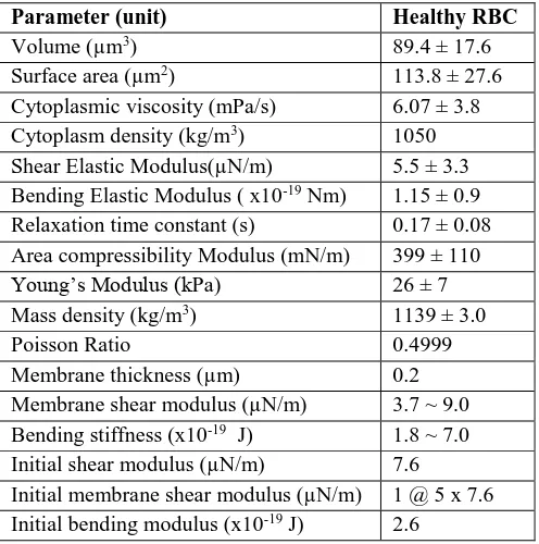
[image:3.595.373.482.223.295.2]During the design stage, important parameters summarized in Table 3 were used to model the healthy RBC.
Table 3:Simulation parameters
Parameter (unit) Healthy RBC
Volume (µm3) 89.4 ± 17.6
Surface area (µm2) 113.8 ± 27.6
Cytoplasmic viscosity (mPa/s) 6.07 ± 3.8 Cytoplasm density (kg/m3) 1050 Shear Elastic Modulus(µN/m) 5.5 ± 3.3 Bending Elastic Modulus ( x10-19 Nm) 1.15 ± 0.9 Relaxation time constant (s) 0.17 ± 0.08 Area compressibility Modulus (mN/m) 399 ± 110
Young’s Modulus (kPa) 26 ± 7
Mass density (kg/m3) 1139 ± 3.0
Poisson Ratio 0.4999
Membrane thickness (µm) 0.2
Membrane shear modulus (µN/m) 3.7 ~ 9.0 Bending stiffness (x10-19 J) 1.8 ~ 7.0 Initial shear modulus (µN/m) 7.6
Initial membrane shear modulus (µN/m) 1 @ 5 x 7.6 Initial bending modulus (x10-19 J) 2.6
RBC model materials definitions
The model was developed using ABAQUS/ CAE Version 6.12. In order to successfully model two components RBC, two different types of shell elements were used. The outer surface of the cell, depicting the membrane employed S4R shell elements (4-node doubly curved shell finite membrane
strains elements). While, the inner surface of the cell was composed of F3D4 shell elements (4-node linear 3-dimensional quadrilateral hydrostatic fluid element), which represented the cytoplasm. This is to mimic the fluidic property of the cytoplasm.
RBC model meshing requirements
As mentioned previously, the shell elements were used while meshing the proposed RBC model. 2964 elements were generated from S4R shell (membrane) and 3000 elements for F3D4 (cytoplasm). Figure 3 shows the meshing.
Figure 3:Mesh of Two components RBC model
Simulation settings for RBC stretching
[image:3.595.40.288.458.708.2]For the purpose to reduce computational complexity, the loading and boundary conditions for the simulation were set in the direction as in Figure 4. The applied force were intended to stretch the RBC in the predefined directions as the displacement in terms of transverse and axial diameter were observed. Furthermore, the flexibility of RBC corresponds to the force applied will indirectly provide hints towards mechanical properties such as bending moduli and health status.
Figure 4:Simulation settings for stretching experiment
RESULTS AND DISCUSSIONS
RBC Hyperelasticity
[image:3.595.376.495.461.640.2]Figure 5:Neo-Hookean RBCHyperelasticity curve
RBC stretching simulation measurements
[image:4.595.62.275.356.464.2]The schematic of measurements during stretching simulation is given by Figure 6 below. It can be seen that the measurements were taken with respect to the direction of cell’s elongation. The elongation with respect to horizontal axis is known as axial diameter, while the elongation along the vertical axis is known as transverse diameter.
Figure 6:Schematic of stretching experiment
RBC stretching simulation results
The following Figures 7(a)-(d) were the examples of simulation results at different tensile force. Greater elongation were observed when larger force were applied. These observations were tabulated and the resultant graph can be seen from Figure 8.
Figure 7: Stretching of RBC when (a) 10pN (b) 20pN (c) 40pN (d) 60pN
Figure 8:Axial and transversal diameter measurement when applied to various stretching force (µo = 7.6 µN/m)
From the graph, it is worthwhile to note that for a given external force applied, the degree of deformation depends largely on the specified initial shear modulus. Furthermore, the coupling between solid membrane and fluidic cytoplasm in this model helps to eliminate the problem concerning accurate prediction for the axial and transverse measurements. The predefined cytoplasm properties in this model also shows that cytoplasm viscosity plays important role in terms of deformation of RBC under loading. Previously, most of researchers neglect this fact in order to simplify computational complexity by assuming the small and insignificant role of cytoplasm viscosity. Another improvement in terms of replicating the actual experimental conditions also can be achieved via this model. An agreement with experimental data coming from (C.T. Lim et al. 2006; Dao et al. 2005) were obtained. Hence, the fluid-interactions model adopted in this paper were able to provide better estimation of RBC deformation as comparable to the experimental approach.
RBC in a microconfined channel
[image:4.595.68.538.610.771.2]To further investigate the deformability limit of the developed RBC model, a microchannel was used. It is equally important to visualize the RBC behavior when undergone a deformation under laminar flow. A constricted hyperbolic microchannel was designed to have the following dimensions (height, width, length) of 5µm, 3µm and 10µm respectively on the contriction area as shown in Figure 9.
As suggested by Figure 10, the ability of RBC to recover its original shape was demonstrated via the simulation. During the simulation, the normal biconcave shaped RBC underwent a severe shape transition from the entrance until an ellipsoidal RBC was observed. Once the RBC exiting the constriction area, RBC slowly recover its original shape. This natural phenomenon explains the capability of RBC to travel within blood capillaries with diameter as small as 3µm.
Figure 10: Recoverability of RBC model in microchannel (a) at the channel entrance, (b) exiting the channel and (c) full recovery
[image:5.595.46.289.192.260.2]Furthermore, Figure 11 below depicts stress distribution in terms of mises experienced by the RBC when travelling in the microchannel. The intensified area (red shaded) normally are due to greater stress experienced with respect to smaller constriction and the edges. With the proposed RBC model, the recoverability rate is improved due to cytoplasm influence which is previously neglected. Theoretically, the averaged value of cytoplasm viscosity is mainly contributed from haemoglobin concentration. It is also temperature dependent (Chee et al. 2008).
Figure 11:Von Mises Stress of RBC model in microchannel (a) at the channel entrance, (b) exiting the channel and (c) full recovery
CONCLUSION
We proposed a two components model of RBC consisting of membrane and cytoplasm adapting Neo-Hookean strain energy potential. The model was carefully evaluated in terms of hyperelasticity and stretching behavior when subjected to applied force. Next, the model was integrated into a microchannel in order to demonstrate the recoverability and in order to achieve better understanding of RBC mechanics. The advantages of the proposed model are: firstly, more accurate stretching simulation results that is comparable to experimental works and secondly, improved recovery rate since RBC uses its own elastic energy with the aid of cytoplasm viscosity.Altogether, the presented results show that the developed RBC model may serve as a quantitative tool for estimating RBC mechanical properties.
ACKNOWLEDGMENT
This work was supported by grants from the Ministry of Education Malaysia (grant nos. 4L640 and 4F351) and Universiti Teknologi Malaysia (grant nos. 02G46, 03H82 and 03H80).
REFERENCES
A. B. A. Q. U. S. (2013). 6.12 Documentation Collection. ABAQUS/CAE User's Manual
Ahmad, I.L. & Ahmad, M.R., 2014. Trends in characterizing single cell ’ s stiffness properties. Micro &
Nano System Letters, 2(8), pp.1–11.
Artmann, G.M. et al., 1997. Micropipette Aspiration of Human Erythrocytes Induces Echinocytes via Membrane Phospholipid Translocation. Biophysical Journal, 72(March), pp.1434–1441.
Bessis, M., Mohandas, N. & Feo, C., 1980. Automated ektacytometry: a new method of measuring red cell deformability and red cell indices. Blood Cells, 6(3), pp.315–327.
Bow, H. et al., 2011. A microfabricated deformability-based flow cytometer with application to malaria. Lab on a Chip, 11(6), pp.1065–1073.
Brandão, M.M. et al., 2003. Optical tweezers for measuring red blood cell elasticity: application to the study of drug response in sickle cell disease. European Journal of
Haematology, 70, pp.207–211.
Bronkhorst, P.J. et al., 1995. A new method to study shape recovery of red blood cells using multiple optical trapping.
Biophysical Journal, 69(5), pp.1666–73.
Byun, S. et al., 2013. Characterizing deformability and surface friction of cancer cells. In Proceedings of the National Academy of Sciences of the United States of
America. pp. 7580–7585.
Chee, C. Y., Lee, H. P., & Lu, C., 2008. Using 3D fluid– structure interaction model to analyse the biomechanical properties of erythrocyte. Phys. Lett. A, vol. 372, no. 9, pp. 1357–1362.
Cooke, B., Coppel, R. & Wahlgren, M., 2000. Falciparum malaria: Sticking up, standing out and out-standing.
Parasitology Today, 16, pp.416–420.
Cross, S.E. et al., 2007. Nanomechanical analysis of cells from cancer patients. Nature Nanotechnology, 2(12), pp.780–3.
[image:5.595.46.287.442.526.2]2259-2280]. Journal of the Mechanics and Physics of Solids, 53(2), pp.493–494.
Evans, E.A., 1983. Bending elastic modulus of red blood cell membrane derived from buckling instability in micropipet aspiration tests. Biophysical Journal, 43(1), pp.27–30.
Evans, E.A., 1973. New membrane concept applied to the analysis of fluid shear- and micropipette-deformed red blood cells. Biophysical Journal, 13(9), pp.941–54.
Glenister, F.K. et al., 2002. Contribution of parasite proteins to altered mechanical properties of malaria-infected red blood cells. Blood, 99, pp.1060–1063.
Guck, J. et al., 2005. Optical deformability as an inherent cell marker for testing malignant transformation and metastatic competence. Biophysical Journal, 88(5), pp.3689–3698.
Hénon, S. et al., 1999. A new determination of the shear modulus of the human erythrocyte membrane using optical tweezers. Biophysical Journal, 76(2), pp.1145–51.
Herricks, T., Antia, M. & Rathod, P.K., 2009. Deformability limits of Plasmodium falciparum-infected red blood cells. Cellular Microbiology, 11(9), pp.1340–1353.
Hochmuth, R.M., 2000. Micropipette aspiration of living cells. Journal of Biomechanics, 33(1), pp.15–22.
Hosseini, S.M. & Feng, J.J., 2012. How malaria parasites reduce the deformability of infected red blood cells.
Biophysical Journal, 103(1), pp.1–10.
Huruta, R.R. et al., 1998. Mechanical Properties of Stored Red Blood Cells Using Optical Tweezers. Blood, 92, pp.2975–2977.
Kim, Y., Kim, K. & Park, Y., 1979. Measurement Techniques for Red Blood Cell Deformability : Recent Advances. , Blood Cell - An Overview of Studies in
Hematology, 2012(1), pp. 167-194.
Kuznetsova, T.G. et al., 2007. Atomic force microscopy probing of cell elasticity. Micron (Oxford, England : 1993), 38(8), pp.824–33.
Lekka, M. et al., 1999. Elasticity of normal and cancerous human bladder cells studied by scanning force microscopy.
European Biophysics Journal, 28, pp.312–316.
Li, X. et al., 2014. Probing red blood cell mechanics , rheology and dynamics with a two-component multi-scale model. Phil. Trans, R. Soc.
Lim, C.T. et al., 2006. Experimental techniques for single cell and single molecule biomechanics. Materials Science
and Engineering: C, 26(8), pp.1278–1288.
Lim, C.T., Zhou, E.H. & Quek, S.T., 2006. Mechanical models for living cells--a review. Journal of Biomechanics, 39(2), pp.195–216.
Liu, X. et al., 2007. The measurement of shear modulus and membrane surface viscosity of RBC membrane with Ektacytometry: a new technique. Mathematical Biosciences, 209(1), pp.190–204.
Luo, Y.N. et al., 2014. A constriction channel based microfluidic system enabling continuous characterization of cellular instantaneous Young’s modulus. Sensors and
Actuators B: Chemical, 202, pp.1183–1189.
Maciaszek, J.L., Andemariam, B. & Lykotrafitis, G., 2011. Microelasticity of red blood cells in sickle cell disease.
Journal of Strain Analysis, 46, pp.368–379.
Mills, J.P. et al., 2004. Nonlinear elastic and viscoelastic deformation of the human red blood cell with optical tweezers. Mechanics & Chemistry of Biosystems, 1(3), pp.169–180.
Park, Y. et al., 2010. Measurement of red blood cell mechanics during morphological changes. Proceedings of the National Academy of Sciences of the United States of
America, 107(15), pp.6731–6.
Quinn, D.J. et al., 2011. Combined simulation and experimental study of large deformation of red blood cells in microfluidic systems. Annals of biomedical engineering, 39(3), pp.1041–1050.
Quinto-Su, P. a et al., 2011. Red blood cell rheology using single controlled laser-induced cavitation bubbles. Lab on a Chip, 11(4), pp.672–8.
Rancourt-Grenier, S. et al., 2010. Dynamic deformation of red blood cell in dual-trap optical tweezers. Optics Express, 18(10), pp.10462–72.
Sakuma, S. et al., 2014. Red blood cell fatigue evaluation based on the close-encountering point between extensibility and recoverability. Lab on a Chip, 14(6), pp.1135–41.
Shelby, J.P. et al., 2003. A microfluidic model for single-cell capillary obstruction by Plasmodium falciparum-infected erythrocytes. In Proceedings of the National
Academy of Sciences of the United States of America. pp.
14618–14622.
Tomaiuolo, G., 2014. Biomechanical properties of red blood cells in health and disease towards microfluidics.
Biomicrofluidics, 8(5), p.051501.
Tomaiuolo, G. et al., 2009. Red blood cell deformation in microconfined flow. Soft Matter, 5(19), p.3736.
Tran-Son-Tay, R., Sutera, S.P. & Rao, P.R., 1984. Determination of red blood cell membrane viscosity from rheoscopic observations of tank-treading motion.
Biophysical Journal, 46(1), pp.65–72.
Tsukada, K. et al., 2001. Direct measurement of erythrocyte deformability in diabetes mellitus with a transparent microchannel capillary model and high-speed video camera system. Microvascular Research, 61(3), pp.231–9.
Turmezei, P. et al., 2003. Low-cost microfilter for red blood cell membrane stiffness measurement using photosensitive BCB. In The 12th International conference on Solid State
Sensors, Actuators and Microsystems. pp. 107–110.
Williamson, J.R. et al., 1985. Microrheologic investigation of erythrocyte deformability in diabetes mellitus. Blood, 65(2), pp.283–8.
Xu, D. et al., 2012. Three-dimensional numerical simulation of the deformation and the aggregation of human red blood cells. Series on Biomechanics, 27(3), pp.11–16.
Yaginuma, T. et al., 2011. Red Blood Cell Deformation in flows through a PDMS Hyperbolic Microchannel X + Y. In