promotes leukemia-initiating cell capacity
Yuki Kagoya, … , Yoichiro Iwakura, Mineo Kurokawa
J Clin Invest.
2014;
124(2)
:528-542.
https://doi.org/10.1172/JCI68101
.
Acute myeloid leukemia (AML) is a heterogeneous hematologic malignancy that originates
from leukemia-initiating cells (LICs). The identification of common mechanisms underlying
LIC development will be important in establishing broadly effective therapeutics for AML.
Constitutive NF-
k
B pathway activation has been reported in different types of AML;
however, the mechanism of NF-
k
B activation and its importance in leukemia progression
are poorly understood. Here, we analyzed myeloid leukemia mouse models to assess
NF-k
B activity in AML LICs. We found that LICs, but not normal hematopoietic stem cells or
non-LIC fractions within leukemia cells, exhibited constitutive NF-
k
B activity. This activity
was maintained through autocrine TNF-
a
secretion, which formed an NF-
k
B/TNF-
a
positive
feedback loop. LICs had increased levels of active proteasome machinery, which promoted
the degradation of I
k
B
a
and further supported NF-
k
B activity. Pharmacological inhibition of
the proteasome complex markedly suppressed leukemia progression in vivo. Conversely,
enhanced activation of NF-
k
B signaling expanded LIC frequency within leukemia cell
populations. We also demonstrated a strong correlation between NF-
k
B activity and TNF-
a
secretion in human AML samples. Our findings indicate that NF-
k
B/TNF-
a
signaling in LICs
contributes to leukemia progression and provide a widely applicable approach for targeting
LICs.
Research Article
Oncology
Find the latest version:
Research article
Positive feedback between NF-
κ
B and TNF-
α
promotes leukemia-initiating cell capacity
Yuki Kagoya,1 Akihide Yoshimi,1 Keisuke Kataoka,1 Masahiro Nakagawa,1 Keiki Kumano,1Shunya Arai,1 Hiroshi Kobayashi,2 Taku Saito,2 Yoichiro Iwakura,3 and Mineo Kurokawa1
1Department of Hematology and Oncology and 2Department of Orthopaedic Surgery, Graduate School of Medicine, The University of Tokyo, Tokyo, Japan. 3Division of Experimental Animal Immunology, Research Institute for Biomedical Sciences, Tokyo University of Science, Chiba, Japan.
Acute myeloid leukemia (AML) is a heterogeneous hematologic malignancy that originates from
leukemia-ini-tiating cells (LICs). The identification of common mechanisms underlying LIC development will be
impor-tant in establishing broadly effective therapeutics for AML. Constitutive NF-
κ
B pathway activation has been
reported in different types of AML; however, the mechanism of NF-
κ
B activation and its importance in
leuke-mia progression are poorly understood. Here, we analyzed myeloid leukeleuke-mia mouse models to assess NF-
κ
B
activity in AML LICs. We found that LICs, but not normal hematopoietic stem cells or non-LIC fractions
within leukemia cells, exhibited constitutive NF-
κ
B activity. This activity was maintained through autocrine
TNF-
α
secretion, which formed an NF-
κ
B/TNF-
α
positive feedback loop. LICs had increased levels of active
proteasome machinery, which promoted the degradation of I
κ
B
α
and further supported NF-
κ
B activity.
Phar-macological inhibition of the proteasome complex markedly suppressed leukemia progression in vivo.
Con-versely, enhanced activation of NF-
κ
B signaling expanded LIC frequency within leukemia cell populations. We
also demonstrated a strong correlation between NF-
κ
B activity and TNF-
α
secretion in human AML samples.
Our findings indicate that NF-
κ
B/TNF-
α
signaling in LICs contributes to leukemia progression and provide
a widely applicable approach for targeting LICs.
Introduction
Acute myeloid leukemia (AML) is a highly aggressive hemato-logic malignancy characterized by a relentless proliferation of immature myeloid blasts. Recent studies have demonstrated that the apparently uniform leukemia cell population is organized as a hierarchy that originates from leukemia-initiating cells (LICs) (1, 2). Although intensive chemotherapy is initially effective in most cases of AML, the surviving LIC clones repopulate the dis-ease, leading to subsequent relapse and an ultimately dismal prognosis (3). Another problem is that AML is a heterogeneous disease with different cytogenetic and molecular abnormalities. This heterogeneity has increasingly been unveiled by recent work involving the screening of recurrent mutations seen in AML cells using high-throughput sequencing technology, which is useful for constructing individualized therapeutics (4, 5). At the same time, however, these findings indicate that it is difficult to develop a treatment strategy in addition to standard chemotherapy that is widely applicable to AML. Therefore, to establish effective treat-ments, it is important to identify the universally essential mech-anisms involved in the LIC phenotype, irrespective of the cells’ diverse genetic abnormalities.
NF-κB is a transcription factor initially discovered in B cells (6). Although well known for its role in controlling various aspects of immune responses, the NF-κB pathway is now also recognized as an important regulator of cell survival, proliferation, and differ-entiation (7–9). Its constitutive activation has been reported in a variety of malignancies and mostly plays a cancer-promoting role (10–12). There is some evidence that this pathway activity is also seen in the AML CD34+CD38– fraction, which is considered
to be enriched for LICs (13, 14). Given that NF-κB activity is not restricted to specific AML subtypes or genetic abnormalities, it is possible that the signaling is universally essential for myeloid leu-kemia progression, and a variety of agents have been reported to induce apoptosis in cultured leukemia cells via NF-κB pathway inhibition (15–19). The effect of specific inhibition of the NF-κB pathway on LICs in vivo, however, has not been sufficiently stud-ied. Furthermore, the mechanism of this pathway’s activation remains to be elucidated. Although several gene mutations found in hematologic malignancies have been reported to be associated with enhanced NF-κB signaling (20–22), these findings do not fully explain why the activation of NF-κB is observed in a num-ber of different types of leukemia. It is more intriguing, as well as reasonable, to consider that NF-κB activation arises from the signaling pathways that are commonly involved in LICs. Another limitation of the previous studies is that LIC-enriched populations in AML are highly heterogeneous among patients and are not nec-essarily confined to the CD34+CD38– fraction, as they are in
nor-mal HSCs. Therefore, it is problematic to strictly define LICs by their surface-marker antigens (23, 24).
To overcome these challenges, we used variable myeloid leukemia mouse models, in which LIC-enriched fractions were well character-ized using a surface marker phenotype and revealed that NF-κB sig-naling is constitutively activated in LICs, but not in normal cells or non-LIC fractions within leukemic BM cells. We also elucidate the mechanism of NF-κB activation in LICs in each model and demon-strate that the inhibition of NF-κB or its upstream machinery in LICs markedly suppresses leukemia progression in vivo.
Results
The NF-κB pathway is activated in LICs of different types of myeloid leu-kemia models. To extensively investigate NF-κB activity in LICs of
Conflict of interest: The authors have declared that no conflict of interest exists.
different types of myeloid leukemia, we used three types of mouse models of myeloid leukemia induced by the retroviral transduction of granulocyte-monocyte progenitors (GMPs) with MLL-ENL and MOZ-TIF2 and the cotransduction of GMPs with BCR-ABL and NUP98-HOXA9 (Supplemental Figure 1; supplemental material available online with this article; doi:10.1172/JCI68101DS1). LIC-enriched populations of these myeloid leukemia models have been investigated in previous studies: GMP-like leukemia cells (L-GMPs) in MLL-ENL and MOZ-TIF2 models and the lin-eage– Sca-1+ fraction in the BCR-ABL/NUP98-HOXA9 model
(Supplemental Figure 2, A–C, and refs. 25–27). In order to obtain cell populations that would barely contain LICs, we also sorted lineage– c-Kit– cells in MLL-ENL and MOZ-TIF2 leukemic mice
and lineage+ cells in a BCR-ABL/NUP98-HOXA9 model. There
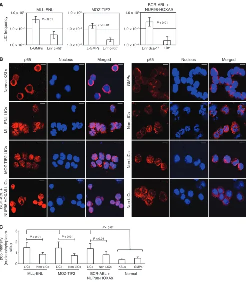
were striking differences in clonogenic potential (Supplemental Figure 3) and LIC frequencies, as determined by in vivo limiting dilution assays in the two populations of each model (Figure 1A and Supplemental Table 1). Therefore, we confirmed that LIC and non-LIC fractions can be clearly isolated through the surface anti-gen profiles of the three leukemia models. Next, we visualized the subcellular distribution of the major NF-κB subunit p65 in LICs, non-LICs, and normal cells by immunofluorescence staining and confocal microscopy. As shown in Figure 1B, prominent nuclear translocation of p65 was observed in the LICs of each model, while it was retained mostly in the cytoplasm in normal lineage–
c-Kit+ Sca-1+ cells (KSLs), which are enriched for HSCs and GMPs.
Interestingly, non-LICs also had relatively reduced p65 nuclear translocation signal compared with that in LICs in all three leu-kemia models. We quantified the nucleus/cytoplasm ratio of p65 staining intensity in these images, which also showed that the LICs in each model had significant nuclear localization compared with that observed in non-LICs, normal KSLs, and GMPs (Figure 1C).
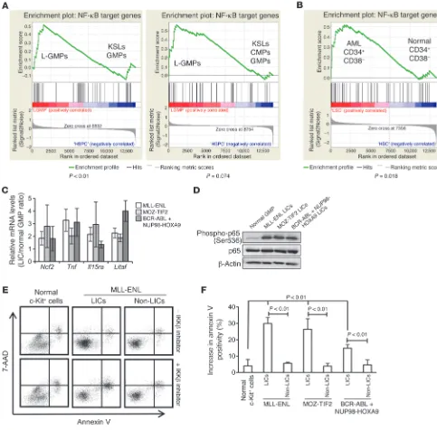
To further test NF-κB transcription activity in LICs, we investi-gated the expression profiles of a subset of genes regulated by the NF-κB pathway. We first used two sets of published gene expres-sion microarray data, which compared the expresexpres-sion profiles of MOZ-TIF2 L-GMPs (26), MLL-AF9 L-GMPs, and HOXA9-MEIS1 L-GMPs (28) with those of normal hematopoietic stem or progen-itor cells (HSPCs). The expression profiles of previously identified NF-κB target genes were assessed by gene set enrichment analysis (GSEA) (Supplemental Table 2 and ref. 29), which showed that L-GMPs had increased expression levels of NF-κB target genes compared with those in normal HSPCs in both sets of gene expres-sion microarray data (Figure 2A). We also compared the expresexpres-sion profiles of the same gene set in CD34+CD38– human AML cells
with those of the equivalent cell population in normal BM cells, which corresponded to the HSC fraction, and observed a similar tendency (Figure 2B and ref. 30). Then, we validated these results using quantitative real-time PCR by comparing the expression levels of several NF-κB target genes in LICs and non-LICs from our three mouse models with those in normal GMPs and found increased expression levels of most of the genes in different types of LICs, but no significant elevation of these levels in non-LICs (Fig-ure 2C and Supplemental Fig(Fig-ure 4). Furthermore, the level of p65 phosphorylation, which is important for enhancing its transcrip-tion activity, was significantly increased in LICs compared with the level observed in normal GMPs (Figure 2D). Consistent with these findings, LICs showed a more prominent increase in apop-tosis than did normal cells or non-LICs when treated with sc-514, a selective inhibitor of IκB kinase β (IKKβ) (Figure 2, E and F,
and ref. 31). Although LICs from BCR-ABL/NUP98-HOXA9– induced leukemia were rather resistant to sc-514 compared with cells from MLL-ENL– and MOZ-TIF2–induced leukemia, they still showed higher sensitivity than non-LICs. Collectively, these data fully support the hypothesis that the NF-κB pathway is constitu-tively activated in the LICs of different types of myeloid leukemia.
LICs maintain their constitutive NF-κB activity via autocrine TNF-α
signaling. In the next step, we addressed the question of how LICs maintain constitutive NF-κB activity in different types of leuke-mia models. In order to investigate genes prevalently dysregulated in LICs, we analyzed the previously published microarray-based gene expression profiles comparing murine and human LICs with normal HSPCs (26, 28, 30). After narrowing down our analysis to the genes commonly upregulated in LICs in three different types of murine leukemia models, we further selected nineteen genes whose expression is elevated in human AML CD34+CD38– cells
(Figure 3A). Among the nineteen genes with typically elevated expression levels in LICs, we focused on Tnf, because it is well known as an activator of NF-κB and as an NF-κB–regulated gene. For the purpose of directly evaluating TNF-α abundance in the BM of leukemic mice, we measured the concentration of TNF-α
in the BM extracellular fluid and confirmed that it was conspic-uously enriched in leukemic BM cells compared with normal BM cells (Figure 3B). We also examined the TNF-α concentration in culture media conditioned by LICs, non-LICs, and normal cells, respectively, to determine whether leukemia cells themselves have the ability to secrete TNF-α. We found that TNF-α secretion was distinctly elevated in LICs, while the normal GMP-conditioned media barely included TNF-α (Figure 3C). Although non-LICs also had TNF-α secretory ability, it was much lower that that of LICs. We therefore reasoned that LICs might maintain their NF-κB pathway activity via autocrine TNF-α signaling. To test this hypothesis, we cultured freshly isolated LICs in serum-free media with a TNF-α–neutralizing antibody or its isotype control and observed p65 subcellular distribution. While LICs treated with isotype control antibodies maintained p65 nuclear translocation even after serum-deprived culture, the p65 translocation signal we observed in three types of LICs was significantly attenuated when these cells were cultured with neutralizing antibodies against TNF-α (Figure 3D). The results were also confirmed by quanti-fication of p65 intensity (Figure 3E). These data strongly suggest that different types of LICs have a similarly increased potential for TNF-α secretion, which maintains constitutive NF-κB activity in an autonomous fashion.
Autocrine TNF-αsignaling promotes leukemia cell progression. We were then interested in exploring the effect of autocrine TNF-α secre-tion on leukemia progression. BM cells derived from WT or Tnf -knockout mice were transplanted into sublethally irradiated WT recipient mice after transduction with MLL-ENL and MOZ-TIF2, and cotransduction with BCR-ABL and NUP98-HOXA9 (Figure 3F). Although several mice did develop leukemia with prolonged latency, Tnf-deficient cells were significantly (P < 0.01) impaired in their ability to initiate leukemia (Figure 3G). We confirmed that
Tnf-deficient LICs show a distinct decrease in nuclear localization of p65 compared with the that in LICs derived from WT BM cells (Supplemental Figure 5, A and B). Next, we examined whether paracrine TNF-α from the BM microenvironment contributes to leukemia progression. When the established leukemia cells were secondarily transplanted into WT or Tnf-knockout recipient mice,
research article
Figure 1
NF-κB pathway is activated in LICs of different murine myeloid leukemia models. (A) LIC frequency in the two fractions of each leukemia model
as determined by limiting dilution assay. See Supplemental Table 1 for detailed transplantation results. (B) Immunofluorescence assessment for
p65 nuclear translocation in KSLs, GMPs, LICs, and non-LICs in three leukemia models. Scale bars: 10 μm. (C) Quantification of p65 nuclear
all three models (Figure 3, H and I). Interestingly, there was no significant difference in leukemogenicity among the recipient genotypes. These results indicate that autocrine TNF-α secretion is important for AML progression and that the contribution of paracrine effects derived from stromal cells is minimal.
[image:5.585.47.530.84.560.2]The impact of specific NF-κB inhibition on leukemia progression. To investigate the influence of specific NF-κB pathway inhibition on leukemia progression in vivo, we transduced MLL-ENL leuke-mia cells with a retroviral vector expressing a dominant-negative form of IκBα (super repressor, referred to herein as IκB-SR) or
Figure 2
NF-κB transcription activity is increased in LICs. (A) GSEA of NF-κB target genes in the published gene expression data comparing LICs in
leukemia mouse models with normal HSPCs. Left panel: comparison of MOZ-TIF2 L-GMP with normal KSLs and GMPs (GSE24797). Right panel: comparison of MLL-AF9 and HOXA9-MEIS1 L-GMPs with normal KSLs, common myeloid progenitors (CMPs), and GMPs (GSE20377).
(B) GSEA of NF-κB target genes in CD34+CD38– fractions in human AML versus healthy controls (GSE24006). (C) Quantitative real-time PCR
analysis of a subset of NF-κB target genes in LICs of MLL-ENL, MOZ-TIF2, and BCR-ABL/NUP98-HOXA9 leukemia models relative to normal
GMPs (n = 4). Error bars indicate SD. (D) Immunoblotting of total and phosphorylated p65 in normal GMPs and LICs in the three leukemia models.
(E) Representative annexin V and 7-AAD profiles of normal c-Kit+ cells, L-GMPs, and Lin–c-Kit– cells in MLL-ENL leukemic mice after a 24-hour
culture with or without 10 μM IKK inhibitor (sc-514). (F) Average percentage increase in apoptotic cells in LICs of the three leukemia models
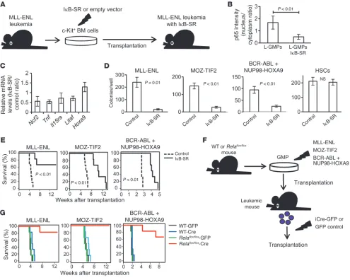
with a control vector, transplanted them into recipient mice, and compared the characteristics of the repopulating cells (Fig-ure 4A). Although the introduction of IκB-SR did not affect the morphology of MLL-ENL leukemia cells (Supplemental Figure 6A), p65 was almost completely sequestered in the cytoplasm of L-GMPs with IκB-SR (Figure 4B and Supplemental Figure 6B), and the expression levels of NF-κB target genes, including Tnf, were substantially decreased (Figure 4C). Considering that the blockage of autocrine TNF-α attenuated NF-κΒ signaling, we hypothesized that NF-κΒ activity and TNF-α secretion form a positive feedback loop in LICs. We therefore established MOZ-TIF2 and BCR-ABL/NUP98-HOXA9 leukemia cells with IκB-SR. The introduction of IκB-SR significantly decreased a proportion of the cells in the S and G2/M phases of the cell cycle and resulted in a substantial growth delay of those cells in liquid culture (Sup-plemental Figure 6, C and D). Moreover, leukemia cells with Iκ B-SR had a reduced colony-forming capacity, while the transduction of IκB-SR into normal HSCs had no significant influence on their colony-forming ability (Figure 4D). Finally, we transplanted leukemia cells with IκB-SR into sublethally irradiated mice and observed a remarkable delay in leukemia progression (Figure 4E). We also confirmed that the developed leukemia cells with IκB-SR had reduced nuclear translocation of p65 compared with that seen in control cells (Supplemental Figure 6E). In contrast, when nor-mal BM cells were transduced with IκB-SR and transplanted into lethally irradiated mice, we observed no significant differences in the reconstitution capacity of the transplanted cells, nor did we find significant differences in peripheral blood cell counts or PBL surface-marker profiles, indicating that NF-κB pathway inhibition exerts a marginal influence on normal hematopoiesis (Supplemen-tal Figure 7, A–C). Collectively, these findings clearly demonstrate that enhanced NF-κB activity in LICs plays a supportive role in leukemia progression and that NF-κB inhibition severely attenu-ates the proliferative ability of these cells.
To further validate the importance of the NF-κB pathway in leukemia progression, we used BM cells from Relaflox/flox mice (32).
We similarly established leukemia cells derived from Relaflox/flox
BM cells. Then, the developed leukemia cells were infected with codon-improved Cre recombinase–IRES-GFP (iCre-IRES-GFP) or GFP empty vector, and GFP-positive cells were isolated and sec-ondarily transplanted into sublethally irradiated mice (Figure 4F). Remarkably, most of the mice transplanted with Rela-deleted leu-kemia cells did not develop leuleu-kemia (Figure 4G). Compared with controls, several mice did develop leukemia after longer latencies, but they did not develop leukemia after tertiary transplantation (data not shown), indicating that the complete ablation of NF-κB drastically reduced leukemogenicity.
High proteasome activity in LICs yields differences in NF-κB activity between leukemia cell populations. We next sought to elucidate the mechanisms underlying the differences in p65 nuclear transloca-tion status between LICs and non-LICs. We confirmed that LICs had substantially lower IκBα protein levels compared with those of non-LICs in all three models (Figure 5, A and B). These results are very consistent with the p65 distribution status of LICs and non-LICs, considering that NF-κB is usually sequestered in the cytoplasm, bound to IκBα, and translocates to the nucleus, where IκBα is phosphorylated and degraded upon stimulation with a variety of agents such as TNF-α (33). We initially tested whether the expression of IκBα is downregulated in LICs at the transcrip-tion level and found that LICs had a tendency toward increased
Nfkbia mRNA expression levels compared with non-LICs (Figure
5C). Moreover, when Nfkbia mRNA translation was inhibited by treatment with cycloheximide, the reduction in IκBα protein levels was more prominent in LICs than in non-LICs (Figure 5, D and E). These data indicate that the differences in IκBα levels are caused by the protein’s predominant degradation in LICs. Since both LICs and non-LICs are similarly exposed to high levels of TNF-α within leukemic BM cells, we considered that there would be differences in response to the stimulus and sequentially examined the down-stream signals. We first hypothesized that there is a difference in TNF-α receptor expression levels between LICs and non-LICs that leads to greater TNF-α signal transmission in LICs. The expression patterns of TNF receptors I and II were, however, almost similar in LICs and non-LICs, although they varied between leukemia models (Supplemental Figure 8A). We next tested the phospho-rylation capacity of IκB kinase (IKK) by examining the ratio of phosphorylated IκBα to total IκBα after treatment with the pro-teasome inhibitor MG132. Contrary to our expectation, a similar accumulation of the phosphorylated form of IκBα was seen in both LICs and non-LICs, implying that they had no significant difference in IKK activity (Supplemental Figure 8B). Another pos-sibility is that the differences in IκBα protein levels are caused by predominant proteasome activity in LICs, because it is required for the degradation of phosphorylated IκBα. We measured 20S proteasome activity in LICs and non-LICs in each leukemia model by quantifying the fluorescence produced upon cleavage of the proteasome substrate SUC-LLVY-AMC and observed a 2- to 3-fold higher proteasome activity in LICs (Figure 5F). Furthermore, the expression of several genes encoding proteasome subunits was ele-vated in LICs compared with that in non-LICs (Figure 5G). Simi-larly, the published gene expression data on human AML samples revealed that CD34+CD38– cells had increased expression levels of
proteasome subunit gene sets compared with those in CD34– cells
(Supplemental Figure 9 and ref. 30). These findings suggest that enhanced proteasome activity in LICs leads to more efficient deg-radation of IκBα in response to TNF-α, thus resulting in elevated NF-κB activity. We then tested the effect of bortezomib, a
well-Figure 3
Autocrine TNF-α secretion maintains constitutive NF-κB activity and
confers proliferative advantage in LICs. (A) Thorough investigation
of genes with elevated expression in murine and human LICs com-pared with that in normal HSPCs in the published gene expression data. (B) TNF-α ELISA in extracellular fluid of normal or leukemic BM (n = 4 each). Error bars indicate SD. (C) TNF-α secretory ability in LICs compared with that of non-LICs and normal GMPs assessed
by ELISA in cultured media (n = 4 each). Error bars indicate SD.
(D) Immunofluorescence assessment for p65 nuclear translocation in
LICs in serum-free culture medium with neutralizing antibody against TNF-α or isotype control. Scale bars: 10 μm. (E) Quantification of p65 nuclear translocation of LICs treated with neutralizing antibody against
TNF-α or isotype control assessed by the mean nucleus/cytoplasm
intensity ratio. More than 50 cells were scored in each specimen, and
the average intensity ratio with SD is shown. (F) Schematic
represen-tation of the experiments. BM cells derived from WT or Tnf-knockout
mice were transduced with MLL-ENL, MOZ-TIF2, and BCR-ABL plus NUP98-HOXA9 and transplanted into sublethally irradiated mice. (G) Survival curves of mice in the experiments shown in F (n = 7 each). (H) Schematic representation of the experiments. WT or Tnf–/–
leuke-mia cells were secondarily transplanted into WT or Tnf–/– recipient mice.
research article
known proteasome inhibitor, on LICs in vivo (Figure 5H). First, we treated mice with full-blown leukemia with a single injection of bortezomib and compared their BM surface-marker profiles with those of the vehicle-treated mice. Notably, bortezomib-treated mice showed a significant decrease in LIC-enriched populations in each type of leukemia (Figure 5, I and J). Finally, we treated mice with bortezomib after LIC transplantation and observed signifi-cant improvement in survival in those treated with bortezomib (Figure 5K). These results are very consistent with the selectively elevated proteasome activity we observed in LICs.
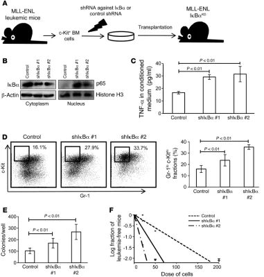
[image:8.585.47.542.83.479.2]Enforced activation of the NF-κB pathway increases LIC frequency in leukemic BM. Given the supportive role of the NF-κB pathway in LIC proliferation as well as the differences in its activation status observed between LICs and non-LICs, we reasoned that the atten-uation of NF-κB activity might be related to the transition from LICs to non-LICs. To test this hypothesis, we transduced MLL-ENL leukemia cells with a retrovirus encoding shRNA against IκBα and transplanted them into sublethally irradiated mice (Fig-ure 6A). Because IκBα works as an inhibitor of NF-κB by hold-ing it in the cytoplasm, its downregulation should function to
Figure 4
Specific inhibition of NF-κB significantly inhibits leukemia progression in vivo. (A) Schematic representation of the following experiments: c-Kit+
BM cells isolated from MLL-ENL leukemic mice were transduced with IκB-SR or control vector and transplanted into sublethally irradiated mice.
(B) Quantification of p65 nuclear translocation assessed by the mean nucleus/cytoplasm intensity ratio by immunofluorescence staining. More
than 50 cells were scored in each specimen, and the average intensity ratio with SD is shown. (C) Relative expression profiles of NF-κB target
genes in MLL-ENL leukemia cells with or without IκB-SR. The change in Hoxa9 expression is shown as a control gene not regulated by NF-κB.
Error bars indicate SD (n = 3 each). (D) CFC assay of leukemia cells and normal HSCS with or without IκB-SR. Cells were seeded at 2,000
cells per well in MLL-ENL or BCR-ABL/NUP98-HOXA9–induced leukemia cells, at 500 cells per well in MOZ-TIF2–induced leukemia cells,
and at 1,000 cells per well in normal HSCs (n = 6 in each experiment). (E) Survival curves of mice transplanted with MLL-ENL, MOZ-TIF2, and
BCR-ABL/NUP98-HOXA9 leukemia cells with or without IκB-SR (n = 6 each). (F) Schematic representation of the following experiments: WT or
Relaflox/flox mice were transduced with MLL-ENL, MOZ-TIF2, or BCR-ABL plus NUP98-HOXA9 and transplanted into sublethally irradiated mice.
The developed leukemia cells were transduced with iCre-IRES-GFP or control GFP, and GFP+ cells were secondarily transplanted into mice.
enhance NF-κB activity, regardless of the basal proteasome activ-ity. We first confirmed that MLL-ENL leukemia cells with shRNA- mediated knockdown of IκBα (MLL-ENL-IκBαKD) showed
decreased IκBα protein levels in the cytoplasm and increased nuclear p65 protein levels, which would indicate that NF-κB sig-nal was enhanced by the reduction of its cytoplasmic inhibitor (Figure 6B). In accordance with this finding, MLL-ENL-IκBαKD
cells had a significantly greater ability to secrete TNF-α than did control cells, reflecting an activated NF-κB/TNF-α signaling loop (Figure 6C). We further investigated the phenotype of leukemic mice with MLL-ENL-IκBαKD. Interestingly, the BM of these
MLL-ENL-IκBαKD mice showed a marked increase in immature Gr-1lo
c-Kithi cell populations (Figure 6D). Consistent with this change,
we found that these leukemic cells had a greater CFC capacity (Figure 6E). Additionally, in order to investigate the frequency of LICs in BM mononuclear cells, we performed limiting dilution analysis by secondary transplantation of leukemia cells. Although the disease latency for leukemia development was not signifi-cantly different among the leukemia cells, MLL-ENL-IκBαKD
leukemia cells had a marked abundance of LICs in the leukemic BM mononuclear cells compared with the control shRNA cells (Figure 6F and Supplemental Figure 10A). These data indicate that enforced NF-κB activation expands the LIC fraction in MLL-ENL leukemic BM cells. We also transduced normal BM cells with shRNAs against IκBα and transplanted them into lethally irradi-ated mice to test whether NF-κB activation by itself can induce leukemia or myeloproliferative-like disease. Over the 4-month follow-up period, the mice exhibited no significant change in peripheral blood values, indicating that NF-κB signal alone is not sufficient for leukemogenesis (Supplemental Figure 10B).
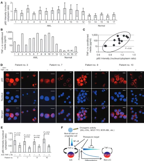
Significant correlation between NF-κB and TNF-αis observed in human AML LICs. Finally, we investigated NF-κB/TNF-α posi-tive feedback signaling in human AML LICs. We analyzed CD34+
CD38– cells derived from 12 patients with previously untreated
or relapsed AML and the same cell population from 5 normal BM specimens (Table 1) and evaluated their NF-κB signal intensity. We also quantified the concentration of TNF-α in the culture media conditioned by CD34+CD38– cells from each patient in
order to measure the TNF-α secretory ability of these cells. As expected, our data from both of these analyses showed a wide vari-ation among patients, one that might reflect a heterogeneous dis-tribution and frequency of the LIC fraction in human AML cells, as was previously described (23). LICs in most of the patients did, however, show increased p65 nuclear translocation and TNF-α
secretory potential compared with normal HSCs (Figure 7, A and B, and Supplemental Figure 11). We plotted these two parame-ters for each patient to compare between patients. Interestingly, a significant positive correlation was demonstrated statistically (P = 0.02), as LICS with enhanced p65 nuclear translocation showed a tendency toward abundant TNF-α secretion (Figure 7C). We also compared p65 intensity between LICs and non- LICs in 2 patients (patients 1 and 3) and found that p65 nuclear translocation was predominant in LICs, which is also consistent with the data obtained in murine AML cells (Supplemental Fig-ure 11). Moreover, we cultFig-ured LICs with or without neutralizing antibodies against TNF-α and assessed p65 nuclear translocation to determine the effect of autocrine TNF-α on NF-κB activity. When incubated in the presence of TNF-α–neutralizing antibod-ies, nuclear translocation of p65 was significantly suppressed in LICs (Figure 7, D and E). These results support our hypothesis
that a positive feedback loop exists between NF-κB and TNF-α in human AML LICs.
Discussion
In the present study, we provide evidence that LICs, but not nor-mal HSPCs or non-LIC fractions within leukemic BM, exhibit constitutive NF-κB pathway activity in different types of myeloid leukemia models. Moreover, we identified the underlying mecha-nism involved in the maintenance of this pathway activity, which had yet to be elucidated. We found that autocrine TNF-α secre-tion, with the support of enhanced proteasome activity, contrib-uted to a constitutive activation of the NF-κB pathway in LICs. Although we observed different sensitivities to the inhibition of these signaling cascades according to the type of leukemia, these cascades play an important role in LIC proliferation, especially considering that the complete ablation of Tnf or Rela distinctly suppressed leukemia progression in vivo. These findings, which we validated in human AML LICs, could translate into improved AML treatment strategies.
The strong connection between inflammation and cancer has been increasingly discussed, and the NF-κB pathway is now rec-ognized as a major regulator bridging the two pathological con-ditions in different types of malignancies. In most of these malig-nancies, aberrant activation of the NF-κB pathway derives from inflammatory microenvironments that are mainly created by proinflammatory immune cells such as tumor-infiltrating mac-rophages, neutrophils, and lymphocytes (34, 35). In this study, however, LICs retained their p65 nuclear translocation even after serum-free culture, suggesting that the constitutive NF-κB activ-ity of LICs is maintained in an autonomous fashion. Through our investigation of gene expression profiles in LICs and normal HSCs, we found that LICs had distinctly elevated TNF-α expres-sion levels that contributed to the maintenance of NF-κB activa-tion in LICs. Conversely, the introducactiva-tion of IκB-SR markedly suppressed TNF-α expression levels, indicating that NF-κB activ-ity and TNF-α secretion create a positive feedback loop in LICs. Moreover, our hypothesis is strongly supported by our findings that a positive correlation exists between NF-κB and TNF-α secre-tory activities in human AML CD34+CD38– cells and that
progression. Unveiling the role of TNF-α as a paracrine mediator would further extend the therapeutic options for AML.
Few studies have compared the NF-κB activity of different frac-tions within leukemia cells, and the mechanism underlying the difference in this activity has not been analyzed (44). We focused on proteasome activity as the essential machinery supporting NF-κB activity in LICs. Although high proteasome activity has been reported in various types of cancers (45, 46), its actual role in the malignant phenotype remained to be elucidated. In this study, we found that proteasome activity was especially high in LICs, which contributed to selective NF-κB activity in LICs via the efficient degradation of IκBα. Conversely, the inefficient NF-κB nuclear translocation we observed in non-LICs, despite TNF-α– enriched leukemic BM cells, could be explained by the low pro-teasome activity in these cells. Therefore, we postulate that both an activating stimulus such as TNF-α and high proteasome activ-ity are required for efficient NF-κB signaling (Figure 7F). Both of these conditions are present exclusively in LICs, which acquire selective NF-κB activation. We also found that the expression lev-els of proteasome subunit genes were elevated in LICs compared with those in non-LICs, genes that could be involved in regulat-ing proteasome function. Because we observed similar expression patterns in LICs and non-LICs in human AML cells, an elevated expression level of proteosome subunit genes might be one of the common characteristics of the LIC phenotype. Further studies will be needed to elucidate the regulatory mechanism of the protea-some gene families.
Our findings provide several advantages when considering their application to the clinical care setting. First, an activated NF-κB/TNF-α feedback loop was seen in AML LICs that had dif-ferent genetic abnormalities. Although the therapeutic strategy of targeting aberrant molecules based on genetic abnormalities such as FLT3-ITD is promising, its application is limited to a particular group of patients. In contrast, inhibition of the NF-κB
signal in addition to standard chemotherapy would show bene-ficial effects in most AML patients. Second, because there was a strong positive correlation between the NF-κB signal and TNF-α
secretion, therapeutic efficacy could easily be inferred from the abundance of TNF-α instead of from evaluation of the activation status of NF-κB. Third, the NF-κB/TNF-α signal and enhanced proteasome activity are selectively seen in LICs, but not in nor-mal HSCs. A recent study has shown that complete ablation of p65 in hematopoietic cells attenuates the long-term capacity for hematopoietic reconstitution (47). However, our data from the experiments in which we introduced IκB-SR into normal BM cells show that partial repression of NF-κB activity exerted minimal influence on normal hematopoiesis, while it markedly inhibited leukemia progression. These results indicate that there is a therapeutic window during which LICs can selectively be killed by NF-κB inhibition without seriously affecting normal hematopoiesis. Alternatively, there is some evidence that TNF-α
has suppressive effects on normal HSCs (48, 49). The oppos-ing role of TNF-α in LICs and HSCs is additionally beneficial, since anti–TNF-α therapy contributes to the recovery of normal hematopoiesis and attenuates LIC proliferation. Now that the TNF-α antagonist etanercept is widely used in inflammatory dis-eases such as rheumatoid arthritis, this drug might be a promis-ing candidate for treatpromis-ing patients with AML.
In summary, the present study shows that blocking the NF-κB pathway offers a promising therapeutic approach for targeting LICs in various types of myeloid leukemia, without disturbing nor-mal hematopoiesis. We further determined that autocrine TNF-α
signaling and enhanced proteasome activity are crucial for main-taining constitutive NF-κB activity in LICs, findings that may also provide a new therapeutic opportunity.
Methods
Animals. C57BL/6 mice and BALB/c mice were purchased from Japan SLC, Inc. Tnf-knockout mice on a BALB/c background were established as described previously (50). Rela-floxed mice on a C57BL/6 background were provided by H. Algül and R.M. Schmid (32). BALB/c mice were used as the controls in the experiments using Tnf-knockout mice, and C57BL/6 mice were used in the other experiments.
Retrovirus production and BM transplantation assays. To obtain retrovi-rus supernatants, platinum-E (Plat-E) packaging cells were transiently transfected with each retrovirus vector, and the viral supernatants were collected 48 hours after transfection and used immediately for infection. To establish each myeloid leukemia mouse model, we used
pMSCV-neo-MLL-ENL; pMSCV-MLL-ENL–internal ribosome entry site–EGFP
(IRES-EGFP); pGCDNsam-MLL-ENL-IRES-Kusabira-Orange;
pGCDN-sam-MOZ-TIF2-IRES-EGFP; pGCDNsam-MOZ-TIF2-IRES-Kusabira-
Orange; pGCDNsam-BCR-ABL-IRES-EGFP; pGCDNsam-BCR-ABL-IRES-
Kusabira-Orange; and pMSCV-neo-NUP98-HOXA9. GMPs isolated from the BM of 8- to 10-week-old mice were transduced with the respective vectors and injected into sublethally irradiated (7.5 Gy) recipient mice. For experiments involving the generation of leukemia cells with IκB-SR, MLL-ENL leukemia cells were transduced with pBabe-GFP or pBabe-GFP-
IκB-SR. MOZ-TIF2, and BCR-ABL/NUP98-HOXA9 leukemia cells were
transduced with pGCDNsam-Kusabira-Orange or pGCDNsam-IκB-
SR-IRES-Kusabira-Orange. For experiments involving the deletion of p65 in Rela-floxed mice, leukemia cells were established using Kusabira- Orange–containing retroviral vectors. The developed leukemia cells were transduced with pGCDNsam-EGFP or pGCDNsam-iCre-EGFP and trans-planted into sublethally irradiated mice.
Figure 5
LICs have higher proteasome activity than non-LICs. (A and B)
Immu-noblotting of IκBα in LICs and non-LICs (A). Protein levels were
quan-tified with ImageJ software (B). Data representative of four
experi-ments with SD are shown. (C) Relative mRNA expression of Nfkbia in
LICs compared with that in non-LICs (n = 4 each). Error bars indicate
SD. (D and E) Immunoblotting of IκBα in LICs and non-LICs. Cells were pretreated with MG132 for 1 hour and incubated for an additional hour with or without cycloheximide (CHX) (D). IκBα protein levels were
quantified with ImageJ software, and the relative decrease in IκBα
after cycloheximide treatment was calculated (n = 3 each). Error bars
indicate SD (E). (F) Analysis of 20S proteasome activity quantified with fluorescence produced upon cleavage of the proteasome substrate
SUC-LLVY-AMC (n = 4 each). Error bars indicate SD. (G) Relative
mRNA expression of proteasome subunits in LICs compared with that in non-LICs (n = 4 each). Error bars indicate SD. (H) Schematic repre-sentation of the experiments. Each type of LIC was secondarily trans-planted into mice. Bortezomib was injected twice weekly or injected
once after incidence of leukemia. (I and J) Comparison of surface
marker profiles in leukemic mice treated with bortezomib or
vehi-cle. Representative FACS data (I) and relative percentages of Gr-1lo
c-Kithi fraction in MLL-ENL– or MOZ-TIF2–induced leukemic mice, and
Gr-1loSca-1hi fraction in BCR-ABL/NUP98-HOXA9–induced leukemic
mice are shown (n = 3 each) (J). Values of control mice were
research article
measurements were performed using FluoView software. The background- subtracted intensity ratio of nucleus/cytoplasm was calculated in more than 50 cells in each specimen, and the average intensity with SD is presented.
Flow cytometry. Isolation of each fraction from normal or leukemic BM cells was performed using a FACSAria II (BD) cell sorter. For isolation of GMPs and KSLs, biotinylated antibodies against Gr-1 (RB6-8C5), CD11b (M1/70), B220 (RA-3-6B2), CD3 (145-2C11), CD4 (GK1.5), CD8 (53-6.7), and TER119 were used for lineage staining. A PerCP-Cy5.5–labeled streptavidin antibody was used for secondary staining, together with APC–anti–c-Kit (2B8), PE-Cy7–anti–Sca-1 (E13-161.7). FITC–anti-CD34 (RAM34), and PE–anti-CD16/32b antibodies (clone 93). The following antibodies were used for isolation of L-GMPs from GFP-containing leu-kemia cells: APC-Cy7–anti–streptavidin, PE-Cy5–anti–c-Kit (2B8), PE-Cy7– anti–Sca-1 (E13-161.7), Alexa Fluor 647–anti–CD34 (RAM34), and PE–anti- CD16/32b (clone 93). APC-antistreptavidin and PE-Cy7–anti–Sca-1 anti-bodies (E13-161.7) were used for sorting LICs and non-LICs in the BCR-ABL plus NUP98-HOXA9 leukemia model. See Supplemental Figures 1 and 2 for detailed FACS plots. For analysis of TNF receptor expression in leuke-mia cells, biotinylated antibodies against TNF receptor I or II (55R-170) and an APC-Cy7–antistreptavidin antibody were used. Analysis was performed using FlowJo software (Tree Star Inc.).
In vivo limiting dilution assays. Varying numbers of cells from different popu-lations were transplanted into sublethally irradiated mice and monitored for disease development (see Supplemental Table 1 for the injected cell numbers).
Immunofluorescence and quantification of p65 nuclear translocation. A total of 1 × 10⁴ to 5 × 10⁴ cells were cytospun onto glass slides. The cells were fixed with 3.7% formaldehyde in PBS for 30 minutes, permeabilized by treatment with 0.2% Triton X in PBS for 10 minutes, and blocked with 1% BSA in PBS for 60 minutes. Then, the slides were incubated with rabbit anti–p65 polyclonal antibody (sc-372; 1:100 dilution; Santa Cruz Biotech-nology Inc.) overnight at 4°C, followed by incubation with Alexa Fluor 555 goat anti-mouse IgG (1:250 dilution; Invitrogen) and TO-PRO3 (1:1,000 dilution; Invitrogen) for 90 minutes. For immunofluorescence staining of Kusabira-Orange+ leukemia cells, Alexa Fluor 647 goat anti-mouse IgG (1:250 dilution; Invitrogen) was used as a secondary antibody, and the nucleus was stained with DAPI. After the cells were washed, they were treated with ProLong Gold Antifade Reagent (Invitrogen). Images were acquired using an Olympus FluoView FV10i confocal microscope with a
[image:12.585.41.422.84.483.2]×60 objective oil immersion lens. The mean intensity of p65 in the nucleus and cytoplasm of each cell was measured within a region of interest (ROI) placed within the nucleus and cytoplasm. Similarly, the background intensity was quantified within an ROI placed outside the cells. All the
Figure 6
Forcible maintenance of NF-κB
activit y in leukemia cells
enhances LIC frequency. (A)
Schematic representation of
the experiments. c-Kit+ BM cells
isolated from MLL-ENL leuke-mic leuke-mice were transduced with
shRNA against IκBα or control
shRNA and transplanted into sub-lethally irradiated mice. (B)
Immu-noblotting of cytoplasmic IκBα
and nuclear p65 in BM mononu-clear cells from MLL-ENL-IκBαKD
mice compared with those from control leukemic mice. (C) TNF-α
secretory ability of MLL-ENL- IκBαKD leukemia cells compared
with that of control leukemia cells (n = 4 each). Error bars indicate SD. (D) Surface marker profiles of MLL-ENL leukemic mice with or without knockdown of IκBα. Rep-resentative FACS plots and mean percentages of Gr-1loc-Kithi
frac-tions (n = 6 each). (E) CFC assay of MLL-ENL leukemia cells with
or without knockdown of IκBα
(n = 6). Cells were seeded at 500 cells per well. Error bars indicate
SD. (F) LIC frequency in BM
mononuclear cells derived from
MLL-ENL-IκBαKD leukemic mice
CFC assays. In each experiment, cells were plated onto MethoCult GF M3434 medium (STEMCELL Tech-nologies). Colony numbers in each dish were scored on day 7.
Measurement of TNF-α levels in BM extracellular fluid and conditioned media. BM extracellular fluid was obtained by f lushing bilateral femurs and tibia of individual mice with 400 μl PBS. The supernatant was collected after centrifugation. To obtain conditioned media, 0.3–1.0 × 10⁶ murine leukemia cells or normal GMPs were cultured in RPMI medium containing 10% FBS and 10 ng/ml IL-3. After a 48-hour incubation, the culture superna-tants were collected. The concentra-tion of TNF-α was measured using
a murine TNF-α ELISA kit
(Gen-Probe Diaclone) according to the manufacturer’s instructions. Simi-larly, 0.5 × 10⁴ to 2.0 × 10⁴ human AML or normal CD34+CD38– cells were cultured for 48 hours in RPMI medium containing 10% FBS and 100 ng/ml SCF, IL-3, and thrombo-poietin. The concentration of TNF-α in the harvested supernatants was measured with a human TNF-α Quantikine ELISA kit (R&D Systems).
20S proteasome activity. A 20S proteasome activity assay kit (Cayman Chemical) was used to analyze proteasome activity. A total of 5 × 10⁴ freshly isolated normal GMPs, LICs, and non-LICs in each model were assayed according to the manufacturer’s protocol. As a control, the pro-teasome activity of each cell was also assayed after the specific protea-some inhibitor epigallocatechin gallate was added. Fluorescence was measured with a Wallac ARVO V (PerkinElmer), and the proteasome activity of each cell type was calculated by subtracting the respective control value.
Bortezomib treatment studies. For in vivo treatment experiments, LICs of each leukemia model were injected into sublethally irradiated mice: 1 × 10³ cells in the MLL-ENL or BCR-ABL/NUP98-HOXA9 models, and 1 × 10⁴ cells in the MOZ-TIF2 model. Bortezomib was administrated i.p. at doses of 1.0 mg/kg twice weekly for 3 weeks. Treatment was started 1 week after transplantation in the MLL-ENL or BCR-ABL/NUP98-HOXA9 mod-els, and 2 weeks after transplantation in the MOZ-TIF2 model. For exper-iments analyzing changes in LIC populations, bortezomib was admin-istrated i.p. at doses of 1.0 mg/kg into fully developed leukemic mice. GFP+ BM cells were collected 24 hours after injection, and surface marker profiles were analyzed.
Analysis of microarray data. We analyzed publicly available gene expres-sion microarray data on murine and human samples from the Gene Expression Omnibus (GEO) database (GEO GSE24797, GSE20377, and GSE24006). A set of CEL files were downloaded from GEO and normal-ized using the JustRMA function from the Affy package 1.22.1 in Bio-conductor. To compare expression profiles of the NF-κB target genes, normalized data were tested for GSEA using previously described NF-κB target gene sets (29), and a nominal P value was calculated. For screening of genes with elevated expression levels in LICs compared with those in normal HSPCs, the expression values of individual genes were compared between groups. Genes significantly elevated in LICs from all three leu-kemia models as determined by an unpaired Student’s t test (P < 0.05)
Real-time quantitative PCR. Real-time quantitative PCR was carried out on the LightCycler480 system (Roche) using SYBR green reagents accord-ing to the manufacturer’s instructions. The results were normalized to
Gapdh levels. Relative expression levels were calculated using the 2-ΔΔCt method (51). The following primers were used for real-time PCR
exper-iments: Gapdh forward, TGGCCTCCAAGGAGTAAGAA, and reverse,
GGTCTGGGATGGAAATTGTG; Ncf2 forward,
CCAGAAGACCTG-GAATTTGTG, and reverse, AAATGCCAACTTTCCCTTTACA; Tnf
for-ward, TCTTCTCATTCCTGCTTGTGG, and reverse,
GGTCTGGGC-CATAGAACTGA; Il15ra forward, TAAGCGGAAAGCTGGAACAT, and
reverse, TGAGGTCACCTTTGGTGTCA; Litaf forward,
CTCCAGGACCT-TACCAAGCA, and reverse, AGGTGGATTCATTCCCTTCC; Hoxa9
for-ward, GGTGCCTGCTGCAGTGTAT, and reverse,
GTTCCAGCCAG-GAGCGCATAT; Psma5 forward, CGAGTACGACAGGGGTGTG, and
reverse, TGGATGCCAATGGCTGTAG; Psmd4 forward,
GTACATGCG-GAACGGAGACT, and reverse, TGTGGTCAGCACCTCACAGT; Psme3
forward, TTTCAGAGAGCGGATCACAA, and reverse, GGTCATGGA-TATTTAGAATTGGTTC.
siRNA interference. Specific shRNAs targeting murine Ikba mRNA were designed and cloned into pSIREN-RetroQ-ZaGreen vectors. Control shRNA is a nonfunctional construct provided by Clontech. The target
sequences, from 5′ to 3′, were: CCGAGACTTTCGAGGAAAT (shIκBα
number 1), and AGCTGACCCTGGAAAATCT (shIκBα number. 2).
[image:13.585.52.402.114.317.2]Immunoblotting. Membranes were probed with the following antibod-ies: anti-IκBα (Cell Signaling Technology), anti–phospho-IκBα (Ser32) (Cell Signaling Technology), anti-p65 (Santa Cruz Biotechnology Inc.), anti–phospho-p65 (Ser536) (Cell Signaling Technology), anti–β-actin (Cell Signaling Technology), and anti–histone H3 (Cell Signaling Tech-nology). Protein levels were quantified with ImageJ software (NIH). To obtain nuclear and cytoplasmic extracts, an Active Motif Nuclear Extract Kit was used according to the manufacturer’s instructions. Cycloheximide treatment assay was performed as described previously, with modification (52). Cells were pretreated with MG132 (20 μM) for 1 hour to initially inhibit the proteasomal degradation of IκBα. Cells were washed twice with medium, then cultured with or without 10 μg/ml of cycloheximide for an additional hour and harvested.
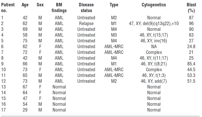
Table 1
Clinical characteristics of the 12 patients with AML and the 5 patients with normal BM findings
Patient Age Sex BM Disease Type Cytogenetics Blast
no. findings status (%)
1 42 M AML Untreated M2 Normal 87 2 62 M AML Relapse M1 47, XY, del(9)(q13q22),+10 96 3 69 M AML Untreated M4 Normal 90 4 58 M AML Untreated M3 46, XY, t(15;17) 63 5 75 M AML Untreated M4 46, XY, inv(16) 27 6 62 F AML Untreated AML-MRC NA 24.8 7 72 F AML Untreated AML-MRC Complex 21 8 42 M AML Untreated M4 46, XY, t(11;17) 25 9 66 M AML Untreated M1 46, XY, t(8;21) 85.4 10 73 F AML Untreated AML-MRC Complex 44.5 11 65 M AML Untreated AML-MRC 46, XY, t(1;3) 53.3 12 73 M AML Untreated M2 46, XY, add(7) 51.5 13 67 F Normal Normal
14 64 F Normal Normal 15 47 F Normal Normal 16 54 M Normal Normal 17 29 M Normal Normal
research article
Figure 7
NF-κB/TNF-α positive feedback loop is activated in human AML LICs. (A) Quantification of p65 nuclear translocation assessed by the mean
nucleus/cytoplasm intensity ratio by immunofluorescence staining. The CD34+CD38– fractions isolated from AML or normal BM were analyzed.
More than 50 cells were scored in each specimen, and the average intensity ratio with SD is shown. (B) TNF-α concentration of culture media
conditioned by human AML LICs and normal HSCs measured by ELISA. ND, not detected. (C) Correlation between p65 nuclear translocation
intensity ratio and TNF-α secretory ability of human AML LICs. (D) Immunofluorescence assessment of p65 nuclear translocation in LICs
puri-fied from 4 patients after serum-free culture with neutralizing antibody against TNF-α or isotype control. Scale bars: 10 μm. (E) Quantification of
p65 nuclear translocation of LICs with or without neutralizing antibody against TNF-α assessed by the mean nucleus/cytoplasm intensity ratio.
(F) Proposed model showing the role of NF-κB signaling in LICs. Positive feedback loop involving NF-κB/TNF-α promotes the maintenance and
Acknowledgments
We thank T. Kitamura for the Plat-E packaging cells; H. Nakauchi and M. Onodera for the pGCDNsam-IRES-EGFP retroviral vector; R. Ono and T. Nosaka for the MLL-ENL cDNA; I. Kitabayashi for the MOZ-TIF2 cDNA; W. Hahn for the GFP and pBabe-GFP-IκB-SR; and H. Algül and R.M. Schmid for providing the
Rela-floxed mice. This work was supported by a Grant-in-Aid for Scientific Research A (KAKENHI 12020240) from the Ministry of Education, Culture, Sports, Science and Technology of Japan. Received for publication December 3, 2012, and accepted in revised form October 17, 2013.
Address correspondence to: Mineo Kurokawa, Department of Hematology and Oncology, Graduate School of Medicine, The University of Tokyo, 7-3-1 Hongo, Bunkyo-ku, Tokyo 113-8655, Japan. Phone: 81.3.5800.9092; Fax: 81.3.5840.8667; E-mail: kurokawa-tky@umin.ac.jp.
were selected, among which genes also elevated in human AML LICs (Stu-dent’s t test set at P < 0.01) were ultimately selected.
Statistics. Statistical significance of differences between groups was assessed with a 2-tailed unpaired Student’s t test. Differences were considered statisti-cally significant at a P value of less than 0.05. LIC frequency was calculated by Poisson statistics. In leukemia cell transplantation experiments, the overall survival of mice in BM transplantation assays is depicted by a Kaplan-Meier curve. Survival between groups was compared using the log-rank test. To measure the correlation between NF-κB intensity and TNF-α secretion in human AML samples, the Spearman’s rank correlation coefficient was used.
Study approval. A total of 12 BM cells derived from patients with AML were obtained from the Department of Hematology and Oncology of the University of Tokyo Hospital. Five BM cells from patients diagnosed with lymphoid neoplasia without BM invasion were used as normal con-trols. The study was approved by the ethics committee of the University of Tokyo, and written informed consent was obtained from all patients whose samples were collected. All animal experiments were approved by the Uni-versity of Tokyo Ethics Committee for Animal Experiments.
1. Bonnet D, Dick JE. Human acute myeloid leuke-mia is organized as a hierarchy that originates from a primitive hematopoietic cell. Nat Med. 1997; 3(7):730–737.
2. Lapidot T, et al. A cell initiating human acute mye-loid leukaemia after transplantation into SCID mice. Nature. 1994;367(6464):645–648.
3. Ishikawa F, et al. Chemotherapy-resistant human AML stem cells home to and engraft within the bone-marrow endosteal region. Nat Biotechnol. 2007; 25(11):1315–1321.
4. Marcucci G, Haferlach T, Döhner H. Molecular genetics of adult acute myeloid leukemia: prognos-tic and therapeuprognos-tic implications. J Clin Oncol. 2011; 29(5):475–486.
5. Mardis ER, et al. Recurring mutations found by sequencing an acute myeloid leukemia genome. N Engl J Med. 2009;361(11):1058–1066.
6. Sen R, Baltimore D. Inducibility of kappa immuno-globulin enhancer-binding protein Nf-κB by a post-translational mechanism. Cell. 1986;47(6):921–928. 7. La Rosa FA, Pierce JW, Sonenshein GE. Differential
regulation of the c-myc oncogene promoter by the NF-κB rel family of transcription factors. Mol Cell Biol. 1994;14(2):1039–1044.
8. Guttridge DC, Albanese C, Reuther JY, Pestell RG, Baldwin AS Jr. NF-κB controls cell growth and dif-ferentiation through transcriptional regulation of cyclin D1. Mol Cell Biol. 1999;19(8):5785–5799. 9. Duckett CS. Apoptosis and NF-κB: the FADD
con-nection. J Clin Invest. 2002;109(5):579–580. 10. Karin M, Greten FR. NF-κB: linking inflammation
and immunity to cancer development and progres-sion. Nat Rev Immunol. 2005;5(10):749–759. 11. Karin M. Nuclear factor-κB in cancer development
and progression. Nature. 2006;441(7092):431–436. 12. Pikarsky E, et al. NF-κB functions as a tumour pro-moter in inflammation-associated cancer. Nature. 2004;431(7007):461–466.
13. Guzman ML, et al. Nuclear factor-κB is constitu-tively activated in primitive human acute myeloge-nous leukemia cells. Blood. 2001;98(8):2301–2307. 14. Guzman ML, et al. Preferential induction of
apop-tosis for primary human leukemic stem cells. Proc Natl Acad Sci U S A. 2002;99(25):16220–16225. 15. Frelin C, et al. Targeting NF-κB activation via
pharmacologic inhibition of IKK2-induced apop-tosis of human acute myeloid leukemia cells. Blood. 2005;105(2):804–811.
16. Carvalho G, et al. Inhibition of NEMO, the regula-tory subunit of the IKK complex, induces apoptosis in high-risk myelodysplastic syndrome and acute myeloid leukemia. Oncogene. 2007;26(16):2299–2307. 17. Guzman ML, et al. An orally bioavailable
parthe-nolide analog selectively eradicates acute myelog-enous leukemia stem and progenitor cells. Blood. 2007;110(13):4427–4435.
18. Jenkins C, et al. Nuclear factor-κB as a potential therapeutic target for the novel cytotoxic agent LC-1 in acute myeloid leukaemia. Br J Haematol. 2008;143(5):661–671.
19. Jin Y, et al. Antineoplastic mechanism of niclosamide in acute myelogenous leukemia stem cells: inactiva-tion of the NF-κB pathway and generation of reactive oxygen species. Cancer Res. 2010;70(6):2516–2527. 20. Takahashi S, et al. Over-expression of Flt3 induces
NF-κB pathway and increases the expression of IL-6. Leuk Res. 2005;29(8):893–899.
21. Liu S, et al. Sp1/NFκB/HDAC/miR-29b regulatory network in KIT-driven myeloid leukemia. Cancer Cell. 2010;17(4):333–347.
22. Nakagawa M, et al. AML1/RUNX1 functions as a cytoplasmic attenuator of NF-κB signaling in the repression of myeloid tumors. Blood. 2011;
118(25):6626–6637.
23. Eppert K, et al. Stem cell gene expression programs influence clinical outcome in human leukemia. Nat Med. 2011;17(9):1086–1093.
24. Sarry JE, et al. Human acute myelogenous leukemia stem cells are rare and heterogenous when assayed in NOD/SCID/IL2Rγc-deficient mice. J Clin Invest. 2011;121(1):384–395.
25. Liu T, et al. Functional characterization of menin-gioma 1 as collaborating oncogene in acute leuke-mia. Leukemia. 2010;24(3):601–612.
26. Kvinlaug BT, et al. Common and overlapping oncogenic pathways contribute to the evolution of acute myeloid leukemias.Cancer Res. 2011; 71(12):4117–4129.
27. Neering SJ, et al. Leukemia stem cells in a genet-ically defined murine model of blast-crisis CML. Blood. 2007;110(7):2578–2585.
28. Wang Y, et al. The Wnt/β-catenin pathway is required for the development ofleukemia stem cells in AML. Science. 2010;327(5973):1650–1653. 29. Hinz M, et al. Nuclear factor κB-dependent gene
expression profiling of Hodgkin’s disease tumor cells, pathogenetic significance, and link to consti-tutive signal transducer and activator of transcrip-tion 5a activity. J Exp Med. 2002;196(5):605–617. 30. Gentles AJ, Plevritis SK, Majeti R, Alizadeh AA.
Association of a leukemic stem cell gene expression signature with clinical outcomes in acute myeloid leukemia. JAMA. 2010;304(24):2706–2715. 31. Kishore N, et al. A selective IKK-2 inhibitor blocks
NF-κB-dependent gene expression in interleukin-1
β-stimulated synovial fibroblasts. J Biol Chem. 2003;278(35):32861–32871.
32. Algül H, et al. Pancreas-specific RelA/p65 trunca-tion increases susceptibility of acini to inflamma-tion-associated cell death following cerulein pan-creatitis. J Clin Invest. 2007;117(6):1490–1501. 33. Beg AA, Finco TS, Nantermet PV, Baldwin AS Jr.
Tumor necrosis factor and interleukin-1 lead to phos-phorylation and loss of IκBα: a mechanism for NF-κB activation. Mol Cell Biol. 1993;13(6):3301–3310. 34. DeNardo DG, Coussens LM. Inflammation
and breast cancer. Balancing immune response: crosstalk between adaptive and innate immune cells during breast cancer progression. Breast Can-cer Res. 2007;9(4):212.
35. McLean MH, et al. The inflammatory microenvi-ronment in colorectal neoplasia. PLoS One. 2011; 6(1):e15366.
36. Charles KA, et al. The tumor-promoting actions of TNF-α involve TNFR1 and IL-17 in ovarian cancer in mice and humans. J Clin Invest. 2009; 119(10):3011–3023.
37. Moore RJ, et al. Mice deficient in tumor necrosis factor-α are resistant to skin carcinogenesis. Nat Med. 1999;5(7):828–831.
38. Popivanova BK, et al. Blocking TNF-α in mice reduces colorectal carcinogenesis associated with chronic colitis. J Clin Invest. 2008;118(2):560–570. 39. Egberts JH, et al. Anti-tumor necrosis factor
ther-apy inhibits pancreatic tumor growth and metas-tasis. Cancer Res. 2008;68(5):1443–1450. 40. Li J, et al. TNF-α induces leukemic clonal evolution
ex vivo in Fanconi anemia group C murine stem cells. J Clin Invest. 2007;117(11):3283–3295. 41. Hoang T, Levy B, Ouetto N, Hainan A, Rodriguez-
Cimadevilla JC. Tumor necrosis factor α stimu-lates the growth of the clonogenic cells of acute myeloblastic leukemia in synergy with granulo-cyte-macrophage colonystimulating factor. J Exp Med. 1989;170(1):15–26.
42. Khoury E, et al. Tumor necrosis factor alpha (TNF
α) downregulates c-kit proto-oncogene product expression in normal and acute myeloid leukemia CD34+ cells via p55 TNF alpha receptors. Blood. 1994;84(8):2506–2514.
43. Zhang B, et al. Altered microenvironmental reg-ulation of leukemic and normal stem cells in chronic myelogenous leukemia. Cancer Cell. 2012; 21(4):577–592.
44. Kerbauy DM, Lesnikov V, Abbasi N, Seal S, Scott B, Deeg HJ. NF-κB and FLIP in arsenic trioxide (ATO)-induced apoptosis in myelodysplasticsyn-dromes (MDSs). Blood. 2005;106(12):3917–3925. 45. Adams J. The development of proteasome inhibitors
research article
ubiquitin-conjugating enzymes, and eEF1A trans-lation factor detected in breast cancer tissue. Cancer Res. 2005;65(13):5599–5606.
47. Stein SJ, Baldwin AS. Deletion of the NF-κB sub-unit p65/RelA in the hematopoietic compartment leads to defects in hematopoietic stem cell func-tion. Blood. 2013;121(25):5015–5024.
48. Iversen PO, Wiig H. Tumor necrosis factor α and adiponectin in bone marrow interstitial fluid
from patients with acute myeloidleukemia inhibit normal hematopoiesis. Clin Cancer Res. 2005; 11(19 pt 1):6793–6799.
49. Pronk CJ, Veiby OP, Bryder D, Jacobsen SE. Tumor necrosis factor restricts hematopoietic stem cell activity in mice: involvement of 2 distinct recep-tors. J Exp Med. 2011;208(8):1563–1570. 50. Taniguchi T, Takata M, Ikeda A, Momotani E,
Sekikawa K. Failure of germinal center formation
and impairment of response to endotoxin in tumor necrosis factor alpha-deficient mice. Lab Invest. 1997; 77(6):647–658.
51. Schmittgen TD, Livak KJ. Analyzing real-time PCR data by the Comparative C(T) method. Nat Protoc. 2008;3(6):1101–1108.