Corrigendum
(July 2012)
Blood flow reprograms lymphatic vessels to
blood vessels
Chiu-Yu Chen, … , Guillermo Oliver, Mark L. Kahn
J Clin Invest.
2012;
122(6)
:2006-2017.
https://doi.org/10.1172/JCI57513
.
Human vascular malformations cause disease as a result of changes in blood flow and
vascular hemodynamic forces. Although the genetic mutations that underlie the formation of
many human vascular malformations are known, the extent to which abnormal blood flow
can subsequently influence the vascular genetic program and natural history is not. Loss of
the SH2 domain–containing leukocyte protein of 76 kDa (SLP76) resulted in a vascular
malformation that directed blood flow through mesenteric lymphatic vessels after birth in
mice. Mesenteric vessels in the position of the congenital lymphatic in mature
Slp76
-null
mice lacked lymphatic identity and expressed a marker of blood vessel identity. Genetic
lineage tracing demonstrated that this change in vessel identity was the result of lymphatic
endothelial cell reprogramming rather than replacement by blood endothelial cells.
Exposure of lymphatic vessels to blood in the absence of significant flow did not alter vessel
identity in vivo, but lymphatic endothelial cells exposed to similar levels of shear stress ex
vivo rapidly lost expression of PROX1, a lymphatic fate–specifying transcription factor.
These findings reveal that blood flow can convert lymphatic vessels to blood vessels,
demonstrating that hemodynamic forces may reprogram endothelial […]
Research Article
Vascular biology
Find the latest version:
http://jci.me/57513/pdf
Blood flow reprograms lymphatic vessels
to blood vessels
Chiu-Yu Chen,1 Cara Bertozzi,1 Zhiying Zou,1 Lijun Yuan,1 John S. Lee,1 MinMin Lu,1
Stan J. Stachelek,2 Sathish Srinivasan,3 Lili Guo,1 Andres Vincente,4 Patricia Mericko,1
Robert J. Levy,2 Taija Makinen,4 Guillermo Oliver,3 and Mark L. Kahn1
1Department of Medicine and Division of Cardiology, University of Pennsylvania, Philadelphia, Pennsylvania, USA. 2Department of Cardiology,
The Children’s Hospital of Philadelphia, Philadelphia, Pennsylvania, USA. 3Department of Genetics, St. Jude Children’s Research Hospital,
Memphis, Tennessee, USA. 4Lymphatic Development Laboratory, Cancer Research UK, London, United Kingdom.
Human vascular malformations cause disease as a result of changes in blood flow and vascular hemodynamic
forces. Although the genetic mutations that underlie the formation of many human vascular malformations
are known, the extent to which abnormal blood flow can subsequently influence the vascular genetic program
and natural history is not. Loss of the SH2 domain–containing leukocyte protein of 76 kDa (SLP76) resulted
in a vascular malformation that directed blood flow through mesenteric lymphatic vessels after birth in mice.
Mesenteric vessels in the position of the congenital lymphatic in mature Slp76-null mice lacked lymphatic
iden-tity and expressed a marker of blood vessel ideniden-tity. Genetic lineage tracing demonstrated that this change in
vessel identity was the result of lymphatic endothelial cell reprogramming rather than replacement by blood
endothelial cells. Exposure of lymphatic vessels to blood in the absence of significant flow did not alter
ves-sel identity in vivo, but lymphatic endothelial cells exposed to similar levels of shear stress ex vivo rapidly
lost expression of PROX1, a lymphatic fate–specifying transcription factor. These findings reveal that blood
flow can convert lymphatic vessels to blood vessels, demonstrating that hemodynamic forces may reprogram
endothelial and vessel identity in cardiovascular diseases associated with abnormal flow.
Introduction
Human vascular malformations are common congenital dis-eases that can result in a variety of clinical disorders later in life. Patients may present with stroke and neurologic impairment from lesions in the central nervous system (1), high-output heart failure from arterio-venous shunting through large malforma-tions (2), and disfigurement due to large or numerous cutaneous lesions (3). In the past two decades, significant progress has been made in identifying the genetic basis for many common human vascular malformations (4). These studies have revealed defects in many of the pathways known to regulate endothelial and ves-sel identity (e.g., NOTCH signaling in Alagille syndrome, ref. 5; TGF-β signaling in hereditary hemorrhagic telangiectasia, ref. 6) and function (e.g., endothelial junction formation in cerebral cavernous malformation, ref. 7), and some human vascular mal-formations have been successfully modeled in genetically altered mice deficient in the same pathways (8, 9).
Despite progress in determining the genetic origins of human vascular malformation, the treatment of these lesions remains primarily mechanical and is limited by a lack of molecular under-standing of the natural history of vascular malformations. A hall-mark of human vascular malformations, especially those with significant clinical consequences, is the shunting of blood away from natural, hierarchical vascular circuits consisting of arteries, arterioles, capillaries, venules, and veins and into abnormal cir-cuits that lack the organization and mechanisms normally used to control blood flow and hemodynamic forces. In the case of
large vascular malformations, such shunts frequently result in hemorrhage or high-output heart failure many years after they first form. Understanding whether and to what extent hemody-namic forces shape the molecular and genetic landscape of these vascular structures would provide much needed insight into how the pathologic syndromes associated with human vascular mal-formations arise and may be treated.
Mice lacking the SLP76 adaptor protein experience abnormal lymphatic vascular development, in which blood-lymphatic vas-cular connections form during embryonic life (10, 11). A fraction of such mice survive to adulthood, at which time they exhibit large arterio-venous malformations that form as a consequence of these connections (10). In contrast to experimental studies of hemodynamic effects on vessel identity and function that utilize surgical interventions, alterations in blood flow in Slp76–/– animals
arise gradually as a consequence of vascular remodeling in much the same way as in human vascular malformations. In the present study, we determined the effect of blood flow and hemodynamic forces on the identity of the lymphatic vessels that constitute part of the efferent loop of the vascular malformation in Slp76–/–
ani-mals. Our studies demonstrate that lymphatic endothelial and vessel identity are negatively regulated by blood flow and that changes in hemodynamic forces can completely reprogram vessel identity in postnatal life. These findings suggest that the genetic basis for human vascular malformations is only the first half of a story that is also written by the molecular and genetic responses to hemodynamic forces.
Results
SLP76-deficient mice undergo late separation of the blood and lymphatic circulation. The hematopoietic signaling proteins SLP76 and SYK have been shown to regulate blood and lymphatic vascular
sepa-Authorship note: Chiu-Yu Chen, Cara Bertozzi, and Zhiying Zou contributed equally to this work.
Conflict of interest: The authors have declared that no conflict of interest exists.
ration during development (10). Animals lacking these proteins develop mixing of blood and lymphatic circulation in the skin and intestine, defects that result in embryonic edema and neo-natal chylous ascites (10, 12, 13). Despite these defects, approxi-mately one-third of SLP76-deficient mice survive to adulthood, when they no longer appear edematous but instead exhibit a high-cardiac-output state due to the presence of an intestinal vascular shunt (discussed below and in ref. 10). These findings suggested that the blood-lymphatic connections that form in SLP76-defi-cient animals might be transient and that vascular remodeling might result in both the resolution of blood-lymphatic mixing and formation of the vascular shunt. To better understand the natural history of the vascular defects in these animals, we ana-lyzed the blood and lymphatic vessels in the skin and intestine of deficient animals and wild-type littermates at various time points during development and postnatal life.
Between E12 and E15, lymphatic vessels invade the skin as they grow by angiogenic sprouting from the central lymph sacs adjacent to the cardinal veins (14, 15). At this time point, Slp76–/– embryos
were identifiable by marked cutaneous edema and the presence of blood-filled cutaneous lymphatics (Figure 1A). By E18, however,
Slp76–/– animals exhibited normal abdominal skin folds, indicating
resolution of the edema, and blood-filled lymphatics appeared less visible or completely resolved (Figure 1A). Resolution of blood-filled lymphatics in the skin of late-gestation Slp76–/– animals was also
detectable histologically by the absence of red blood cells in cutane-ous LYVE1+ lymphatic vessels (Figure 1B). A similar rapid
resolu-tion of the cutaneous vascular phenotype has also been reported for SYK-deficient embryos between E16.5 and E18.5, although the basis for this observation was not investigated in detail (12). Lymphatic vessels first appear in the mouse intestine after E15, a time point considerably later than their appearance in the skin (16). Signifi-cantly, in SLP76-deficient mice, blood-filled intestinal lymphatics and intestinal edema were observed after E18, a time point at which the skin phenotype was noted to have resolved (Figure 1B).
These findings suggested that blood-lymphatic connections in SLP76-deficient mice are transient and resolve at one site even as they form at another site in the same animal. To map the formation and resolution of connections between the blood and lymphatic circulations of these animals, we injected biotin-conjugated lectin into the blood to label endothelial cells in contact with circulat-ing blood. Subsequent staincirculat-ing with FITC-streptavidin and anti-LYVE1 antibodies was used to functionally identify lymphatic endothelium exposed to circulating blood. LYVE1– blood vessels
but not LYVE1+ lymphatic vessels in the skin, intestine, and
mesen-tery of wild-type neonates were positive for lectin staining (Figure 1C), confirming that circulating blood does not normally come into contact with lymphatic vessels. In contrast, in neonatal SLP76-deficient animals, the LYVE1+ lymphatic vessels of the intestinal
wall and mesentery were positive for lectin staining even though LYVE1+ lymphatic vessels in the skin were lectin negative (Figure
1C). Thus, blood-lymphatic mixing was present in the intestine but not the skin of neonatal SLP76-deficient mice. When blood-lym-phatic mixing was studied using this approach in mature, 12-week-old littermates, however, the LYVE1+ intestinal vessels of
SLP76-deficient mice were no longer lectin+ (Figure 1D). These findings
demonstrate that blood-lymphatic connections in SLP76-deficient mice ultimately resolve and suggest that the intestinal shunts pre-viously described in mature animals form as a consequence of the vascular remodeling that resolves those connections.
Mesenteric vessels in the anatomic position of lymphatics lack lymphatic identity after exposure to blood flow in Slp76–/– mice. Mature
SLP76-deficient mice develop a high-cardiac-output state due to the pres-ence of intestinal shunts in which afferent arterial blood is directly returned to the heart through efferent mesenteric vessels, bypass-ing the intestinal microvasculature (10). Previous angiographic studies have shown that the efferent shunt vessels (SVs) corre-spond anatomically to the mesenteric veins and collecting lym-phatic vessels (10), suggesting that mesenteric lymlym-phatics become annexed to the blood vascular system during the remodeling pro-cess that achieves vascular separation in the intestine. The results of the studies of blood-lymphatic mixing shown in Figure 1 were notable for indicating an absence of large LYVE1+ lymphatic
ves-sels in the mesentery of mature SLP76-deficient mice, even though these animals had characteristic mesenteric vascular bundles con-taining a thick-walled artery and two thin-walled vessels consistent with the congenital vein and lymphatic vessels (Figure 1D). This finding suggested that the mesenteric lymphatic vessels exposed to flowing blood due to blood-lymphatic vascular mixing in the intestine of SLP76-deficient animals lose lymphatic identity.
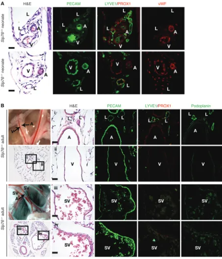
To better define the identity of the endothelial cells lining the effer-ent SVs, we examined expression of prospero homeobox 1 (PROX1), a transcription factor that specifies and maintains lymphatic endothelial identity (17, 18); podoplanin (PDPN), a glycoprotein expressed by lymphatic but not blood endothelial cells (19, 20); the lymphatic marker LYVE1 (21); and vWF, a procoagulant protein syn-thesized by blood but not by lymphatic endothelial cells (LECs) (22, 23); in addition to the pan-endothelial protein PECAM. Mesentery from wild-type and SLP76-deficient neonates and wild-type 12-week-old animals contained thick-walled, PECAM1+vWF+PROX1–
LYVE1– arteries, thin-walled PECAM1+vWF+PROX1–LYVE1– veins,
and thin-walled PECAM1+PROX1+PDPN+LYVE1+vWF–
lym-phatic vessels (Figure 2, A and B). At age 12 weeks, the artery and vein were visible as two adjacent blood-filled vessels in the mes-entery of wild-type mature animals. In contrast, the mesmes-entery of 12-week-old SLP76-deficient mice contained a third blood-containing vessel in the anatomic position of the congenital lym-phatic that participates in the intestinal shunt (Figure 2B and see below; and ref. 10). These vascular bundles contained thick-walled, PECAM1+vWF+PROX1–PDPN–LYVE1– arteries and large,
thin-walled PECAM1+vWF+PROX1–PDPN–LYVE1– SVs (Figure 2B).
Previous studies have shown that SLP76 is required in platelets to prevent the formation of blood-lymphatic connections in the intestine (10) and that these connections send blood flow through mesenteric collecting lymphatic vessels that ultimately carry effer-ent blood from a vascular shunt that forms in the intestine proper (11). To test whether the observed loss of lymphatic mesenteric col-lecting vessels in SLP76-deficient animals could result from loss of SLP76 in endothelial cells themselves, we next assessed the natural history of these vessels in Vav-Cre;Slp76fl/– mice. Previous lineage
tracing experiments performed using both Vav-Cre;R26RYFP and
Vav-Cre;R26RYFP;Slp76fl/– mice have revealed high-level
Cre-medi-ated recombination in hematopoietic cells, with no detectable recombination in either blood or LECs of the intestine or else-where (11, 24). Mature Vav-Cre;Slp76fl/– mice developed vascular
Finally, to determine whether the SVs that arise in Slp76–/–
and Vav-Cre;Slp76fl/– mice acquire arterial or venous identity,
we stained for EPHB4, a venous marker, and CX40, an arterial marker. SV endothelium was EPHB4+ and CX40–, while that
of mesenteric arteries was EPHB4– and CX40+, a result
[image:4.585.48.537.82.578.2]consis-tent with venous identity (Figure 3A). These findings suggested that the mesenteric lymphatics of SLP76-deficient animals lose lymphatic vessel identity and acquire venous identity as they
Figure 1
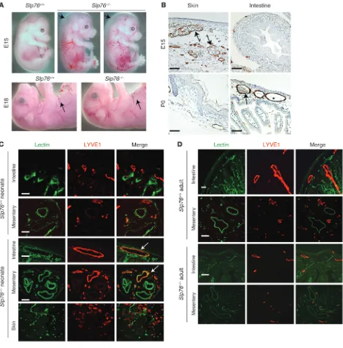
Late separation of the blood and lymphatic circulations in SLP76-deficient mice. (A) Cutaneous edema resolves in Slp76–/– embryos between E15
and E18. Arrows indicate sites of raised skin indicative of edema at E15 (top) and the presence of normal skin folds indicating a lack of edema at E18 (bottom). (B) Blood-filled lymphatics resolve in the skin of Slp76–/– embryos at the same time that they appear in the intestine. LYVE1
stain-ing (brown) identifies lymphatic vessels in the skin and intestine of E15 and neonatal Slp76–/– animals. Arrows indicate blood-filled lymphatics in
E15 skin and in neonatal intestine. (C) Functional identification of blood-filled lymphatics in neonatal Slp76–/– animals using intravenous injection
of biotinylated lectin. Blood-perfused vessels are identified by FITC-streptavidin binding to biotin-lectin after intravenous injection (green), and lymphatic vessels are visualized by LYVE1 immunostaining (red). Co-stained vessels are present in the intestine and mesentery but not skin of Slp76–/– neonates (arrows). (D) Late resolution of mesenteric and intestinal blood-lymphatic mixing in Slp76–/– mice demonstrated using staining
become exposed to blood flow from blood-lymphatic connec-tions in the intestine (Figure 3B).
Prox1CreERT2 lineage tracing reveals that LECs give rise to venous
endo-thelial cells in the mesenteric vessels of Slp76–/– mice. The hypothesis
that mesenteric lymphatic vessels in SLP76-deficient mice alter
[image:5.585.65.518.82.611.2]identity in response to blood flow is based on their characteris-tic anatomical position within the mesenteric vascular bundle. It is possible, however, that SVs do not derive from the congeni-tal lymphatic due to a complex remodeling process. In addition, even if correctly identified, the endothelial cells lining this vessel
Figure 2
Mesenteric SVs in adult Slp76–/– mice lose lymphatic identity. (A) Neonatal mesenteric lymphatics in Slp76–/– mice express the lymphatic
may have been molecularly reprogrammed or replaced to confer blood vessel identity. To determine whether LECs give rise to the blood endothelial cells that line efferent SVs, we bred SLP76-deficient mice onto a Prox1CreERT2;Rosa26RYFP background in
which tamoxifen induction of CRE activity in Prox1-expressing cells activates permanent expression of YFP in LECs (15). Slp76–/–;
Prox1CreERT2;Rosa26RYFP animals and control littermates were
treated with tamoxifen from P1 to P5, a time point prior to intesti-nal vascular separation and the appearance of the intestiintesti-nal shunt,
and YFP was detected by immunohistochemistry at 12 weeks of age. Neonatal tamoxifen induction conferred mosaic YFP expres-sion in the endothelial cells of lymphatic but not blood vessels in the mesentery of 12 week-old Slp76+/–;Prox1CreERT2;Rosa26RYFP
control animals (Figure 4A). YFP+ endothelial cells were identified
in the large mesenteric SVs that lack lymphatic identity in Slp76–/–;
Prox1CreERT2;Rosa26RYFP littermates (Figure 4B). YFP+ endothelial
[image:6.585.51.535.79.514.2]cells in these vessels were negative for PROX1, PDPN, and LYVE1 but expressed vWF (Figure 4B). These results suggested that
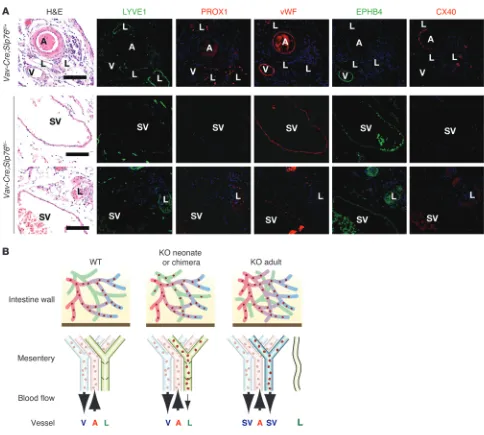
Figure 3
Mesenteric SVs in adult Vav-Cre;Slp76fl/– mice lose lymphatic identity and acquire venous identity. (A) Mesenteric SVs that form in Vav-Cre;Slp76fl/–
mice lose expression of lymphatic molecular markers and acquire expression of the blood vessel marker vWF and venous marker EPHB4 but not the arterial marker CX40. Scale bars: 50 μm. (B) Model of vascular remodeling in SLP76-deficient mice. Shown are the vascular anatomy and flow through the intestinal and mesenteric vessels of neonatal wild-type, neonatal Slp76–/– (KO neonate), and mature Slp76–/– (KO adult) animals.
LECs were reprogrammed from blood endothelial cells in vessels exposed to flowing blood, but only a small number of such cells could be identified using this Prox1CreERT2 allele.
The small number of YFP+ endothelial cells detected in the
mesenteric SVs of Slp76–/–;Prox1CreERT2;Rosa26RYFP could indicate
that most LECs are simply replaced by blood endothelial cells, or it could reflect inefficient labeling due to reduced levels of Prox1
expression in postnatal animals compared with embryos (25) and/ or low levels of CreERT2 expressed behind Prox1 using an IRES in the knock-in Prox1CreERT2 allele. To distinguish between these
possi-Figure 4
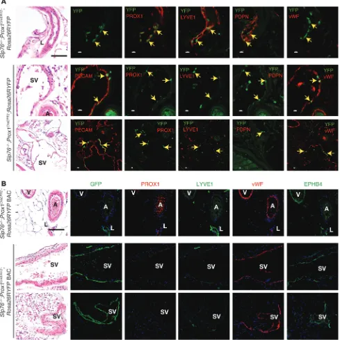
Genetic lineage tracing demonstrates that the blood endothelial cells lining mesenteric SVs in Slp76–/– mice arise from LECs. (A) Lineage tracing
studies performed using a Prox1CreERT2 knock-in line. Antibody staining of mesentery from 12-week-old Slp76+/–;Prox1CreERT2;Rosa26RYFP animals
exposed to tamoxifen as neonates reveals YFP only in PROX1+LYVE1+PDPN+vWF–LECs (top). Antibody staining of mesentery from 12-week-old
Slp76–/–;Prox1CreERT2;Rosa26RYFP animals exposed to tamoxifen as neonates reveals YFP in PROX1–LYVE1–PDPN–vWF+ blood endothelial cells
that line large SVs (bottom). Black scale bars: 50 μm; white scale bars: 10 μm. (B) Lineage tracing studies using a Prox1CreERT2 BAC transgenic line.
Studies were performed as described in A using a single tamoxifen injection at P14. Note that with this Prox1CreERT2 line, virtually all the endothelial
[image:7.585.51.540.88.577.2]bilities, we repeated this experiment using a recently described BAC
Prox1CreERT2 transgene that labels postnatal LECs highly efficiently
(26). In contrast to lineage tracing studies using the Prox1CreERT2
knock-in animals, a single tamoxifen injection at P14 resulted in virtually 100% labeling of LECs in Prox1CreERT2;Rosa26RYFP BAC
transgenic mice, although sparse labeling of venous endothelial cells could also be detected with this regimen (Figure 4B). Analysis of Slp76–/–;Prox1CreERT2;Rosa26RYFP BAC transgenic mice revealed
that in approximately half of SVs, virtually all endothelial cells were YFP+, indicating that these vessels were of lymphatic origin
(Figure 4B). The YFP+ endothelium of these vessels was uniformly
PROX1–LYVE1–vWF+EPHB4+, consistent with a venous blood
ves-sel identity (Figure 4B). These genetic studies demonstrate that the LECs of mesenteric lymphatic vessels of SLP76-deficient mice are neither lost nor replaced during exposure to blood flow, but are instead reprogrammed to venous blood endothelial cells.
Lymphatic endothelial identity is maintained in the presence of blood. Molecular reprogramming of the LECs lining SLP76-deficient mes-enteric lymphatic vessels may be a response to factors in the blood that are not present in lymph, to hemodynamic shear forces present in blood vessels but not in lymphatic vessels, or to both. Lethally irradiated wild-type mice that are reconstituted with SLP76-defi-cient hematopoietic cells develop blood-filled mesenteric lymphat-ics (10, 27), but these mature animals do not undergo subsequent vascular separation and fail to develop the shunts or cardiomegaly observed in Slp76–/– animals (10, 27). Previous angiographic studies
of SLP76-deficient mice revealed that paired mesenteric SVs in the anatomic position of the congenital vein and lymphatic are exposed to high levels of blood flow (10). In contrast, angiographic studies performed in SLP76-deficient radiation chimeras revealed minimal blood flow in blood-filled mesenteric lymphatics compared with adjacent veins (Supplemental Videos 1 and 2; supplemental materi-al available online with this article; doi:10.1172/JCI57513DS1), and Doppler ultrasound confirmed that there was minimal blood flow in these vessels (Figure 5, A–D). Unlike the endothelial cells lining
Slp76–/– SVs, the endothelial cells lining blood-filled lymphatic
ves-sels in Slp76–/– radiation chimeras exhibited persistent expression of
PROX1, PDPN, and LYVE1 and no detectable vWF 16 weeks after reconstitution (Figure 5E). These studies demonstrate that expo-sure to blood is not sufficient to convert lymphatic vessels to blood vessels in vivo and suggest that fluid flow is necessary.
The fluid shear forces experienced by Slp76–/– mesenteric lymphatics are
sufficient to downregulate PROX1 expression in LECs ex vivo. To define a potential link between blood flow and the loss of lymphatic vessel identity in vivo, we calculated the hemodynamic forces in SLP76-deficient mesenteric SVs using high-frequency ultrasound to measure vessel size and blood flow (Figure 6). Two-dimensional (2D) ultrasound of wild-type mesentery revealed a small-caliber artery and a large-caliber vein (Figure 6A). The inability to visual-ize the wild-type lymphatic vessel was likely due to compression of this very low-pressure vessel by the ultrasound transducer. In contrast, the 3 blood-filled vessels that constitute a typical Slp76–/–
mesenteric vascular bundle were easily visualized using 2D ultra-sound, including a small-caliber artery and two large-caliber SVs (Supplemental Videos 1 and 2, and Figure 6B). Flow profiles in the vessels visualized by 2D ultrasound were obtained using pulsed-wave Doppler ultrasound (Figure 6, C and D). The small-caliber arteries of both wild-type and SLP76-deficient mice displayed high-velocity pulsatile flow in the direction of the intestine (i.e., afferent blood flow) (Figure 6C). The larger-caliber veins in
wild-type animals and SVs in SLP76-deficient animals were character-ized by non-pulsatile, lower-velocity flow away from the intestine (i.e., efferent blood flow) (Figure 6D). The wall shear stress in each vessel was next calculated using the measured mean flow veloc-ity and vessel diameter, assuming a constant blood viscosveloc-ity η of 7 mPa*s (28–30). Shear stresses in wild-type veins and the SVs of SLP76-deficient animals were similar in magnitude, although SVs exhibited greater heterogeneity (Figure 6E and Supplemental Table 1). These studies suggest that mesenteric lymphatic vessels in SLP76-deficient animals become exposed to fluid shear stresses similar to those experienced by mesenteric veins, a result consis-tent with their molecular venous identity (Figure 3A).
To determine whether fluid shear stress in this range might be sufficient to negatively regulate lymphatic endothelial identity, we subjected primary human LECs to either pulsatile or steady flow with a shear stress of 20 dynes/cm2. Following 8 hours of shear
stress, LECs exhibited a greater than 6-fold reduction in the level of PROX1 mRNA expression compared with static cultured cells (Figure 6F). Exposure to fluid shear forces did not alter expression of the pan-endothelial gene PECAM1 (Figure 6F and Supplemental Figure 1). Expression of the secondary lymphatic markers LYVE1
and PDPN was not significantly reduced in response to shear forces (Figure 6F). Real-time PCR assessment of other endothelial identity markers and known shear-responsive genes revealed little change in the venous markers COUP-TFII and EPHB4, and increas-es in the arterial markers HEY1, HEY2, and EFNB2, as well as the shear-responsive gene KLF2 (Supplemental Figure 2). The gener-ally arterial shift in gene expression observed in these cells is in contrast to the venous identity observed in vivo and may reflect the difference in the way these forces are exerted, i.e., rapid and full onset in vitro versus slow and gradual onset in vivo. Consis-tent with the drop in PROX1 mRNA levels, LECs demonstrated dramatically reduced PROX1 protein levels after exposure to shear stress (Figure 6G). Finally, loss of PROX1 mRNA was transient in this system, and levels recovered after 24 hours without shear (Fig-ure 6H). These studies confirm that fluid shear forces like those experienced by lymphatic vessels in postnatal SLP76-deficient animals are sufficient to negatively regulate the expression of the lymphatic endothelial fate regulator PROX1.
Discussion
lin-ing these vessels as blood endothelial cells. Similar changes are observed in LECs exposed to fluid shear ex vivo, suggesting that hemodynamic forces can reprogram lymphatic vessels to blood vessels through transcriptional pathways that establish and main-tain endothelial identity.
The key finding in this study is the demonstration that lym-phatic vessels exposed to blood flow in vivo after birth are repro-grammed to acquire blood vessel identity. Studies performed more than 30 years ago revealed that endothelial cell turnover in large vessels such as the aorta is low but non-uniform and higher in areas of turbulent blood flow where hemodynamic forces are more varied (34, 35). Thus, the onset of significant fluid shear forces in the lymphatic vessels of SLP76-deficient mice could stimulate endothelial turnover and the replacement of LECs by either
circu-lating blood endothelial cells or circucircu-lating blood endothelial pre-cursor cells believed to contribute to the endothelium of injured or new vessels (36–39). Alternatively, the gradual rise in hemodynam-ic shear forces may alter the gene expression of the LECs lining these vessels and reprogram them to a blood endothelial identity. Our genetic lineage tracing experiments demonstrate that virtu-ally all of the PROX1-negative endothelial cells that line the SVs of surviving SLP76-deficient animals derive from PROX1-expressing cells. These studies therefore provide definitive evidence of molec-ular reprogramming of endothelial and vessel identity in response to blood flow in vivo.
Our findings provide strong evidence that hemodynamic forces underlie the reprogramming of lymphatic vessels to blood vessels in response to blood flow. Studies using Vav-Cre;Slp76fl/– mice
dem-Figure 5
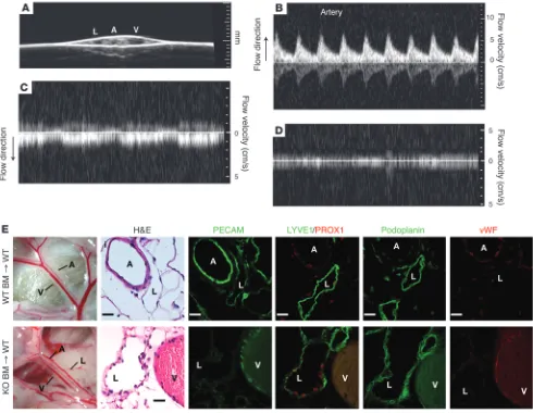
Lymphatic endothelial identity is maintained in the presence of blood in Slp76–/– radiation chimeras. (A) The mesenteric vessels of Slp76–/–
radia-tion chimeras were visualized using 2D ultrasound. (B–D) Pulsed-wave Doppler signals indicative of blood flow detected in the mesenteric artery, vein, and lymphatic are shown. Note that arterial flow is directed opposite to that of venous flow and that arterial flow is pulsatile (B, corresponding to heart rate), while venous flow is phasic (C, corresponding to respiration). No significant signal in either direction was obtained from the lym-phatic (D). (E) Blood-filled mesenteric lymphatics retain lymphatic identity in Slp76–/– radiation chimeras. Mesenteric vessels in wild-type lethally
irradiated mice reconstituted with Slp76+/+ (+/+) or Slp76–/– (–/–) bone marrow are shown 16 weeks after reconstitution (left). Analysis of serial
sections reveals that mesenteric lymphatics in Slp76–/– radiation chimeras that are exposed to blood but not flow retain expression of LYVE1,
[image:9.585.46.536.84.464.2]onstrate that the endothelial and vessel identity changes observed do not reflect an unexpected role for SLP76 in endothelial cells, but instead arise due to changes in the vascular environment that result from blood-lymphatic vascular connections in the intestine. Radiation chimeras reconstituted with SLP76-deficient hemato-poietic cells develop blood-filled mesenteric lymphatics but have
[image:10.585.56.523.82.539.2]little or no flow in those vessels, most likely because these mature, irradiated animals do not remodel their vasculature to create arterio-venous-lymphatic shunts like neonatal animals. These ani-mals demonstrate that the formation of blood-lymphatic connec-tions and contact with blood is not sufficient to alter lymphatic endothelial and vessel identity. In contrast, LECs exposed to fluid
Figure 6
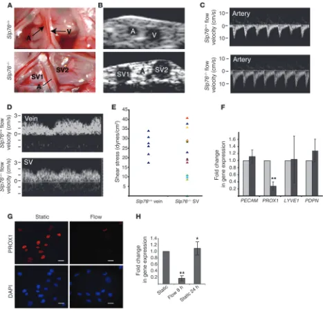
Fluid shear forces equivalent to those in Slp76–/– mesenteric lymphatics drive loss of PROX1 expression in LECs. (A) Representative mesenteric
vessels in 12-week-old Slp76+/+ and Slp76–/– animals that were studied using Doppler ultrasound. The dilated, blood-containing vein and
congeni-tal lymphatic are denoted as SV1 and SV2. (B) The vessels shown in A were visualized using 2D ultrasound. (C and D) The pulsed-wave Doppler signals and measured flow velocity of blood in the vessels indicated in A and B are shown. Note the difference in direction of flow between the arteries and veins or efferent SVs. (E) Calculated shear stresses for Slp76+/+ veins and Slp76–/– SVs. Slp76–/– data points in the same color indicate
values of paired SVs from the same mesenteric bundle. (F) LECs exposed to flow downregulate PROX1. LECs were subjected to a shear stress of 20 dynes/cm2 for 8 hours, and the expression of the indicated mRNAs measured using qPCR. n = 7. **P < 0.01. (G) Loss of PROX1 protein in
shear forces in the absence of blood ex vivo exhibit rapid downregu-lation of PROX1. Loss of PROX1 in LECs has been shown to result in the loss of other lymphatic identity markers such as LYVE1 and PDPN (17, 20), and genetic deletion of Prox1 has recently been shown to be sufficient to convert LECs to blood endothelial cells in vivo (18). Thus, our studies support the concept that expression of the PROX1 transcription factor is required to maintain lymphatic endothelial and vessel identity (18) and that loss of PROX1 expres-sion in LECs exposed to fluid shear forces associated with blood flow is a likely mechanism by which lymphatic vessels are repro-grammed to blood vessels in SLP76-deficient mice.
What are the molecular pathways through which hemody-namic forces negatively regulate PROX1 and endothelial lym-phatic identity? We found that HEY1 and HEY2 are strongly upregulated in coordination with downregulation of PROX1 in LECs exposed to fluid shear forces (Supplemental Figure 2), and HEY1/2 or NOTCH expression was recently demonstrated to negatively regulate lymphatic endothelial identity in cultured LECs by reducing expression of PROX1 (40). To test the role of HEY1/2 expression and loss of lymphatic endothelial identity in SLP76-deficient mice in vivo, we have examined Slp76–/–;Hey2LacZ/+
mice, in which LacZ is expressed in place of Hey2 (41). However, we did not detect LacZ expression in the endothelium of mesen-teric arteries or SVs, suggesting that the NOTCH/HEY signaling is not the basis for this endothelial reprogramming event in vivo. It is possible that flow-dependent activation of HEY1/2 signaling plays a transient role in this process that was not detected by these studies, but it seems more likely that ex vivo studies of cultured endothelial cells performed over hours do not fully model molec-ular changes that take place over weeks in vivo. Flow chamber experiments have identified large numbers of endothelial genes that are upregulated and downregulated by fluid flow, includ-ing many involved in endothelial identity (31, 42, 43), but there is no definitive means of testing whether and to what extent these genes mediate endothelial responses to hemodynamic forces in vivo. Transiently blocking blood flow in very young embryos can distinguish between programmed endothelial identity gene expression and gene expression driven by fluid forces (44, 45), but this approach is not feasible when hemodynamic changes arise more gradually, as they do in patients with congenital vascular and cardiac defects. Thus, the identity of the molecular signals that downregulate PROX1 expression in response to fluid flow in vivo is not yet defined.
An important implication of this study is that vessel identity remains plastic after vascular development is complete and may be radically altered by hemodynamic forces later in life. The healthy mature vasculature is thought to be very quiescent, but many con-genital and acquired human cardiovascular diseases are associated with persistent changes in blood flow and fluid shear forces, e.g., the left-to-right shunting of blood to the low-pressure pulmonary vasculature from the high-pressure arterial system in congenital heart disease. Molecular changes in endothelial and vessel identity are very likely to accompany the hemodynamic alterations in these diseases. Defining these molecular changes is expected to provide new insight into the pathogenesis and treatment of human cardio-vascular diseases such as cardio-vascular malformations.
Methods
Histology and immunohistochemistry. Intestine and mesentery were dissected from murine neonates and adults and fixed in 4% paraformaldehyde
for 24–48 hours, dehydrated in 100% ethanol, and embedded in
paraf-fin. Serial 8-μm-thick sections were subjected to hematoxylin and eosin
and/or immuno histo chemical staining as detailed by the University of Pennsylvania Molecular Cardiology Research Center Histology and Gene Expression Core (http://www.med.upenn.edu/mcrc/histology_core/). Antibodies used were MEC13.3 rat anti–mouse CD31 (BD Biosciences — Pharmingen) at 1:500, rabbit anti-LYVE1 at 1:1,000, rabbit anti-Prox1 (Abcam) at 1:100, CX40 (Alpha Diagnostics) at 1:100, and anti-EPHB4 (Cell Sciences) at 1:50.
Injection of biotinylated lectin. Intracardiac injection of 50 μg of biotinylated
Lycopersicon esculentum (tomato) lectin (Vector Laboratories) was performed
in neonatal Slp76–/– mice and wild-type littermate controls. Co-injection
with FITC-dextran (Sigma-Aldrich) was used to determine successful
perfusion. Biotinylated tomato lectin (300 μg; Vector Laboratories) was
injected intravenously into 10- to 12-week-old Slp76–/– mice and wild-type
littermate controls via tail vein. The mice were then euthanized and fixed by perfusion with 4% paraformaldehyde. Tissues were then processed for immunohistochemical analysis as described above.
Hemodynamic studies. A Visual Sonics Vevo 770 Imaging System with single-element mechanical transducers was used to image the mesenteric vascular structures and blood flow spectra. The center frequency was set at 40 MHz (for lateral and axial resolutions of 68 and 38 nm, respectively). Twelve-week-old mice were anesthetized with Avertin, and their intestine and mesentery were dissected and exposed. Body temperature was moni-tored via a rectal thermometer and maintained between 36°C and 37°C using a heating pad and lamp, and heart rate and electrocardiogram were also continuously monitored. To eliminate interference from structures underlying the mesentery, the mesenteric and intestinal areas of interest were spread on a slide glass. A thin layer of prewarmed ultrasound gel was applied to the slide glass and covered with a plastic membrane before tissue exposure. A second membrane was placed on the tissue surface to keep it clean for later dissection. Another layer of prewarmed thick ultrasound gel was placed on this membrane to provide a coupling medium for the trans-ducer. Two mesenteric vascular bundles from each mouse were selected for imaging. Cross-sectional images of the vessels were recorded and the lumen diameters measured by 2D ultrasonography. The transducer was then rotated 90° to obtain a longitudinal view of the vessels. For Doppler spectral analysis, the transducer was adjusted for an angle of insonation less than 60°. The mean blood flow velocities were measured and used to calculate wall shear stress for the vessels of interest.
The wall shear stress in each vessel was next calculated using the mea-sured mean flow velocity Vmean and vessel diameter ID, assuming a constant blood viscosity η of 7 mPa*s (28–30): τ (shear stress) = γ • η = (4Vmeanη)/ID.
Although the wall shear rate γ was not directly measured, it was calculated
from the measured parameters by using Poiseuille’s parabolic model of velocity distribution.
Cell culture under shear stress. Passage 4 LECs were cultured to confluence on gelatin-coated glass slides for use in a parallel plate flow chamber.
Pul-satile flow with shear stress of 20 dynes/cm2 was achieved using a
Master-flex peristaltic pump and a parallel plate flow chamber provided by J.A. Frangos (CytoDyne, San Diego, California, USA). Non-pulsatile flow with shear stress of 20 dynes/cm2 was achieved using a 6-slide parallel plate flow
chamber designed by Flexcell International Corp. The medium reservoir was maintained at 37°C using an incubator or water bath. Cells were incu-bated in the flow chamber for 8 hours. Static control cells were fed with fresh medium at the beginning of the experiment and maintained in the incubator for the duration of the experiment.
Real-time PCR analysis. Total RNA from LECs (primary isolation described above) and human umbilical vein endothelial cells (Lonza) was isolated using an RNeasy Mini Kit (QIAGEN) according to the manufacturer’s
protocols. For reverse transcriptase reactions, 1 μg of total RNA and 100
ng of random hexamers was used to generate cDNA with the First-Strand cDNA Synthesis Kit (Invitrogen) according to the manufacturer’s proto-cols. Real-time semi-quantitative PCR and relative quantitation normal-ized to GAPDH was performed using SYBR Green Master Mix reagent (Applied Biosystems) on the ABI Prism 7900HT Sequence Detection System (Applied Biosystems). Real-time PCR primers were designed and obtained from IDT. Primer sequences are listed in Supplemental Methods.
Immunocytochemistry. Cells were fixed in 4% paraformaldehyde for 15 min-utes at room temperature and permeabilized with 0.2% Triton X-100/PBS. Samples were blocked with 2% bovine serum albumin in PBS and stained with rabbit anti-Prox1 antibody (Abcam), followed by Alexa Fluor 488 secondary antibody (Molecular Probes, Invitrogen), and mounted with DAPI-containing Vectashield mounting medium (Vector Laboratories) to visualize cell nuclei.
Genetic reconstitution with SLP76-deficient bone marrow and angiography
stud-ies. Wild-type mice were lethally irradiated and reconstituted with
SLP76-deficient bone marrow according to published methods (10, 27). Recipient mice were sacrificed for study 8–10 weeks after transplantation. FITC-dextran angiography with real-time video microscopy was performed on anesthetized animals as previously described (10). Tissues were harvested, fixed, and processed for immunohistochemical analysis.
Statistics. P values were calculated using an unpaired, 1-tailed Student’s
t test. A P value less than 0.05 was considered significant. All bar graphs
and error bars represent mean values and SEM.
Study approval. Animal studies were approved by the Institutional Animal Care and Use Committee at the University of Pennsylvania, Philadelphia, Pennsylvania, USA.
Acknowledgments
This work was supported by an American Heart Association (AHA) award (Scientist Development Award 0730286N to S.J. Stachelek), Cancer Research UK (to T. Makinen), and the National Heart, Lung, and Blood Institute (NHLBI; HL073402 to G. Oliver and HL072798 to M.L. Kahn).
Received for publication February 20, 2012, and accepted in revised form April 5, 2012.
Address correspondence to: Mark L. Kahn, University of Pennsyl-vania, 952 BRB II/III, 421 Curie Blvd., Philadelphia, Pennsylvania 19104, USA. Phone: 215.898.9007; Fax: 215.573.2094; E-mail: markkahn@mail.med.upenn.edu.
1. Friedlander RM. Clinical practice. Arteriovenous malformations of the brain. N Engl J Med. 2007; 356(26):2704–2712.
2. Peery WH. Clinical spectrum of hereditary hemor-rhagic telangiectasia (Osler-Weber-Rendu disease).
Am J Med. 1987;82(5):989–997.
3. Garzon MC, Huang JT, Enjolras O, Frieden IJ. Vas-cular malformations: Part I. J Am Acad Dermatol. 2007;56(3):353–370.
4. Brouillard P, Vikkula M. Genetic causes of vas-cular malformations. Hum Mol Genet. 2007; 16(spec no. 2):R140–R149.
5. Oda T, et al. Mutations in the human Jagged1 gene are responsible for Alagille syndrome. Nat Genet. 1997;16(3):235–242.
6. McAllister KA, et al. Endoglin, a TGF-beta binding protein of endothelial cells, is the gene for heredi-tary haemorrhagic telangiectasia type 1. Nat Genet. 1994;8(4):345–351.
7. Sahoo T, et al. Mutations in the gene encoding KRIT1, a Krev-1/rap1a binding protein, cause cere-bral cavernous malformations (CCM1). Hum Mol Genet. 1999;8(12):2325–2333.
8. McCright B, Lozier J, Gridley T. A mouse model of Alagille syndrome: Notch2 as a genetic modi-fier of Jag1 haploinsufficiency. Development. 2002; 129(4):1075–1082.
9. Bourdeau A, Dumont DJ, Letarte M. A murine model of hereditary hemorrhagic telangiectasia.
J Clin Invest. 1999;104(10):1343–1351.
10. Abtahian F, et al. Regulation of blood and lym-phatic vascular separation by signaling proteins SLP-76 and Syk. Science. 2003;299(5604):247–251. 11. Bertozzi CC, et al. Platelets regulate lymphatic
vas-cular development through CLEC-2-SLP-76 signal-ing. Blood. 2010;116(4):661–670.
12. Turner M, et al. Perinatal lethality and blocked B-cell development in mice lacking the tyrosine kinase Syk. Nature. 1995;378(6554):298–302. 13. Clements JL, et al. Fetal hemorrhage and platelet
dysfunction in SLP-76-deficient mice. J Clin Invest.
1999;103(1):19–25.
14. Sabin FR. On the origin of the lymphatic system from the veins, and the development of the lymph hearts and thoracic duct in the pig. Am J Anat. 1902;1(3):367–389.
15. Srinivasan RS, et al. Lineage tracing demonstrates the venous origin of the mammalian lymphatic vasculature. Genes Dev. 2007;21(19):2422–2432. 16. Kim KE, Sung HK, Koh GY. Lymphatic
develop-ment in mouse small intestine. Dev Dyn. 2007; 236(7):2020–2025.
17. Wigle JT, et al. An essential role for Prox1 in the induction of the lymphatic endothelial cell pheno-type. Embo J. 2002;21(7):1505–1513.
18. Johnson NC, et al. Lymphatic endothelial cell iden-tity is reversible and its maintenance requires Prox1 activity. Genes Dev. 2008;22(23):3282–3291. 19. Kriehuber E, et al. Isolation and characterization
of dermal lymphatic and blood endothelial cells reveal stable and functionally specialized cell lin-eages. J Exp Med. 2001;194(6):797–808.
20. Petrova TV, et al. Lymphatic endothelial repro-gramming of vascular endothelial cells by the Prox-1 homeobox transcription factor. EMBO J. 2002; 21(17):4593–4599.
21. Banerji S, et al. LYVE-1, a new homologue of the CD44 glycoprotein, is a lymph-specific receptor for hyaluronan. J Cell Biol. 1999;144(4):789–801. 22. Nelson GM, Padera TP, Garkavtsev I, Shioda T,
Jain RK. Differential gene expression of primary cultured lymphatic and blood vascular endothelial cells. Neoplasia. 2007;9(12):1038–1045.
23. Jiang S, et al. Hematopoietic stem cells contribute to lymphatic endothelium. PLoS One. 2008;3(11):e3812. 24. Stadtfeld M, Graf T. Assessing the role of hemato-poietic plasticity for endothelial and hepatocyte development by non-invasive lineage tracing. Devel-opment. 2005;132(1):203–213.
25. Bazigou E, et al. Integrin-alpha9 is required for fibronectin matrix assembly during lymphatic valve morphogenesis. Dev Cell. 2009;17(2):175–186.
26. Bazigou E, et al. Genes regulating lymphangiogen-esis control venous valve formation and mainte-nance in mice. J Clin Invest. 2011;121(8):2984–2992. 27. Abtahian F, et al. Evidence for the requirement of
ITAM domains but not SLP-76/Gads interaction for integrin signaling in hematopoietic cells. Mol Cell Biol. 2006;26(18):6936–6949.
28. Vogel J, et al. Transgenic mice overexpressing erythro-poietin adapt to excessive erythrocytosis by regulat-ing blood viscosity. Blood. 2003;102(6):2278–2284. 29. Gnasso A, et al. Association between intima-media
thickness and wall shear stress in common carot-id arteries in healthy male subjects. Circulation. 1996;94(12):3257–3262.
30. Irace C, Cortese C, Fiaschi E, Carallo C, Farinaro E, Gnasso A. Wall shear stress is associated with inti-ma-media thickness and carotid atherosclerosis in subjects at low coronary heart disease risk. Stroke. 2004;35(2):464–468.
31. Buschmann I, et al. Pulsatile shear and Gja5 mod-ulate arterial identity and remodeling events dur-ing flow-driven arteriogenesis. Development. 2010; 137(13):2187–2196.
32. le Noble F, et al. Flow regulates arterial-venous dif-ferentiation in the chick embryo yolk sac. Development. 2004;131(2):361–375.
33. Kwei S, et al. Early adaptive responses of the vascu-lar wall during venous arterialization in mice. Am J Pathol. 2004;164(1):81–89.
34. Caplan BA, Schwartz CJ. Increased endothelial cell turnover in areas of in vivo Evans Blue uptake in the pig aorta. Atherosclerosis. 1973;17(3):401–417. 35. Schwartz SM, Benditt EP. Clustering of replicating
cells in aortic endothelium. Proc Natl Acad Sci U S A. 1976;73(2):651–653.
36. Walter DH, et al. Statin therapy accelerates reen-dothelialization: a novel effect involving mobiliza-tion and incorporamobiliza-tion of bone marrow-derived endothelial progenitor cells. Circulation. 2002; 105(25):3017–3024.
nitric oxide synthase-dependent mobilization of bone marrow-derived endothelial progenitor cells contributes to reendothelialization after arterial injury. Circulation. 2003;108(25):3115–3121. 38. Ii M, et al. Endothelial progenitor
thrombospon-din-1 mediates diabetes-induced delay in reendo-thelialization following arterial injury. Circ Res. 2006;98(5):697–704.
39. Sahara M, et al. Comparison of various bone mar-row fractions in the ability to participate in vascu-lar remodeling after mechanical injury. Stem Cells. 2005;23(7):874–878.
40. Kang J, et al. An exquisite cross-control mecha-nism among endothelial cell fate regulators directs
the plasticity and heterogeneity of lymphatic endothelial cells. Blood. 2010;116(1):140–150. 41. Fischer A, Schumacher N, Maier M, Sendtner M,
Gessler M. The Notch target genes Hey1 and Hey2 are required for embryonic vascular development.
Genes Dev. 2004;18(8):901–911.
42. Jones EA, le Noble F, Eichmann A. What deter-mines blood vessel structure? Genetic prespecifi-cation vs. hemodynamics. Physiology (Bethesda). 2006;21:388–395.
43. Parmar KM, et al. Integration of flow-dependent endothelial phenotypes by Kruppel-like factor 2.
J Clin Invest. 2006;116(1):49–58.
44. Jones EA, Yuan L, Breant C, Watts RJ, Eichmann A.
Separating genetic and hemodynamic defects in neuropilin 1 knockout embryos. Development. 2008;135(14):2479–2488.
45. Nicoli S, Standley C, Walker P, Hurlstone A, Fog-arty KE, Lawson ND. MicroRNA-mediated integra-tion of haemodynamics and Vegf signalling during angiogenesis. Nature. 2010;464(7292):1196–1200. 46. Veikkola T, et al. Intrinsic versus