Corrigendum
(August 2016)
4-Dimensional light-sheet microscopy to
elucidate shear stress modulation of cardiac
trabeculation
Juhyun Lee, … , Rongsong Li, Tzung K. Hsiai
J Clin Invest.
2016;
126(5)
:1679-1690.
https://doi.org/10.1172/JCI83496
.
Hemodynamic shear forces are intimately linked with cardiac development, during which
trabeculae form a network of branching outgrowths from the myocardium. Mutations that
alter Notch signaling also result in trabeculation defects. Here, we assessed whether shear
stress modulates trabeculation to influence contractile function. Specifically, we acquired
4D (3D + time) images with light sheets by selective plane illumination microscopy (SPIM)
for rapid scanning and deep axial penetration during zebrafish morphogenesis. Reduction
of blood viscosity via
gata1a
morpholino oligonucleotides (MO) reduced shear stress,
resulting in downregulation of Notch signaling and attenuation of trabeculation. Arrest of
cardiomyocyte contraction either by troponin T type 2a (
tnnt2a
) MO or in weak atrium
m58(
wea
) mutants resulted in reduced shear stress and downregulation of Notch signaling and
trabeculation. Integrating 4D SPIM imaging with synchronization algorithm demonstrated
that coinjection of neuregulin1 mRNA with
gata1
MO rescued trabeculation to restore
contractile function in association with upregulation of Notch-related genes. Crossbreeding
of
Tg(flk:mCherry)
fish, which allows visualization of the vascular system with the
Tg(tp1:gfp)
Notch reporter line, revealed that shear stress–mediated Notch activation
localizes to the endocardium. Deleting endocardium via the
cloche
sk4mutants
downregulated Notch signaling, resulting in nontrabeculated […]
Technical Advance
Cardiology
Development
Find the latest version:
Introduction
Hemodynamic forces such as shear stress are intimately linked with cardiac morphogenesis and function (1, 2). In addition to cardiogenic differentiation and transcription factors, intracar-diac fluid forces are essential for embryonic cardiogenesis (3–9). Occlusion of flow at either the cardiac inflow or outflow tract resulted in hearts with an abnormal third chamber, diminished looping, and impaired valve formation (4). The myocardium differentiates into 2 layers, an outer compact zone and an inner trabeculated zone. The trabeculae form a network of branching outgrowths from the myocardial wall (10), and both trabeculation and compaction are essential for normal cardiac contractile func-tion. A significant reduction in trabeculation is usually associ-ated with ventricular compact zone deficiencies, whereas hyper-trabeculation (noncompaction) is closely associated with left ventricular noncompaction (LVNC) (11). LVNC is the third most common cardiomyopathy after dilated and hypertrophic cardio-myopathy in the pediatric population (12). Its prevalence was estimated from at 4.5 to 26 per 10,000 adult patients referred for echocardiographic diagnosis (12, 13). However, the
mechano-transduction mechanisms underlying trabeculation during car-diac development remain elusive (11).
We have demonstrated that peristaltic contractions of the embryonic heart tube produce time-varying shear stress (∂τ/∂t) and pressure gradients (∇P) across the atrioventricular (AV) canal in a
zebrafish model of cardiac development (14). The advent of genet-ic manipulation in zebrafish has enabled the applgenet-ication of the fli1 promoter to drive expression of EGFP throughout the vasculature during embryogenesis (Tg[fli1a:EGFP]y1) (15), thereby allowing for
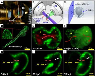
3D visualization of the moving boundary conditions (2D + time) for computational fluid dynamics (CFD) simulation. However, 4D (3D + time) imaging throughout the cardiac cycle requires fast tissue scan-ning and deep axial penetration. For these reasons, a laser light-sheet approach known as selective plane illumination microscopy (SPIM) coupled with nongated 4D synchronization algorithm enabled us to capture live 3D cardiac morphogenesis. By illuminating with a thin plane of light and detecting with a high-speed sCMOS camera, we were able to acquire a stack of cardiac sections with both high axial and temporal resolution (Figure 1 and Supplemental Figures 1 and 2; supplemental material available online with this article; doi:10.1172/ JCI83496DS1). The 4D SPIM imaging technique revealed dynamic architecture in response to changes in hemodynamic forces, cardio-myocyte contraction, and Notch signaling.
To elucidate hemodynamic forces underlying the initiation of trabeculation, we lowered hemodynamic shear forces via (a)
Hemodynamic shear forces are intimately linked with cardiac development, during which trabeculae form a network of branching outgrowths from the myocardium. Mutations that alter Notch signaling also result in trabeculation defects. Here, we assessed whether shear stress modulates trabeculation to influence contractile function. Specifically, we acquired 4D (3D + time) images with light sheets by selective plane illumination microscopy (SPIM) for rapid scanning and deep axial penetration during zebrafish morphogenesis. Reduction of blood viscosity via gata1a morpholino oligonucleotides (MO) reduced shear stress, resulting in downregulation of Notch signaling and attenuation of trabeculation. Arrest of cardiomyocyte contraction either by troponin T type 2a (tnnt2a) MO or in weak atriumm58 (wea) mutants resulted in reduced
shear stress and downregulation of Notch signaling and trabeculation. Integrating 4D SPIM imaging with synchronization algorithm demonstrated that coinjection of neuregulin1 mRNA with gata1 MO rescued trabeculation to restore contractile function in association with upregulation of Notch-related genes. Crossbreeding of Tg(flk:mCherry) fish, which allows visualization of the vascular system with the Tg(tp1:gfp) Notch reporter line, revealed that shear stress–mediated Notch activation localizes to the endocardium. Deleting endocardium via the clochesk4 mutants downregulated Notch signaling, resulting in nontrabeculated ventricle. Subjecting endothelial cells to pulsatile flow in the presence of the ADAM10 inhibitor corroborated shear stress–activated Notch signaling to modulate trabeculation.
4-Dimensional light-sheet microscopy to elucidate
shear stress modulation of cardiac trabeculation
Juhyun Lee,1 Peng Fei,2,3 René R. Sevag Packard,4 Hanul Kang,4,5 Hao Xu,6 Kyung In Baek,1 Nelson Jen,1 Junjie Chen,1 Hilary Yen,1
C.-C. Jay Kuo,6 Neil C. Chi,7 Chih-Ming Ho,3 Rongsong Li,4,5 and Tzung K. Hsiai1,4,5,8
1Department of Bioengineering, UCLA, Los Angeles, California, USA. 2School of Optical and Electronic Information, Huazhong University of Science and Technology, Wuhan, China.
3Department of Mechanical Engineering and 4Division of Cardiology, Department of Medicine, UCLA, Los Angeles, California, USA. 5Division of Cardiology,
Veterans Affairs Greater Los Angeles Healthcare System, Los Angeles, California, USA. 6Department of Electrical Engineering, University of Southern California, Los Angeles, California, USA. 7Department of Medicine, Institute of Genomic Medicine, UCSD, La Jolla, California, USA. 8California NanoSystem Institute, UCLA, Los Angeles, California, USA.
Authorship note: J. Lee and P. Fei contributed equally to this work.
Conflict of interest: The authors have declared that no conflict of interest exists.
Submitted: June 30, 2015; Accepted: February 9, 2016.
crossbred Tg(flk:mCherry) with the Tg(tp1:gfp) Notch reporter line to localize shear stress–mediated endocardial Notch activation. In addition, we demonstrated using a dynamic flow system that pul-satile shear stress (PSS) upregu-lated Notch ligands and target genes in human aortic endothelial cells (HAEC). Overall, interfacing 4D light-sheet imaging with the zebrafish system opens a funda-mental direction for demonstrat-ing shear stress modulation of tra-beculation to influence contractile function via Notch signaling.
Results
Trabeculation formation.
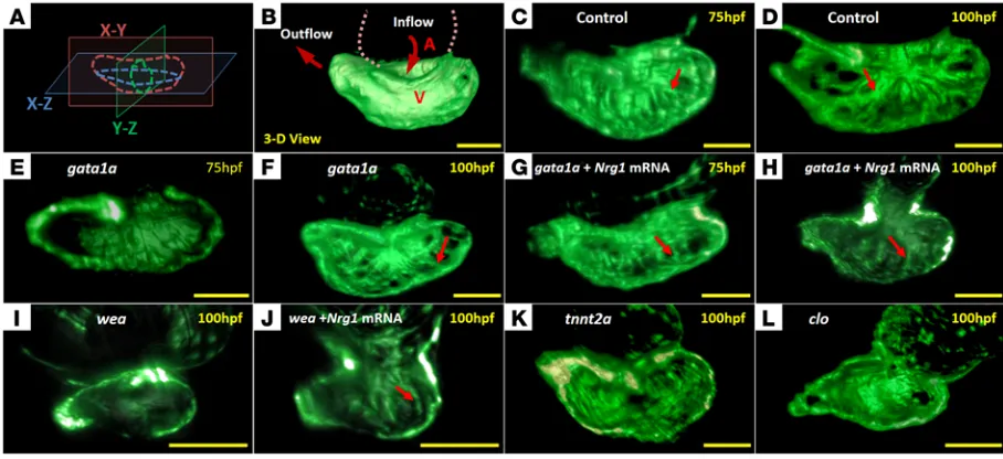
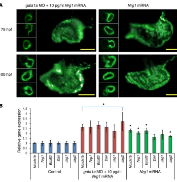
Trabecu-lation was visualized with the trans-genic zebrafish line, Tg(cmlc2:gfp), in which a cardiac-specific pro-moter drives the expression of GFP in cardiomyocytes. Trabeculation starts to appear after cardiac loop-ing (21). At approximately 50 hours post fertilization (hpf), 3D SPIM imaging revealed a nontrabecu-lar myocardium (Figure 1G). At approximately 60 hpf, trabecular ridges started to form in the region exposed to ventricular inflow across the AV canal (Figure 1H). At approximately 70 hpf, these ridges developed into a network of trabecular myocardium (Figure 1I and ref. 22).
Reduced hemodynamic shear stress attenuated trabecular forma-tion. The gata1a MO microinjection reduced hematopoiesis and
viscosity by 90% (16, 17), leading to a reduction in hemodynamic shear stress and a delayed initiation of trabecular network at 75 and 100 hpf when compared with control Tg(cmlc2:gfp) zebrafish (Figure 2, C–F, and Supplemental Videos 1 and 2). Furthermore,
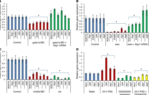
gata1a MO injection significantly downregulated Notch ligands
(delta-like 4 [Dll4], jagged 1 [Jag1], and Jag2), receptor (Notch1b), and downstream signaling components (Nrg1 and ErbB2) (P < 0.05, n = 5) (Figure 3A and refs. 10, 23, 24), whereas coinjection with Nrg1 mRNA (5 pg/nl) rescued trabecular formation at 75 and 100 hpf in association with upregulation of Notch signaling–relat-ed genes (Figure 2, G and H; Figure 3A; and Supplemental Video 3). Tg(tp1:gfp) zebrafish allow assessment of Notch activation in response to shear stress (25). Crossbreeding Tg(flk:mCherry) with
Tg(tp1:gfp) lines further localized altered shear stress–activated
Notch signaling in endocardium in response to gata1a MO injec-tion (Figure 4), whereas tp1-gfp signal was present outside of the endocardium in response to coinjection of Nrg1 mRNA with gata1a MO. Thus, gata1a MO injection reduced hemodynamic shear forc-microinjection of gata1a morpholino oligonucleotides (MO) at the
1- to 4-cell stage to reduce hematopoiesis and viscosity by 90% (16, 17), (b) microinjection of troponin T type 2a (tnnt2a) MO to arrest cardiomyocyte contraction in embryos (18, 19), and (c) genetic mutation of the weak atriumm58 (wea) to inhibit atrial
con-traction (10, 20). A myocardium-specific GFP transgene in zebra-fish allowed for visualization of the ventricular wall. SPIM imag-ing characterized the attenuated trabecular myocardial network in response to both gata1a MO and tnnt2a MO injection. Both the
wea mutants with noncontractile atrium and clochesk4 (clo) mutants
[image:3.585.38.402.55.348.2]with deletion of endocardium expressed substantially lower levels of Notch-related genes as compared with those of WT, and both developed nontrabeculated myocardium. Integrating computa-tion with quantitative analyses, we demonstrated that attenuacomputa-tion of trabeculation in response to gata1a MO, erb-b2 receptor tyro-sine kinase 2 (ErbB2) inhibitor (AG1478), and wea mutation was associated with decreased ventricular strain and ejection fraction (EF). The aforementioned loss-of-function was rescued with neu-regulin1 (Nrg1) mRNA to restore trabeculation and contraction in association with upregulation of Notch-related genes. We further
mutants (26, 27). At 100 hpf, clo mutants displayed a small and thin ventricle as previously described (refs. 14, 27, Figure 2L, and Supplemental Video 5), accompanied by a reduction in cardiac mRNA for Notch ligands, receptor, and target genes as compared with those of WT (Figure 3C). Using the well-calibrated in vitro flow system, HAEC were exposed to PSS with the time-averaged shear stress (τavg) at 23 dyn × cm–2 and 1 Hz cycle (28–31). We
were able to recapitulate PSS-mediated upregulation in endothe-lial Notch signaling–related genes, which were inhibited in the presence of the ADAM10 inhibitor, GI254023X, which blocks proteolytic cleavage of Notch and formation of Notch intracel-lular domain (NICD) (Figure 3D). In addition, we demonstrated Notch signaling–mediated trabecular formation by inhibiting the translocation of NICD to the nuclei with the γ-secretase inhibitor (DAPT) (32, 33), where we observed downregulation of cardiac Notch-related gene expression and absence of trabecular for-mation (Supplemental Figure 3). Taken together, hemodynamic forces were implicated in the initiation of trabeculation via endo-cardial-dependent Notch signaling.
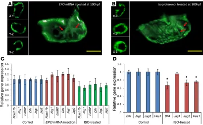
EPO-augmented viscosity induced no additional trabecular net-work. We further cloned erythropoietin (EPO) mRNA, which was
injected at the 1- to 2-cell stage at 20 pg/nl to increase hemato-poiesis and, thus, blood viscosity (Supplemental Videos 6 and 7, and ref. 34). Since heart rates remained regular, assuming that pressure gradients are not affected by injection of EPO mRNA, shear stress increased by keeping the tangential velocity gradient normal. Despite an EPO-augmented time-averaged shear stress, the Notch-related genes were not upregulated, and the trabecular network appeared to be the same as that of WT (Figure 5, A and C). We further used isoproterenol (ISO), a β1- and β2-
adrenore-es, leading to downregulation of Notch signaling, whereas coinjec-tion with Nrg1 mRNA restored trabecular formacoinjec-tion and resulted in the upregulation of Notch-related genes.
To further elucidate hemodynamic modulation of trabecula-tion via Notch signaling, we introduced the wea mutants to reduce atrial contraction, leading to reduced ventricular inflow. The wea mutants developed downregulation of cardiac mRNA levels of Notch signaling–related genes as compared with those of WT and developed small, nontrabeculated ventricles (Figure 2I and Fig-ure 3B). However, microinjection of Nrg1 mRNA (10 pg/nl) to wea mutants at the 1- to 4-cell stage partially rescued trabecular ridges to enhance contractile ventricular function, accompanied with upregulation of Notch signaling genes (Figure 2J and ref. 10). In this context, the wea mutants demonstrated that a noncontractile atrium engendered a nontrabeculated ventricle, underscoring the need for synchronized alternating atrial and ventricular contrac-tion to generate hemodynamic forces to upregulate Notch signal-ing for trabeculation.
The tnnt2a MO inhibited trabecular formation. The tnnt2a MO
microinjection inhibited cardiac tnnt2a, leading to the arrest of myocardial contraction and the absence of hemodynamic forces (18, 19). Microinjection of tnnt2a MO significantly downregulated Notch signaling (P < 0.05 for all comparisons, n = 5) (Figure 3C). The tnnt2a MO–injected zebrafish persistently harbored a nontra-beculated and thin ventricular wall at 100 hpf (Figure 2K and Sup-plemental Video 4). These observations further support hemody-namic forces underlying the initiation of trabeculation.
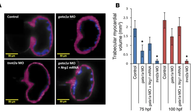
The clo mutants attenuated notch signaling and trabeculation.
[image:4.585.67.521.56.264.2]To demonstrate hemodynamic force–mediated Notch signaling in the endocardium, we abolished endothelial lining with the clo
Nrg1 mRNA rescue. At 75 hpf, the volume of trabecular myocardial
ridges was reduced by approximately 2.7 ± 0.4-fold in response to gata1a MO (0.7 × 10⁻⁴ ± 0.3 × 10⁻⁴μm3), by approximately 19.0
± 0.3-fold in response to tnnt2a MO (0.1 × 10⁻⁴ ± 0.1 × 10⁻⁴μm3),
and by approximately 1.7 ± 0.4-fold in response to coadministra-tion of Nrg1 mRNA with gata1a MO (1.1 × 10⁻⁴ ± 0.4 × 10⁻⁴μm3) as
compared with the WT (1.9 × 10⁻⁴ ± 0.4 × 10⁻⁴μm3) (Figure 7). At
100 hpf, the volume of trabecular myocardial ridges was reduced by approximately 1.5 ± 0.4-fold in response to gata1a MO (1.5 × 10⁻⁴ ± 0.3 × 10⁻⁴μm3), by approximately 23.0 ± 0.3-fold in response
to tnnt2a MO (0.1 × 10⁻⁴ ± 0.1 × 10⁻⁴μm3), and by approximately
1.0 ± 0.5-fold in response to coadministration of Nrg1 mRNA with
gata1a MO (2.0 × 10⁻⁴ ± 0.5 × 10⁻⁴μm3) as compared with the WT
(2.3 × 10⁻⁴ ± 0.4 × 10⁻⁴μm3) (Figure 7). This reduction in volume
remained persistent in the tnnt2a MO–injected group at 100 hpf. However, Nrg1 mRNA coinjection partially rescued trabecular for-mation in the gata1a MO group.
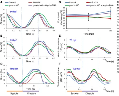
To assess contractile function, we analyze time-dependent changes in ventricular strain in terms of circumferential displace-ment (D) during cardiac cycles in response to gata1a MO and ErbB signaling inhibitor (AG1478) at 50 hpf, 75 hpf, and 100 hpf (Fig-ure 8, A–C). Treatment with AG1478 significantly reduced strain ceptor agonist, at 50 μM to increase myocardial contractility and
heart rate. However, neither Notch-related genes nor trabecular network was significantly affected (Figure 5, B and C, and ref. 35). Moreover, ISO treatment moderately reduced Notch activation. We further corroborated the effect of ISO in HAEC, where treat-ment at 24 hours resulted in reduced Notch signaling (Figure 5D).
Overexpression of Notch signaling induced abnormal ventricular morphogenesis. We showed that coinjection of Nrg1 mRNA (10 pg/
nl) with gata1a MO and Nrg1 mRNA alone engendered thickened ventricular wall and disrupted trabeculation (Figure 6A). Further-more, both treatments increased Notch signaling and Notch-related genes (Figure 6B). Zhao et al. reported that, while Notch signaling in both endo- and epicardium is considered important in cardiac regeneration in response to ventricular amputation, hyperactiva-tion of Notch signaling is found to suppress cardiomyocyte prolif-eration and heart regenprolif-eration in zebrafish (36). Therefore, our data also suggested that a well-defined level of Notch signaling is indi-cated as inducing trabeculation during cardiac morphogenesis.
Trabeculation contributed to contractile function. To
demon-strate the role of trabeculation on ventricular function, we quan-tified the volume of trabecular myocardial ridges in response to
[image:5.585.44.541.57.375.2]gata1a MO and tnnt2a MO injection and assessed the effects of
nontrabeculated myocardium (Supplemental Figure 4, A, C, and D). In the AG1478 group, trabeculation was absent, and the local strain was similar to that of the nontrabeculated regions of control zebrafish (Supplemental Figure 4, B–D).
The combination of reduced atrial contractility and absent active ventricular filling in the wea mutants resulted in a significant reduc-tion in ventricular volume (Table 1 and Supplemental Figure 5). Injec-tion of Nrg1 mRNA to the wea mutants at the 1- to 4-cell stage partially restored trabecular formation (Figure 2J) and partially improved the strain, stroke volume, and ventricular contraction (Supplemental during ventricular diastole at 100 hpf (Figure 8C), which was
[image:6.585.43.330.56.266.2]accompanied by a decreased fractional shortening (Figure 8D). Both gata1a MO and AG1478 treatment reduced ventricular EF, as assessed by the changes in 4D SPIM-acquired ventricular volume during the cardiac cycle (Figure 8, E and F). Both groups devel-oped an increased end systolic volume (ESV) and end diastolic vol-ume (EDV) (Table 1). Nrg1 mRNA rescue to gata1a MO injection normalized the ventricular strain, fractional shortening, and EF. Interestingly, trabeculated myocardium in the control zebrafish demonstrated higher local strain and local deformation than the
Figure 4. Endocardial Notch activation in
Tg(cmlc2:mCherry;tp1:gfp) zebrafish. (A) Tp1-gfp signal was present throughout the ventricle. (B) Following gata1a MO injection, Notch signaling was diminished in the endocar-dium, as demonstrated by reduced tp1 signals. (C) Nrg1 mRNA coinjection restored Notch signaling signals. (D–F) flk:mCherry transgene was used to delineate the endocardium. (G–I) The merged gfp and mCherry channels revealed endocardial Notch signaling. Scale bars: 50 μm. Original magnification, ×20.
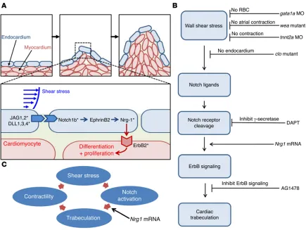
[image:6.585.90.500.440.693.2]Figure 5B). Thus, the reduced-contractile atrium in the wea mutants is associated with (a) an absence of hemodynamic force to generate active ventricular filling, (b) downregulation of Notch-related genes, and (c) absence of trabeculation to contribute to contractile function. Taken together, hemodynamic shear force–mediated trabeculation contributed to cardiac contractile function (Figure 9).
Discussion
The main contribution of integrating 4D SPIM technique with hemodynamic forces resides in the elucidation of the interplay between shear stress and Notch activation in initiating trabecula-tion during cardiac morphogenesis. By implementing the nongated 4D synchronization algorithm with SPIM, we have unraveled the trabecular network in association with ventricular contractile func-tion. While the development of trabeculation is conserved between the zebrafish and chick, mouse, and human embryos (3, 23, 37–39), there are numerous layers of cardiomyocytes in mice (23), and there is a thin layer of cardiomyocytes for trabeculation in zebra-fish embryos. For this reason, 4D SPIM imaging enables uniform illumination to uncover shear stress–induced endocardial Notch signaling to form a trabecular network for contractile function with high axial, spatial, and temporal resolution. We demonstrated that (a) gata1a MO decreased hematopoiesis to reduce shear stress, (b) tnnt2a MO inhibited ventricular contractile function to reduce hemodynamic shear forces, and (c) wea mutants lacked atrial
con-traction, resulting in a reduction of hemodynamic forces to the ven-tricle. All 3 conditions resulted in downregulation of Notch-related genes and attenuation of trabeculation. As a corollary, clo mutants lacked endocardium, resulting in the absence of trabeculation. Subjecting endothelial cells to PSS in the presence of the ADAM10 inhibitor corroborated that shear stress activated Notch signaling.
[image:7.585.41.400.51.421.2]The use of the Tg(flk:mCherry;tp1:gfp) line further localized and corroborated Notch signaling in endocardium (Figure 4). In addition, Notch activation was present beyond endocardium in both the WT and Nrg1 mRNA–rescued groups (Figure 4, A, C, G, and I). Notch1a is recognized as present in the endothelium dur-ing angiogenesis (40) and vascular development (41). Endothelial cells lining the coronary arteries communicate from endocardium to myocardium to epicardium (42). As development proceeds, the trabecular myocardium collapses toward the myocardial wall, forming a thick and compact ventricular wall (43). Recently, Tian et al. reported that trabecular compaction traps endocardial cells to mature into the inner-wall vasculature (44). Furthermore, Notch signaling in both endo- and epicardium is considered important in cardiac regeneration in response to ventricular amputation (36). In this context, we speculate that Notch signaling is present from endocardium to myocardium and epicardium via the coronary vasculature. Nevertheless, there remains a complicated role of Notch signaling beyond endocardium to activate Nrg1, and the precise mechanism remains to be investigated.
Figure 6. High-dose Nrg1 mRNA (10
μM) did not restore trabeculation in
the gata1a MO–injected zebrafish.
Current imaging techniques, including confocal microscopy, are limited by their intrinsic depth penetration, axial resolution, and long scanning time (45). Confocal microscopy is oftentimes limited from capturing the entire live zebrafish embryo and the beating hearts due to its small working distance for the objective lens and the long acquisition time. Digital particle image velocimetry (DPIV) is inherently limited in analyzing 3D cardiac mechanics due to its time-dependent 2D image (2D + time domain) and assumption needed for volumetric analysis. Images acquired with the conven-tional microscopy techniques incur (a) serious background noise due to out-of-focus illumination and (b) low axial resolution due to a large depth of field, whereas our SPIM-based imaging applies 2 sep-arate sets of lenses for illumination and detection through selective plane excitation via laser light sheet, which greatly reduces back-ground noise with the use of long working distance objectives (Sup-plemental Figure 2 and refs. 46, 47). Furthermore, the SPIM-based system is able to visualize the neonatal mouse hearts, following euthanasia and optical clearing (48). SPIM is superior to ultra–high frequency ultrasound (U-HFU) for its capability in tracking fluores-cently labeled proteins, cellular structures, and cells of interest with superior spatial resolution (0.6 μm for SPIM vs. ~30 μm for U-HFU) as well as deep penetration into tissues cleared by ClARITY (49, 50). Thus, we integrated 4D SPIM (3D + time domain) with a postimag-ing synchronization algorithm to address the irregular periodicity of zebrafish heart rhythms, thereby providing a basis to overcome time-dependent 3D CFD simulation (14). In this context, our SPIM system has advanced optical imaging for (a) deep axial resolution, (b) large dynamic ranges, (c) fast acquisition, and thus, (d) reduced photo-bleaching/toxicity (Supplemental Figure 1).
During heart development, the myocardium differentiates into 2 layers: an outer compact zone and an inner trabeculated zone. In the developing nonzebrafish embryonic hearts, trabeculation facil-itates oxygenation and nutrition to the myocardium and enhances cardiac contractile function (37). As development proceeds, the trabecular myocardium collapses toward the myocardial wall, forming a thick and compact ventricular wall (43). The formation of a multilayered spiral myocardium during the late fetal and neo-natal stage is essential for cardiac contractile function (51). A recent study of the highly trabeculated zebrafish heart demonstrates that
Nrg1 and ErbB2, in addition to their role in promoting cell
prolifera-tion, have another important function in regulating cardiomyocyte delamination to initiate ventricular trabeculation (23).
Biomechanical forces upregulate Notch ligands and receptors, and Notch upregulation establishes force-induced proteolysis as a mechanism of cellular mechanotransduction (52, 53). We dem-onstrated that PSS (23 dyne × cm–2 at 1 Hz for 24 hours)
upregu-lated Notch activation in HAEC in vitro (Figure 3D). We further recapitulated hemodynamic forces–mediated endocardial Notch activation by genetic manipulation of hematopoiesis (gata1a MO), cardiac contraction (tnnt2a Mo), atrial contraction (wea), endocar-dial endothelial deletion (clo mutants), and localization of Notch activation in endocardium (crossbreeding Tg[flk:mCherry] with
Tg[tp1:gfp] Notch reporter line). We further demonstrated that Nrg1 mRNA rescue restored trabeculation in association with an
improved ventricular strain and EF. Concomitant upregulation of Notch-related genes, including Dll4, Jag1, and Jag2, suggested that Nrg1 mRNA–restored trabeculation and contractile function is implicated in activation of Notch ligands (Jag1, Jag2, and Dll4) and receptor (Notch 1b) to promote proteolytic cleavage of Notch (52, 53). Masumura et al. have demonstrated that shear stress further induced time-dependent Notch signaling in murine embryonic stem cell–derived vascular endothelial cells (54). de la Pompa et al. reported a positive feedback loop between Notch ligands and target genes (55), and inactivation of Notch in embryonic endocar-dium results in a decrease in Dll4 expression (24, 56).
Notch activation by Dll1 or Dll4 in the endocardium results in the transcription of EphrinB2, which in turn regulates Nrg1 (57). As a secreted factor, Nrg1 signals to the adjacent cells to promote their differentiation into trabecular myocytes. In a parallel path-way, Notch activity in the endocardium activates BMP10 expres-sion in the adjacent myocytes to promote proliferation (57). Unlike mouse and chick development, ErbB2 contributes to both prolif-eration and differentiation in zebrafish cardiomyocytes to form a relatively thin layer of myocardium (23).
Nrg1 contributes to cardiac contractility (57, 58). Disruption of Nrg1 expression after ischemic insult impairs cardiac
contractil-ity (59), whereas Nrg1 preconditioning confers cardiac protection from ischemic injury (60). Nrg1 mRNA injection rescued
[image:8.585.49.377.52.242.2]Methods
4D cardiac SPIM imaging with synchronization algorithm. We have
integrated our in-house 4D SPIM imaging system with postprocess-ing synchronization (Supplemental Methods) to visualize the dynamic cardiac architecture with high axial resolution (Supplemental Figure 1). Using the SPIM technique, we scanned 300 sections from the ros-tral to the caudal end of the zebrafish heart. Each section was captured with 300 x-y planes (frames) at 10-ms exposure time per frame via a sCMOS camera (Hamamatsu Photonics). The thickness of the light
sheet was tuned to approximately 5 μm to provide a high axial (z axis)
resolution for adequate reconstruction of the 3D cardiac
morphol-ogy, and the Z scanning was set to 1 μm for lossless digital sampling
according to the Nyquist sampling principle. To synchronize with the cardiac cycle, we determined the cardiac periodicity on a frame-to-frame basis by comparing the pixel intensity from the smallest during peak systole to the largest ventricular volume during end diastole (61, 62). The reconstructed 4D images were processed by Amira software (Supplemental Video 8).
Zebrafish embryos. Zebrafish were bred and maintained at the
UCLA Core Facility, and experiments were performed in compli-ance with UCLA IACUC protocols (63). The transgenic Tg(cmlc2:gfp),
Tg(flk:mCherry;tp1:gfp), and Tg(cmlc2:mCherry;tp1:gfp) zebrafish lines
were used under the following conditions: (a) control (WT), (b) gata1a MO, and (c) tnnt2a MO microinjection (64). The gata1a MO reduced hematopoiesis and blood viscosity by 90%, as previously reported (16, 17). Fluid shear stress (τ) is characterized as the frictional force that acts tangentially on the surface of endothelial cells (65). For Newtonian
flu-ids, shear stress is defined as follows: τ = μ × du/dy, where μ represents
lation to restore cardiac strain (Figure 8). This gain of function is suggested by the increase in contractile function–mediated hemo-dynamic forces to activate Notch signaling (Figure 3). We further demonstrated that shear stress–activated Notch signaling is endo-cardium dependent. Coinjection of Nrg1 mRNA with gata1a MO restored both trabecular formation (Figure 2) and contractile func-tion (Figure 8). Although increasing Nrg1 mRNA injecfunc-tion from 5 to 10 pg/nl increased Notch-related gene expression by approxi-mately 2-fold, it led to abnormal ventricular morphogenesis, sug-gesting that a well-defined level of Notch signaling is required for trabeculation (Figure 6). In response to gata1a MO, tnnt2a MO,
wea mutation, or AG1478 treatment, Notch-related genes were
significantly downregulated, and trabeculation was attenuated or inhibited (Figures 2 and 3). However, in response to EPO mRNA– augmented shear stress, neither Notch-related genes nor trabecu-lation were altered (Figure 5).
gata1a MO and AG1478 treatment reduced strain and
fraction-al shortening, leading to decreased EF. Interestingly, both gata1a MO– and AG1478-treated embryos developed large ventricular volumes. However, the wea mutants developed small ventricles and low ventricular volumes (Table 1).
[image:9.585.39.448.54.384.2]Overall, we provide new mechanotransduction insights into shear stress modulation of cardiac trabeculation via endocardial Notch signaling (Figure 9). Interfacing 4D light-sheet imaging with the genetically engineered zebrafish system introduces a dynamic model to establish the significance of blood flow under-lying cardiac architecture and function with clinical relevance to noncompaction cardiomyopathy.
E3 medium at 30 hpf. Similarly, DAPT (Sigma-Aldrich) at 100 μM in 1% DMSO was diluted in E3 medium to inhibit Notch signaling at 40
hpf. GI254023X, an ADAM10 inhibitor at 5 μM, was added in HAEC
culture medium and incubated for 30 minutes. 5 μM of premixed
GI254023X was added to the culture medium for HAEC subject to PSS in the ensuing experiments. To increase shear stress in the
ven-tricular cavity, 50 μM of isoprenaline hydrochloride (DL-ISO) (I5627,
Sigma-Aldrich) was applied at 50 hpf, thereby increasing heart rate and contractility (35).
Blood shear stress modulation. Shear stress (τ) is characterized as dynamic viscosity (μ) of fluid multiplied by shear rate (r), defined as a gradient of velocity between 2 adjacent fluid layers (68).
viscosity, and du/dy is the velocity gradient along the y axis perpendicu-lar to the wall (66). In Newtonian fluid mechanics, reduction in viscos-ity (μ) by 90% resulted in a proportional reduction in wall shear stress (τ) (22, 67). The tnnt2a MO inhibited cardiac tnnt2a to prevent cardio-myocyte contraction, leading to noncontractile atrium and ventricle (18, 19). The clo mutants with specific deletion of endocardium were used to verify endocardium-dependent Notch signaling in response to shear stress. The noncontractile wea mutants (a gift from Deborah Yelon at UCSD) were used to inhibit the development of AV gradients. Tricaine mesylate (0.5%) was used to humanely sacrifice the embryos.
Chemical treatment to modulate trabeculation. ErbB signaling
[image:10.585.42.554.86.149.2]inhibitor AG1478 at 5 μM (Sigma-Aldrich) in 1% DMSO was diluted in
Table 1. Ventricular volume and EF in response to gata1a MO– and AG1478-treated zebrafish
Control AG1478 gata1a MO gata1a MO + Nrg1 mRNA wea wea + Nrg1 mRNA
EDV (105μm3) 5.5 ± 0.5 6.9 ± 0.5 8.5 ± 0.8 7.3 ± 0.6 0.8 ± 0.2 1.0 ± 0.2
ESV (105μm3) 1.5 ± 0.6 2.7 ± 0.4 2.9 ± 0.6 2.2 ± 0.6 0.7 ± 0.2 0.7 ± 0.1
Stroke volume (105μm3) 4.0 ± 0.5 4.2 ± 0.5 5.4 ± 0.8 5.2 ± 0.6 0.1 ± 0.1 0.3 ± 0.1
EF (%) 72.2 ± 4.2 60.8 ± 3.5 66.2 ± 3.1 69.8 ± 4.0 12.5 ± 2.8 30.0 ± 3.6
[image:10.585.45.480.356.683.2]employed Amira 3D software to reconstruct the cardiac volume. The ventricular volume without trabeculation was simulated by removing trabecular ridges. The volume of trabecular myocardial ridges was derived by subtracting the volume of smooth curve, ignoring trabecu-lar ridges from the total myocardial volume.
Stroke volume and EF. Based on SPIM images with nongated 4D
synchronization computational algorithm (61, 62), the time-depen-dent changes in ventricular chamber volume throughout the cardiac cycle were measured by Amira. ESV and EDV were obtained by deter-mining the ventricular volume during systole and diastole, respective-ly. EF was calculated using ESV and EDV (69).
Preparation of Nrg1 and EPO mRNA for rescue. Human Nrg1 cDNA
(a gift from William Talbot, Stanford University, Stanford, Califor-nia, USA) was amplified from a donor plasmid (purchased from GE
Health) and cloned into the plasmid pCS2+ at the BamHI and EcoRI
sites (Supplemental Figure 6A). Clones with the human Nrg1 cDNA insert were selected by PCR screening. Two clones with human Nrg1 cDNA insert were verified by transfecting the plasmids into HEK-293 cells, and Nrg1 protein expression was detected by Western blot with anti-Nrg1 antibody (Supplemental Figure 6B). mRNA was made from the clone 1 plasmid using the mMessage SP6 Kit (Invitrogen) follow-ing the manufacturer’s instructions. In vitro–transcribed Nrg1 mRNA was purified with Bio-Rad’s Total RNA Isolation Kit for in vivo rescue experiments. Zebrafish EPO cDNA was amplified from a donor
plas-mid and cloned into pCS2+ at the EcoRI and XhoI sites (Creative
Bio-gene). Zebrafish EPO mRNA was prepared and purified with the same procedure as described above.
Genes knocked down by morpholinos and rescued by Nrg1 mRNA.
MO (GeneTools) were designed against the ATG of gata1a (5′
-CTG-CAAGTGTAGTATTGAAGATGTC-3′) and tnnt2a (5′
-CGCGTGGA-CAGATTCAAGAGCCCTC-3′). Morpholinos were resuspended in
nuclease-free water and injected at 8 ng/nl and 4 ng/nl at 1- to 4-cell stages, respectively. Nrg1 mRNA at 5 or 10 pg/nl was coinjected at 1- to 4-cell stages with gata1a MO to overexpress Notch target genes and restore trabeculation. 20 pg/nl of EPO mRNA was also injected at the 1- to 4-cell stage to increase hematopoiesis (34).
Dynamic shear stress model. Confluent HAEC were exposed to PSS
at 23 dyn × cm–2 at 1 Hz at a slew rate (∂τ/∂t) of 71 dyn × cm–2s–1 for 24
hours (29) in a well-defined dynamic flow system (29). The pulsatile flow simulates hemodynamic shear stress in the arterial system (70, 71). Notch ligands (Dll4, Jag1, and Jag2) and target genes (hairy and enhancer of split [Hes]) were quantified by quantitative real-time PCR (qRT-PCR) as previously described (31).
Zebrafish heart RNA isolation for Notch ligands, receptor, and target gene expression. Zebrafish embryos were humanely sacrificed
by overdosing with tricaine methylate. The embryonic hearts were isolated under a dissecting microscope as previously described (72). Total RNA was isolated from the extracted hearts using Aurum Total RNA Mini Kit (Bio-Rad), and cDNA was synthesized using iScript cDNA Synthesis Kit (Bio-Rad). PCR primers for Notch ligands (Jag1 and Jag2, Dll4), receptor (Notch1b), and signaling related genes (Nrg1 and ErbB2) were designed (Supplemental Tables 2–4). The mRNA expression levels were determined by qRT-PCR and normalized to
zebrafish α-actin.
Statistics. All the values were expressed as mean ± SD. For statistical
comparisons between 2 experimental conditions, unpaired 2-tailed t test was used. P < 0.05 was considered significant. Comparisons of multiple
(Equation 1)
where (∂ux/∂y) is the tangential velocity gradient between 2 adjacent
fluid layers. Since shear stress is a function of velocity (∂ux), injection
of tnnt2a MO and use of wea mutants reduced ventricular contractil-ity, which in turn decreased shear stress to the endocardium. In addi-tion, we applied ISO treatment to increase heart rate and contractil-ity, thereby augmenting shear stress. Furthermore, shear stress is also
interpreted as time rate of momentum change (ṗ) as the rate of fluid
mass (ṁ) acting on the surface per unit area (A) as follows:
(Equation 2)
(Equation 3)
(Equation 4)
where ρ is the density of blood, ū is the average molecular speed,
⟨ux⟩ is the mean velocity along the x axis of fluid molecule hitting the
unit area, and λ is the mean free path defined as the average
travel-ing distance of movtravel-ing particles between collisions. Injecttravel-ing gata1a MO inhibited hematopoiesis to decrease the hematocrit, leading to a decrease in viscosity, whereas injecting EPO mRNA increased hema-topoiesis, leading to an increase in viscosity and shear stress to the endocardium (Supplemental Videos 7 and 8).
Quantification of strain and fractional shortening. To measure 2D
ventricular diameter change, we used SPIM imaging to follow the ventricular developmental stages at 50, 75, and 100 hpf via a sCMOS camera. The captured images were segmented to create the 2D mov-ing boundary conditions with 600 nodes (14). The nodes were guided to replicate cardiac wall motion captured by SPIM segmentation as described previously (14). Matlab (Mathworks) was employed to cal-culate the strain as defined by the time-dependent changes in dis-placement (D) between the individual time steps:
(Equation 5)
The changes in displacement between end diastole and systole were used to calculate the fractional shortening (69):
(Equation 6)
where Do denotes the initial displacement at time = 0, and D at time =
Δt. Accuracy of repetition error was tested by calculating the strain of
coefficient of variation at end diastole (Supplemental Table 1).
3D quantification of the volume of trabecular ridges. To
Acknowledgments
The authors would like to express gratitude to William Talbot from Stanford University for providing the human Nrg1 plasmid and to Deborah Yelon from UCSD for providing the wea mutants. In addi-tion, the authors also thank David Traver at UCSD and Nathan Law-son at the University of Massachusetts Medical School (Worcester, Massachusetts, USA) for generously providing the Tg(tp1:gfp) line. This study was supported by grants NIH HL118650 (to T.K. Hsiai), HL083015 (to T.K. Hsiai), HD069305 (to N.C. Chi and T.K. Hsiai.), HL111437 (to T.K. Hsiai and N.C. Chi), HL129727 (to T.K. Hsiai), T32HL007895 (to R.R. Sevag Packard), and American Heart Asso-ciation Pre-Doctoral Fellowship 15PRE21400019 (to J. Lee). Address correspondence to: Tzung K. Hsiai, Department of Medicine (Cardiology) and Bioengineering, University of California, Los Ange-les, 10833 Le Conte, CHS 17-054, Los AngeAnge-les, California 90095-1691, USA. Phone: 310.268.3839; E-mail: [email protected].
mean values were performed by 1-way ANOVA, and statistical signifi-cance among multiple groups was determined using Tukey’s method.
Study approval. Zebrafish were maintained in accordance with
UCLA Institutional Animal Care and Use Committee (IACUC) proto-cols under a project license also approved by the UCLA IACUC (ARC no. 2015-055).
Author contributions
JL and PF set up the SPIM system and imaging. JL, PF, RRSP, and TKH wrote the manuscript. JL and PF performed postimaging processing. JL, PF, HX, and RRSP performed 4D beating zebra-fish heart imaging and analysis. HK and RL prepared DNA clones and in vitro–transcribed RNAs. JL, HK, and KIB performed gene-expression studies. JL, KIB, and NJ performed in vitro experi-ments. JL, KIB, JC, and HY performed microinjections. CCJK, NCC, CMH, RL, and TKH designed, supervised, revised, and sup-ported the study.
1. Auman HJ, Coleman H, Riley HE, Olale F, Tsai HJ, Yelon D. Functional modulation of cardiac form through regionally confined cell shape changes. PLoS Biol. 2007;5(3):e53. 2. Hove JR, Koster RW, Forouhar AS,
Acevedo-Bolton G, Fraser SE, Gharib M. Intracardiac fluid forces are an essential epigenetic fac-tor for embryonic cardiogenesis. Nature. 2003;421(6919):172–177.
3. Chen H, et al. BMP10 is essential for maintain-ing cardiac growth durmaintain-ing murine cardiogenesis. Development. 2004;131(9):2219–2231. 4. Hove JR, Köster RW, Forouhar AS,
Acevedo-Bolton G, Fraser SE, Gharib M. Intracardiac fluid forces are an essential epigenetic fac-tor for embryonic cardiogenesis. Nature. 2003;421(6919):172–177.
5. Li J, et al. Piezo1 integration of vascular archi-tecture with physiological force. Nature. 2014;515(7526):279–282.
6. Banjo T, et al. Haemodynamically dependent valvulogenesis of zebrafish heart is mediated by flow-dependent expression of miR-21. Nat Com-mun. 2013;4:1978.
7. Ten Dijke P, Egorova AD, Goumans MJ, Poel-mann RE, Hierck BP. TGF-β signaling in endothelial-to-mesenchymal transition: the role of shear stress and primary cilia. Sci Signal. 2012;5(212):pt2.
8. Santhanakrishnan A, Miller LA. Fluid dynam-ics of heart development. Cell Biochem Biophys. 2011;61(1):1–22.
9. Lucitti JL, Jones EA, Huang C, Chen J, Fraser SE, Dickinson ME. Vascular remodeling of the mouse yolk sac requires hemodynamic force. Development. 2007;134(18):3317–3326. 10. Peshkovsky C, Totong R, Yelon D. Dependence
of cardiac trabeculation on neuregulin sig-naling and blood flow in zebrafish. Dev Dyn. 2011;240(2):446–456.
11. Zhang W, Chen H, Qu X, Chang CP, Shou W. Molecular mechanism of ventricular trabecula-tion/compaction and the pathogenesis of the left ventricular noncompaction cardiomyopathy (LVNC). Am J Med Genet C Semin Med Genet. 2013;163C(3):144–156.
12. Nugent AW, et al. The epidemiology of child-hood cardiomyopathy in Australia. N Engl J Med. 2003;348(17):1639–1646.
13. Hoedemaekers YM, et al. The importance of genetic counseling, DNA diagnostics, and cardiologic family screening in left ventricular noncompaction cardiomyopathy. Circ Cardiovasc Genet. 2010;3(3):232–239.
14. Lee J, et al. Moving domain computational fluid dynamics to interface with an embryonic model of cardiac morphogenesis. PLoS One. 2013;8(8):e72924.
15. Lawson ND, Weinstein BM. In vivo imaging of embryonic vascular development using trans-genic Zebrafish. Dev Biol. 2002;248(2):307–318. 16. Vermot J, et al. Reversing blood flows act
through klf2a to ensure normal valvulo-genesis in the developing heart. PLoS Biol. 2009;7(11):e1000246.
17. Galloway JL, Wingert RA, Thisse C, Thisse B, Zon LI. Loss of gata1 but not gata2 converts erythro-poiesis to myeloerythro-poiesis in zebrafish embryos. Dev Cell. 2005;8(1):109–116.
18. Arnaout R, et al. Zebrafish model for human long QT syndrome. Proc Natl Acad Sci U S A. 2007;104(27):11316–11321.
19. Chi NC, et al. Genetic and physiologic dissection of the vertebrate cardiac conduction system. PLoS Biol. 2008;6(5):e109.
20. Berdougo E, Coleman H, Lee DH, Stainier DY, Yelon D. Mutation of weak atrium/atrial myosin heavy chain disrupts atrial function and influ-ences ventricular morphogenesis in zebrafish. Development. 2003;130(24):6121–6129. 21. Hu N, Sedmera D, Yost HJ, Clark EB. Structure
and function of the developing zebrafish heart. Anat Rec. 2000;260(2):148–157.
22. Liu E, et al. [Hyperconjugation, characteristic infrared absorption of methylsulfones and crystal structures of selected aromaticsulfones]. Guang Pu Xue Yu Guang Pu Fen Xi. 2000;20(1):31–39. 23. Liu J, et al. A dual role for ErbB2
signal-ing in cardiac trabeculation. Development. 2010;137(22):3867–3875.
24. Grego-Bessa J, et al. Notch signaling is essential for ventricular chamber development. Dev Cell.
2007;12(3):415–429.
25. Parsons MJ, et al. Notch-responsive cells initiate the secondary transition in larval zebrafish pan-creas. Mech Dev. 2009;126(10):898–912. 26. Liao W, et al. The zebrafish gene cloche acts
upstream of a flk-1 homologue to regulate endothelial cell differentiation. Development. 1997;124(2):381–389.
27. Stainier DY, Weinstein BM, Detrich HW 3rd, Zon LI, Fishman MC. Cloche, an early acting zebrafish gene, is required by both the endothe-lial and hematopoietic lineages. Development. 1995;121(10):3141–3150.
28. Li R, et al. Disturbed flow induces autophagy, but impairs autophagic flux to perturb mito-chondrial homeostasis. Antioxid Redox Signal. 2015;23(15):1207–1219.
29. Jen N, et al. Atrial fibrillation pacing decreases intravascular shear stress in a New Zealand white rabbit model: implications in endothelial function. Biomech Model Mechanobiol. 2013;12(4):735–745. 30. Takabe W, et al. Oscillatory shear stress induces
mitochondrial superoxide production: impli-cation of NADPH oxidase and c-Jun NH2-terminal kinase signaling. Antioxid Redox Signal. 2011;15(5):1379–1388.
31. Li R, et al. Shear stress-activated Wnt-angiopoi-etin-2 signaling recapitulates vascular repair in zebrafish embryos. Arterioscler Thromb Vasc Biol. 2014;34(10):2268–2275.
32. Geling A, Steiner H, Willem M, Bally-Cuif L, Haass C. A gamma-secretase inhibitor blocks Notch signaling in vivo and causes a severe neurogenic phenotype in zebrafish. EMBO Rep. 2002;3(7):688–694.
33. Mumm JS, Kopan R. Notch signaling: from the outside in. Dev Biol. 2000;228(2):151–165. 34. Paffett-Lugassy N, et al. Functional conservation
of erythropoietin signaling in zebrafish. Blood. 2007;110(7):2718–2726.
35. De Luca E, et al. ZebraBeat: a flexible platform for the analysis of the cardiac rate in zebrafish embryos. Sci Rep. 2014;4:4898.
2014;111(4):1403–1408.
37. Sedmera D, Pexieder T, Vuillemin M, Thompson RP, Anderson RH. Developmental patterning of the myocardium. Anat Rec. 2000;258(4):319–337. 38. Chen H, Zhang W, Li D, Cordes TM, Mark Payne
R, Shou W. Analysis of ventricular hypertra-beculation and noncompaction using genetically engineered mouse models. Pediatr Cardiol. 2009;30(5):626–634.
39. Ben-Shachar G, Arcilla RA, Lucas RV, Manasek FJ. Ventricular trabeculations in the chick embryo heart and their contribution to ventricu-lar and muscuventricu-lar septal development. Circ Res. 1985;57(5):759–766.
40. Ramasamy SK, Kusumbe AP, Wang L, Adams RH. Endothelial Notch activity promotes angiogenesis and osteogenesis in bone. Nature. 2014;507(7492):376–380.
41. Hofmann JJ, Iruela-Arispe ML. Notch signaling in blood vessels: who is talking to whom about what? Circ Res. 2007;100(11):1556–1568. 42. Brutsaert DL. Cardiac endothelial-myocardial
signaling: its role in cardiac growth, contractile performance, and rhythmicity. Physiol Rev. 2003;83(1):59–115.
43. Risebro CA, Riley PR. Formation of the ventri-cles. ScientificWorldJournal. 2006;6:1862–1880. 44. Tian X, et al. Vessel formation. Science.
2014;345(6192):90–94.
45. Huisken J, Swoger J, Del Bene F, Wittbrodt J, Stelzer EH. Optical sectioning deep inside live embryos by selective plane illumination micros-copy. Science. 2004;305(5686):1007–1009. 46. Engelbrecht CJ, Stelzer EH. Resolution
enhance-ment in a light-sheet-based microscope (SPIM). Opt Lett. 2006;31(10):1477–1479.
47. Verveer PJ, Swoger J, Pampaloni F, Greger K, Marcello M, Stelzer EH. High-resolution three-dimensional imaging of large specimens with light sheet-based microscopy. Nat Methods. 2007;4(4):311–313.
48. Dodt HU, et al. Ultramicroscopy: three-dimension-al visuthree-dimension-alization of neuronthree-dimension-al networks in the whole
mouse brain. Nat Methods. 2007;4(4):331–336. 49. Chung K, Deisseroth K. CLARITY for
map-ping the nervous system. Nat Methods. 2013;10(6):508–513.
50. Yang B, et al. Single-cell phenotyping within transparent intact tissue through whole-body clearing. Cell. 2014;158(4):945–958. 51. Rumyantsev PP, Krylova MI. Ultrastructure of
myofibers and cells synthesizing DNA in the developing and regenerating lymph-heart mus-cles. Int Rev Cytol. 1990;120:1–52.
52. Shergill B, Meloty-Kapella L, Musse AA, Wein-master G, Botvinick E. Optical tweezers studies on Notch: single-molecule interaction strength is independent of ligand endocytosis. Dev Cell. 2012;22(6):1313–1320.
53. Gordon WR, et al. Mechanical allostery: evidence for a force requirement in the proteolytic activa-tion of Notch. Dev Cell. 2015;33(6):729–736. 54. Masumura T, Yamamoto K, Shimizu N, Obi S, Ando
J. Shear stress increases expression of the arterial endothelial marker ephrinB2 in murine ES cells via the VEGF-Notch signaling pathways. Arterioscler Thromb Vasc Biol. 2009;29(12):2125–2131. 55. de la Pompa JL, Epstein JA. Coordinating tissue
interactions: Notch signaling in cardiac develop-ment and disease. Dev Cell. 2012;22(2):244–254. 56. Timmerman LA, et al. Notch promotes
epithelial-mesenchymal transition during cardiac develop-ment and oncogenic transformation. Genes Dev. 2004;18(1):99–115.
57. High FA, Epstein JA. The multifaceted role of Notch in cardiac development and disease. Nat Rev Genet. 2008;9(1):49–61.
58. Ge W, Ren J. mTOR-STAT3-notch signalling contributes to ALDH2-induced protection against cardiac contractile dysfunction and autophagy under alcoholism. J Cell Mol Med. 2012;16(3):616–626.
59. Hedhli N, et al. Endothelium-derived neuregulin protects the heart against ischemic injury. Circu-lation. 2011;123(20):2254–2262.
60. Fang SJ, et al. Neuregulin-1 preconditioning
pro-tects the heart against ischemia/reperfusion inju-ry through a PI3K/Akt-dependent mechanism. Chin Med J (Engl). 2010;123(24):3597–3604. 61. Liebling M, Forouhar AS, Gharib M, Fraser SE,
Dickinson ME. Four-dimensional cardiac imag-ing in livimag-ing embryos via postacquisition synchro-nization of nongated slice sequences. J Biomed Opt. 2005;10(5):054001.
62. Mickoleit M, et al. High-resolution reconstruc-tion of the beating zebrafish heart. Nat Methods. 2014;11(9):919–922.
63. Westerfield M. The Zebrafish Book: A Guide For The Laboratory Use Of Zebrafish (Danio Rerio). Corvallis, Oregon, USA: University of Oregon Press; 2000.
64. Choi WY, et al. In vivo monitoring of cardio-myocyte proliferation to identify chemical modifiers of heart regeneration. Development. 2013;140(3):660–666.
65. Fung Y, Liu S. Elementary mechanics of the endothelium of blood vessels. J Biomech Eng. 1993;115(1):1–12.
66. Fung YC. Biomechanics: Motion, Flow, Stress, and Growth. New York, New York, USA: Springer; 1990. 67. Kay JM, Nedderman RM. Fluid Mechanics And
Transfer Processes. Cambridge, United Kingdom: Cambridge University Press; 1985.
68. Lee J, Packard RR, Hsiai TK. Blood flow modula-tion of vascular dynamics. Curr Opin Lipidol. 2015;26(5):376–383.
69. Mayet J, et al. Improvement in midwall myocar-dial shortening with regression of left ventricular hypertrophy. Hypertension. 2000;36(5):755–759. 70. Hwang J, et al. Pulsatile versus oscillatory shear
stress regulates NADPH oxidase subunit expres-sion: implication for native LDL oxidation. Circ Res. 2003;93(12):1225–1232.
71. Hsiai TK, et al. Monocyte recruitment to endo-thelial cells in response to oscillatory shear stress. FASEB J. 2003;17(12):1648–1657.