R E S E A R C H A R T I C L E
Open Access
Berberine inhibits NLRP3 Inflammasome
pathway in human triple-negative breast
cancer MDA-MB-231 cell
Mingjiang Yao
1,2†, Xiaodi Fan
1,2†, Bo Yuan
3, Norio Takagi
4, Sai Liu
1,2, Xiao Han
1,2, Junguo Ren
1,2and Jianxun Liu
1,2*Abstract
Background:Breast cancer is still the most common malignant tumor that threatens the female’s life in the world, especially triple-negative breast cancer (TNBC), one of the most difficult subtypes. Lack of targeted therapies brings about urgent demand for novel treatments. In this study we aim to investigate the anti-tumor activity of Berberine (BBR), a Chinese plant-derived alkaloid, against the TNBC cell line MDA-MB-231 and elucidate its mechanism referring to anti-inflammation.
Methods:Cell inhibition rate was measured by Cell Proliferation Assay, the cytotoxic effects was detected by Lactate dehydrogenase (LDH) leakage assay, the colony formation and migration potential were evaluated by colony formation assay and wound healing assay, the release of inflammatory cytokines was detected by EMD multifactor detection, and alterations of proteins and genes related to the NLR family pyrin domain containing 3 (NLRP3) inflammasome pathway were analyzed using western blotting and real-time Polymerase Chain Reaction (PCR). Results:BBR reduce the viability of MDA-MB-231 cells and increased the release of LDH from the cells in a dose-dependent manner, with and inhibition of colony formation potential and migration of the cells. BBR also caused a marked reduction in the secretion of proinflammatory cytokines, Interleukin-1α(IL-1α), Interleukin-1β(IL-1β), Interleukin-6 (IL-Interleukin-6), and tumor necrosis factor-α(TNF-α). Besides, a down-regulated behavior was observed with the expression of P2X purinoceptor 7 (P2X7), NLRP3, pro-1, apoptosis-associated speck-like protein containing a caspase-activation and recruitment domain (ASC), caspase-1 p20, Interleukin-18 (IL-18), IL-1βproteins and NLRP3, Caspase-1 and ASC mRNAs in the NLRP3 inflammasome cascade.
Conclusions:Our results confirmed that BBR can effectively affect both tumor outgrowth and spontaneous metastasis in TNBC, and that we identified a new mechanism associated with inhibition the NLRP3 inflammasome pathway, suggesting its potential therapeutic relevance in clinical use.
Keywords:Berberine, Triple-negative breast cancer, NLRP3 inflammasome, Anti-inflammation
Background
Breast cancer is reported to be one of the most prevalent malignancies among women throughout the world, with a youth oriented tendency and continuously increasing mortality rate every year [1–3]. Despite considerable
developments in anti-cancer therapies such as hormone therapy, chemotherapy, and targeted therapy that have improved the outcomes of breast cancer patients dis-tinctly [4], there are still hurdles and challenges we should face in order to promote therapeutic efficacy, overcome side-effects and drug resistance, especially in triple-negative breast cancer (TNBC), a very aggressive subtype of breast cancer, possesses less than 30% of five-year survival rate in cases with metastasis [5,6]. Herein, looking for efficient and safer therapeutic agents and thus investigate the pharmacological mechanism is critically needed for the treatment of TNBC.
© The Author(s). 2019Open AccessThis article is distributed under the terms of the Creative Commons Attribution 4.0 International License (http://creativecommons.org/licenses/by/4.0/), which permits unrestricted use, distribution, and reproduction in any medium, provided you give appropriate credit to the original author(s) and the source, provide a link to the Creative Commons license, and indicate if changes were made. The Creative Commons Public Domain Dedication waiver (http://creativecommons.org/publicdomain/zero/1.0/) applies to the data made available in this article, unless otherwise stated.
* Correspondence:liujx0324@sina.com
†Mingjiang Yao and Xiaodi Fan contributed equally to this work.
1Xiyuan Hospital of China Academy of Chinese Medical Sciences, Institute of Basic Medical Sciences, No.1 Xiyuan Caochang, Haidian District, Beijing 100091, China
2Key Laboratory of Pharmacology of Chinese Materia Medica, Beijing 100091, China
Accumulating data indicate that inflammation is closely related with tumor initiation, progression and metastasis, and to a great extent due to its participating in the interaction between malignant cells and their microenvironment [7,8]. On the one hand, there would be a series of pro-inflammatory mediators, for instance, the cytokines, chemokines and transcriptional factors se-creted from the cancerous cells, on the other hand, these critical inflammation-related components, in turn, were proved to be the core molecular players in regulating signaling pathways and processes involved in oncogen-esis [9]. Thus, regulation of inflammatory reaction is a promising anti-tumor strategy that should be paid more attention to.
The NLR family pyrin domain containing 3 (NLRP3) inflammasome, composed by NLRP3 oligomers and apoptosis-associated speck-like (ASC) adapter protein, is a key innate immune pathway through triggering the ac-tivation of caspase-1, leading to the processing of inter-leukin-1 beta (IL-1β) and IL-18, as well as inducing of pyroptosis and malignant transformation [10, 11], and have been proved to take part in the genesis and devel-opment of several inflammatory disorders, including in-flammatory bowel disease (IBD) [12], pancreatitis [13], and may also increase the risk of cancer [14,15]. There-fore, targeting the NLRP3 inflammasome pathway has opened the door for novel strategy in cancer prevention and treatment.
Berberine (BBR) is a kind of organic heteropentacyclic compound and natural isoquinoline alkaloid isolated from several traditional Chinese herbal plants as Coptis chinensis (Huanglian)and Cortex Phellodendri Chinensis (Huangbai), which have been used to treat gastroenter-itis caused diarrhea for more than one thousand years. BBR is an odorless yellow crystalline powder with char-acteristic alkaloid bitterness. Because of its poor solubil-ity in water, the chloride or sulfate of BBR are more commonly used in practice due to the relatively water-soluble of the salt form [16]. A substantial body of evidence supports the multiple novel biological roles of BBR in anti-pathogenic microorganism [17], anti-diabetes [18], cerebrovascular protection [19, 20], and blood lipid regulation [21], among which the anti-in-flammation effect is considered to be the most represen-tative one and its molecule mechanisms have been revealed referring to certain signaling pathways and in-flammatory factors [22, 23]. In our previous study [23], BBR has been found to inhibit oxidized low-density lipo-protein (ox-LDL) induced inflammation in J774A.1 cells by activating autophagy via the 5′ AMP-activated pro-tein kinase (AMPK) / mechanistic Target of Rapamycin (mTOR) signaling pathway, also further proved the anti-inflammation effect of BBR. Recently, the anti-tumor ac-tivities of BBR have been revealed in various human
cancer cells [24–26], and a broad range of mechanism are involved as inducing autophagy, apoptosis [27, 28], generation of reactive oxygen species [29] and cell cycle arrest [28]. Moreover, several studies have revealed that BBR could regulate NLRP3 inflammasome pathway in different types of macrophages, such as THP-1, J774A.1 and RAW264.7 [30–32]. However, whether BBR can affect the human breast cancer cell through inflamma-tion related pathways has rarely been investigated before.
In this study, we identified a new potential mechanism by which BBR inhibits the growth of human breast cell line MDA-MB-231 associated with inhibition of the NLRP3 inflammasome pathway. This suggests that BBR may regulate inflammation in breast cancer, and could therefore have potential therapeutic relevance.
Materials and methods Materials
Berberine Hydrochloride was purchased from National Institute for Food and Drug Control (China, Lot: 110713–201,212), dissolved in distilled water and filtered by a 0.22μm filter. DMEM medium and Fetal Bovine Serum (FBS) were purchased from GIBCO® by Life Technologies (Carlsbad, CA, USA). The CellTiter 96® AQueous One Solution Cell Proliferation Assay (MTS) (Lot: 0000185040) was purchased from Promega Corp. (Madison, WI, USA). The LDH Cytotoxicity Assay Kit (Lot: 072418181030) was acquired from Beyotime Biotechnology Co., Ltd. (Shanghai, China). The Giemsa stain solution was provided by Solarbio Life Sciences (Beijing, China). The Cytokine Magnetic Bead assay kit (#RECYTMAG-65 K) was purchased from Millipore Corp. (Billerica, MA, USA). Antibodies used in this study were P2X7 from Bioss (Beijing, China); pro-Cas-pase-1, ASC, NLRP3, Caspase-1 p20 and IL-18 from Wanleibio (Shenyang, Liaoning, China); IL-1β from Proteintech (Wuhan, Hubei, China); β-actin from Cell Signaling Technology (Boston, MA, USA). ReverTra Ace qPCR RT Master Mix were purchased from Toyobo CO., LTD. (Osaka, Japan). TRIzol Reagent and ABI Power SYBR Green PCR Master Mix were purchased from Thermo Fisher Scientific, China (Shanghai, China). Primers used in this study were NLRP3, Caspase-1, and IL-1β, GAPDH from iGenebio CO., LTD. (Guangzhou, Guangdong, China).
Cell culture and BBR treatment
air) and allowed to attach for 24 h. Then the cells were treated with indicated concentrations of BBR for another 48 h.
Cell viability assay
After treatment with various concentrations of BBR (2.5, 5, 10, 20, 40, 60, 80, 100μg/ml) for 48 h, the cell viability was measured by the CellTiter 96® AQueous One Solu-tion Cell ProliferaSolu-tion Assay (MTS) according to the method previously described with mild modification [33]. Briefly, the MTS solution was added into 5 times volume of serum-free medium to get reaction solution, the supernatants in each well was discarded and the cells were gently rinsed once by phosphate-buffered saline (PBS), and then 120μl of reaction solution was added into each well, after incubation at 37 °C for 2 h, the ab-sorbance at 490 nm was measured with a microplate reader (Synergy™ 4, BioTek). The relative cell viability was expressed as the ratio of the absorbance of each treatment group against those of the corresponding un-treated control group. The results were representative of three independent experiments and the IC50 values of
BBR were calculated by GraphPad Prism® 6 software.
Lactate dehydrogenase (LDH) cytotoxicity assay
MDA-MB-231 cells were cultured in 96-well plates, and the culture medium containing various concentration of BBR (10, 20, 40μg/ml) were added to each well and in-cubated for 48 h. Then LDH leakage was measured using Cytotoxicity Detection Kit according to the manufac-turer’s procedure. Briefly, 120μl culture supernatants were collected from each well and then 60μl freshly prepared reaction buffer supplied in the kit was added. 30 min after gentle mixing at room temperature, the ab-sorbance at 490 nm for each sample was measured using a microplate reader (Synergy™4, BioTek). The release of LDH was represented as the folds of control group. The results were from three independent experiments.
Colony formation assay
The reproductive viability of MDA-MB-231 cells was measured by colony formation assay as described previ-ously [34, 35]. Briefly, cells were seeded into 6-well plates with the density of 1000cells/well, 24 h after cells had attached, the medium was changed by different con-centrations of BBR contained medium and treated for another 48 h, then the medium was again replaced by fresh drug free medium and followed by another 12 days at 37 °C in a humidified atmosphere (5% CO2 in air).
After that, the colonies were fixed with 100% methanol for 2 min and stained with Giemsa stain solution for 20 min in room temperature. Stained cell colonies in each well were flat scanned and analyzed, and the results were representative of three independent experiments.
Wound healing assay
An in vitro wound healing assay was performed to evalu-ate the effect of BBR on cell migration followed by the methods previously described [36]. Briefly, MDA-MB-231 cells were seeded at a density of 1 × 10⁵cells/ml in 24-well plates and allowed to form a confluent mono-layer after 24 h. The mono-layer of cells was then scraped with a 20-200μL micropipette tip to create a wound of about 500μm width, and then the cells were gently rinsed twice by PBS, followed by the treatment with indicated concentrations of BBR for 48 h. The cells were photo-graphed at 0 h, 24 h and 48 h using an inverted micro-scope (IX51, Olympus, Japan) mounted with a digital camera (E330, Olympus, Japan). The distance between the edges of the cell-free areas was measured and the cell migration was calculated using the following equa-tion: %R = [1-(wound length at T24h (or 48h)/wound
length at T0h)] × 100% where %R is the recovery
percent-age, T0h is the wound length at 0 h, T24hand T48his the
wound length at 24 h and 48 h after injury, respectively. The results were representative of three independent experiments.
Inflammatory cytokines detection
The concentration of cytokines, TNF-α, IL-1α, IL-1β, and IL-6, in the supernatant of MDA-MB-231 cells after 48 h of BBR treatment was quantified by the EMD Milli-pore’s MILLIPLEX MAP Human Cytokine/Chemokine Magnetic Bead assay according to the manufacturer’s instructions. The median fluorescence intensity was assayed on a FLEXMAP 3D™system, and a five-parameter logistic method was performed to obtain the fitting curve and thus calculate the concentration of cytokines in the supernatant samples.
Quantitative real-time PCR
TGCCCAAGTTTGAAGG-3′ and reverse 5′- AGCATC ATCCTCAAACTCTTCTG-3′), and IL-1β (forward 5′ -TCTGTACCTGTCCTGCGTGT-3′and reverse 5′-ACTG GGCAGACTCAAATTCC-3′). The amplification condi-tions were: 10 min at 95 °C for initial denaturation; 40 reac-tion cycles including a denaturareac-tion step at 95 °C for 15 s with an annealing/extension step at 60 °C for 1 min; followed with an instrument preset melting curve analysis process. A fold change in relative expression of target gene was calculated using the comparative Ct (2-ΔΔCt) method.
Western blot analysis
Total protein extraction and western blot analysis meet the previously described methods [35]. The collected cell pellets of each treated group were lysed in Leammli buf-fer with protease inhibitor, and then were centrifuged at 12000 rpm for 12 min at 4 °C. The supernatant was then extracted, of which the concentration was determined by Bradford (BIO-RAD, USA). Whole cell lysate were elec-trophoresed on 10% or 12% SDS-PAGE, and then the proteins were transferred to a polyvinylidene difluoride (PVDF) membrane (Millipore Corp, MA, USA), the levels of targeted proteins were detected using the fol-lowing primary antibodies and dilution ratios: P2X7 (1: 1000), NLRP3(1:500), pro-caspase-1(1:500), ASC (1:500), caspase-1 p20(1:500), IL-18(1:500), IL-1β(1:1000), and β-actin (1:2000). Blotted protein bands were detected with respective horseradish peroxidase-conjugated secondary antibody (1:20000) and an enhanced chemiluminescence (ECL) reagent (Thermo Fisher Scientific Inc., USA). Im-ages were generated using ChemiDoc XRS+ System (BIO-RAD, USA). The visualized protein bands were quantified with Gel-Pro 4.0 software.
Statistical analysis
All experiments were performed at least three times, the data were represented as means ± SD, and the statistical significance was analyzed by SPSS 19.0 using one-way ANOVA followed with Tukey’s Post Hoc analysis, the level ofp< 0.05 was considered significant.
Results
Antiproliferative and cytotoxic effects of BBR
First, we examined the growth inhibitory effects of BBR using the MTS cell proliferation assay in human breast cancer cell line MDA-MB-231. A significant decrease in cell viability was observed after treatment with various concentrations of BBR (2.5, 5, 10, 20, 40, 60, 80, 100μg/ ml) for 48 h in a dose-dependent manner (96.98 ± 6.98%, 99.80 ± 3.69% 93.05 ± 6.38%, 81.43 ± 7.09%, 47.47 ± 6.79%, 32.38 ± 1.84%, 22.20 ± 2.75% and 13.67 ± 1.07%, respectively) with an IC50 of 40.06μg/ml (Fig. 1a). We
further evaluated the cytotoxic activity of BBR using LDH measurement assay. An increased LDH leakage in
cell culture medium with increasing concentration of BBR was observed (1.00 ± 0.08 VS 1.42 ± 0.11, 1.54 ± 0.14, and 2.16 ± 0.24, respectively) (Fig. 1b), indicating BBR damaged the integrity of cell plasma membrane.
BBR decreases colony formation potentials of MDA-MB-231 cells
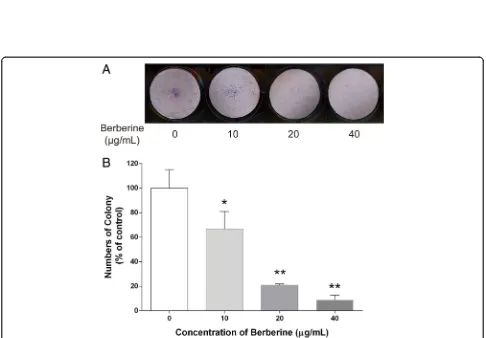
The ability of cultured cancer cells to proliferate and divide into groups to form colony was considered to have the potential of causing cancer. To explore whether exposure to BBR could suppress the surviving fraction of MDA-MB-231 cells, colony formation assay was con-ducted. Our results demonstrated that treatments with 10, 20, and 40μg/ml of BBR significantly reduced the colony numbers of MDA-MB-231 cells by 33.35, 79.23, and 91.24%, respectively (Fig.2a, b).
BBR suppresses the migration of MDA-MB-231 cells
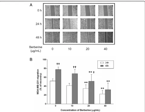
We next examined the effect of BBR on cell migration by performing wound healing assay. Our results showed the gaps scraped by a 20-200μl micropipette tip that re-main unfiled by the migrated cells in the 20 and 40μg/ ml BBR-treated groups were significantly wider than those of the untreated group at 24 h (34.98 ± 8.31% and 22.41 ± 8.52%) and 48 h (51.10 ± 15.45% and 32.12 ± 14.84%), furthermore, the gaps in the control group as well as in the 10 and 20μg/ml BBR-treated groups at 48 h (68.48 ± 9.33% and 51.10 ± 15.45%) were significantly wider than those at 24 h (41.60 ± 4.72% and 34.98 ± 8.31%), respectively (Fig.3a, b).
BBR reduces secretion of inflammatory cytokines from MDA-MB-231 cells
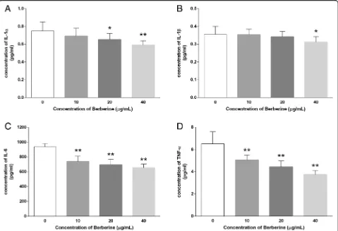
Concentrations of inflammatory cytokines secreted into the supernatant reflected the extent of the inflammatory response. After treatment with various concentrations of BBR (0, 10, 20, 400μg/ml) for 48 h, the supernatant was collected and analyzed by the Cytokine Magnetic Bead assay to examine whether the treatments affect the release of inflammatory cytokines. As shown in Fig.4, the level of IL-1α (0.75 ± 0.10 VS 0.69 ± 0.09, 0.65 ± 0.07 and 0.59 ± 0.05 pg/mL), IL-1β(0.36 ± 0.04 VS 0.35 ± 0.03, 0.34 ± 0.03 and 0.31 ± 0.03 pg/mL), IL-6 (938.9 ± 43.26 VS 739.1 ± 71.88, 693.8 ± 75.63 and 652.4 ± 51.13 pg/mL), and TNF-α (6.51 ± 1.08 VS 5.06 ± 0.43, 4.43 ± 0.56 and 3.74 ± 0.36 pg/ mL) in the supernatant of MDA-MB-231 cells were re-duced in a dose-dependent manner, respectively, indicat-ing the suppressive effect by BBR treatment against excessive secretion of inflammatory cytokines.
BBR regulates the expression profile of NLRP3 inflammasome related gene in MDA-MB-231 cells
Fig. 1Effect of BBR on MDA-MB-231 viability and LDH release. Cells were seeded in 96-well plates at 5000 cells per well and treated with various concentrations of BBR for 48 h, the control wells were treated with equivalent amount of medium alone. The effect of BBR on MDA-MB-231 (a) viability and (b) LDH release was determined by the assays described in the Methods section. The relative cell viability and LDH leakage was calculated as the ratio of the absorbance at 490 nm of each treatment group against those of the corresponding untreated control group. Each value represents the mean ± S.D. (n= 6) of three independent experiments. *p< 0.05, **p< 0.01, compared to the control
Fig. 2Effect of BBR on colony formation potential of MDA-MB-231 cells. Cells were seeded in 6-well plates at 1000 cells per well following treatment with indicated concentrations of BBR for 24 h. The medium was then replaced with fresh media, and the cells were allowed to grow for 12 days at 37 °C in a humidified atmosphere (5% CO2in air) before staining with Giemsa stain solution.aTypical results of the clonogenic
[image:5.595.57.545.88.255.2] [image:5.595.55.541.335.673.2]immunotherapy [3, 10, 11, 14], gene expression of re-lated targets in the NLRP3 cascade was detected by real-time PCR to clarify the mechanisms underlying this signaling pathway in MDA-MB-231 cells after treatment with BBR (0, 10, 20, 400μg/ml) for 48 h. As shown in Fig. 5, the expression profile of NLRP3 inflammasome related gene, NLRP3 mRNA (1.00 ± 0.10 VS 0.98 ± 0.14, 0.71 ± 0.33 and 0.27 ± 0.01), Caspase-1 mRNA (1.00 ± 0.05 VS 1.11 ± 0.31, 0.90 ± 0.19 and 0.61 ± 0.08), and IL-1β mRNA (1.00 ± 0.05 VS 0.83 ± 0.08, 0.59 ± 0.05 and 0.09 ± 0.01), were significantly suppressed in a dose-dependent manner, and glyceraldehyde-3-phosphate dehydrogenase (GAPDH) was served as an endogenous control to normalize the expression. Results were representative of three biological replicates and three technical duplications.
BBR modulates the NLRP3 inflammasome signaling in MDA-MB-231 cells
Consistent with that observed in the gene expression profile, after treatment with various concentrations of BBR (0, 10, 20, 400μg/ml) for 48 h, the expression level of P2X7, considered as sensor of cell damage and a trig-ger of the NLRP3 inflammasome [38], was significantly down-regulated by BBR in a dose-dependent manner (1.00 ± 0.00 VS 0.68 ± 0.11, 0.59 ± 0.08 and 0.36 ± 0.12). Subsequently, the expressions of major components of NLRP3 inflammasome complex, NLRP3 (1.00 ± 0.00 VS 0.99 ± 0.15, 0.77 ± 0.33 and 0.41 ± 0.28), pro-caspase-1 (1.00 ± 0.00 VS 0.62 ± 0.14, 0.55 ± 0.26 and 0.29 ± 0.13) and ASC (1.00 ± 0.00 VS 0.58 ± 0.16, 0.44 ± 0.27 and 0.12 ± 0.10), were remarkably down-regulated by BBR. Fur-thermore, the expressions of caspase-1 p20 (1.00 ± 0.00 VS
Fig. 3Effect of BBR on MDA-MB-231 cell migration. Cells were seeded into 12-well plates at 1 × 10⁵cells per well and cultured to near confluence. The wounded monolayer was incubated in culture medium containing various concentrations of BBR for 24 and 48 h.aRepresentative images of wound healing assays for cells treated with various concentrations of BBR, for 24 and 48 h.bThe percent recovery as determined in the scratch wound healing assay. Data are presented as the mean ± S.D. (n= 3) of three independent experiments. *,p< 0.01, compared to the control;†,p< 0.05,
[image:6.595.59.538.85.454.2]0.45 ± 0.23, 0.40 ± 0.31 and 0.05 ± 0.03), IL-18 (1.00 ± 0.00 VS 0.51 ± 0.21, 0.18 ± 0.06 and 0.15 ± 0.06) and IL-1β (1.00 ± 0.00 VS 0.66 ± 0.02, 0.55 ± 0.03 and 0.06 ± 0.03) proteins were also decreased after treating with BBR (Fig.6). These results indicated that BBR probably inhibits P2X7-mediated NLRP3 inflammasome activation.
Discussion
It has been reported that BBR, single use or in combin-ation with other compound, contributes to the inhibition of breast cancer cells, and the mechanism mainly focus on inducing apoptosis though mitochondrial or caspase-dependent pathway [29, 39–42]. The main purpose of
Fig. 4Effect of BBR on inflammatory cytokines in the supernatant of MDA-MB-231 cells. Cells were seeded into 12-well plates at 1 × 10⁵cells
per well following treatment with indicated concentrations of BBR for 48 h, and the supernatant of MDA-MB-231 cells was collected and the cytokines (a) IL-1α, (b) IL-1β, (c) IL-6, and (d) TNF-αwere analyzed. Data were presented as mean ± S.D. of six duplicated wells. *p< 0.05, **p< 0.01, compared to the control
[image:7.595.59.538.88.415.2] [image:7.595.59.540.572.684.2]present work was to investigate whether other mechan-ism, especially the anti-inflammatory effect, was involved in the anti-tumor activity of BBR. Our results clearly in-dicated that BBR dose-dependently reduce the viability and increase the LDH leakage of MDA-MB-231 cells after 48 h exposure (Fig.1a, b). In addition, inhibition of colony formation potential and migration of the cells by treatments with different concentrations of BBR have also been observed in this study (Figs.2and3), these re-sults were in the tendencies consistent with previous studies about anti-tumor activity of BBR on the same TNBC cell line [39, 40], although there was slightly difference in the efficacy of BBR. Our results suggested that BBR can effectively affect both tumor proliferation and spontaneous metastasis, and the mechanism is ne-cessarily to be further explored.
In recent years, inflammation has been reported as one of the major risk factors for the development and metastatic process of breast cancer [7, 8], thus anti-in-flammation treatment should be a novel strategy against breast cancer. It has been reported that treatment with
nonsteroidal anti-inflammatory drugs (NSAIDs), including aspirin and ibuprofen, is bound up with a reduced risk of breast cancer, though the biological mechanisms remain to be elucidated [43]. Celecoxib, a cyclooxygenase-2 (COX-2) selective inhibitor, also exerted anti-tumor effect in primary breast cancer tissue during a clinical trial [44]. As one of the most classical anti-inflammatory agents originated from Chinese Medicine, and although the anti-cancer effects have been proven, however, the insight mechanisms of BBR in inhibiting inflammatory response were still not well known on breast cancer. Therefore, we then focused on the effect of BBR on the release of inflam-matory cytokines and the expression of proteins and mRNAs in the NLRP3 Inflammasome signaling pathways in triple-negative breast cancer cell line MDA-MB-231.
Uncontrolled and sustained generation of cytokines may affect the growth, differentiation and apoptosis of cells [9]. Both IL-6 and TNF-αare considered to be the best characterized tumorigenic cytokines that involved in the promotion, progression and metastasis of tumor [45], and also, co-expression of these two cytokines
[image:8.595.58.538.88.410.2]determines the extension and outcome of breast cancer [46]. The IL-1 family was reported to preferentially ex-press in TNBC and be involved in the development of breast cancer, while inhibition of interleukin 1 receptor (IL-1R) affected the proliferation, prevented the tumor progression and metastasis [47, 48]. More critically, IL-1β is the product of self-activated caspase-1 derived from NLRP3 inflammasome formation. Our results dem-onstrated that BBR caused a marked reduction in the se-cretion of proinflammatory cytokines that implicated in carcinogenesis, such as IL-1α, IL-1β, IL-6, and TNF-α, from MDA-MB-231 cells (Fig. 4), indicating that BBR treatment inhibited the maturation and secretion of cy-tokines and changed the tumor microenvironment.
Besides, a down-regulated behavior was observed with the expression of mRNAs and proteins in the inflamma-some cascade in MDA-MB-231 cells treated with differ-ent concdiffer-entrations of BBR. The P2X7 receptor (P2X7R), trigger of the NLRP3 inflammasome, was pivotal in the cancer cell invasion associated with metastasis; there-upon by antagonizing the P2X7R specifically could in-hibit the invasiveness of human cancer cell [49]. In previous studies [32], BBR was reported to interfere with the P2X7 signaling in a methionine- and choline-defi-cient diet induced mouse liver injury model. Our results found that BBR could significantly down-regulate the ex-pression of P2X7 in MDA-MB-231 cells (Fig.6, a and c), confirming its ability in inhibiting the formation of inflammasome as well as tumor metastasis. Several stud-ies have elucidated the relationship between NLRP3 inflammasome signaling and carcinogenesis [15,50], that NLRP3 inflammasome inhibition is responsible for can-cer prevention. Our results revealed that the mRNA and protein expression of Nod-like receptor protein NLRP3, ASC and pro-caspase-1, components in the multiprotein platform of inflammasome, were down-regulated after BBR treatment (Figs. 5 a and6, a, d, e and f), and then directly resulted in the decreased activity of caspase-1 p20 and low expression of IL-1βand IL-18 (Figs. 5 b, c and 6, b, g, h and i). These findings suggested the BBR treatment associated with inhibiting the NLRP3 inflam-masome signaling pathway and prevent the process of tumor development at both the mRNA and protein levels.
Conclusion
In conclusion, we confirmed the cytotoxic capability of BBR on TNBC MDA-MB-231, manifested as de-creased cell growth, LDH releasing, cloning formation ability. Concomitantly, the migration capability of MDA-MB-231 cells was decreased by BBR. More crit-ically, we identified a new potential mechanism asso-ciated with the NLRP3 inflammasome pathway inhibition. Our current study revealed that BBR may
exert the inhibition of cell growth and migration in breast cancer associated with the regulation of inflam-masome pathway, and could therefore have potential clinical therapeutic relevance. However, the definite mechanism of BBR on breast cancer should be further investigated.
Abbreviations
AMPK:5′AMP-activated protein kinase; ASC: Apoptosis-associated speck-like protein containing a caspase-activation and recruitment domain;
BBR: Berberine; COX-2: Cyclooxygenase-2; DMEM: Dulbecco’s Modified Eagle Medium; FBS: Fetal bovine serum; GAPDH: Glyceraldehyde-3-phosphate dehydrogenase; IBD: Inflammatory bowel disease; 18: Interleukin-18; IL-1α: Interleukin-1α; IL-1β: Interleukin-1β; IL-6: Interleukin-6; LDH: lactate dehydrogenase; mTOR: Mechanistic target of rapamycin; NLRP3: Nucleotide-binding oligomerization domain-like receptor containing pyrin domain 3; NSAIDs: Nonsteroidal anti-inflammatory drugs; ox-LDL: Oxidized low-density lipoprotein; P2X7: P2X purinoceptor 7; PBS: Phosphate-buffered saline; PCR: Polymerase chain reaction; TNBC: Triple-negative breast cancer; TNF-α: Tumor necrosis factor-α
Acknowledgements
Not applicable.
Authors’contributions
MJY, XDF, BY designed the study; MJY, XDF, SL and XH performed the experiments; MJY, BY, NT, JGR and JXL contributed to manuscript preparation. All authors have read and approved the final manuscript.
Funding
This study was supported by National Natural Science Foundation of China (No.81403141, No.81873040) and Nursery Project of Xiyuan Hospital, CACMS (No.XYKY-MP2013–33). The funding bodies have no role in the design of the study and collection, analysis, and interpretation of data and in writing the manuscript.
Availability of data and materials
The datasets used and/or analyzed during the current study available from the corresponding author on reasonable request.
Ethics approval and consent to participate
Not applicable.
Consent for publication
Not applicable.
Competing interests
The authors declare that they have no competing interests.
Author details
1
Xiyuan Hospital of China Academy of Chinese Medical Sciences, Institute of Basic Medical Sciences, No.1 Xiyuan Caochang, Haidian District, Beijing 100091, China.2Key Laboratory of Pharmacology of Chinese Materia Medica, Beijing 100091, China.3Laboratory of Pharmacology, School of Pharmacy, Faculty of Pharmacy and Pharmaceutical Sciences, Josai University, 1-1 Keyakidai, Sakado, Saitama 350-0295, Japan.4Department of Applied Biochemistry, School of Pharmacy, Tokyo University of Pharmacy & Life Sciences, 1432-1 Horinouchi, Hachioji, Tokyo 192-0392, Japan.
Received: 4 May 2019 Accepted: 23 July 2019
References
1. DeSantis C, Ma J, Bryan L, Jemal A. Breast cancer statistics, 2013. CA Cancer J Clin. 2014;64(1):52–62.
2. Global Burden of Disease Cancer C, Fitzmaurice C, Dicker D, Pain A, Hamavid H, Moradi-Lakeh M, MacIntyre MF, Allen C, Hansen G, Woodbrook R, et al. The global burden of Cancer 2013. JAMA Oncol. 2015;1(4):505–27. 3. Guo B, Fu S, Zhang J, Liu B, Li Z. Targeting inflammasome/IL-1 pathways for
4. Siegel R, DeSantis C, Virgo K, Stein K, Mariotto A, Smith T, Cooper D, Gansler T, Lerro C, Fedewa S, et al. Cancer treatment and survivorship statistics, 2012. CA Cancer J Clin. 2012;62(4):220–41.
5. Thummuri D, Kumar S, Surapaneni SK, Tikoo K. Epigenetic regulation of protein tyrosine phosphatase PTPN12 in triple-negative breast cancer. Life Sci. 2015;130:73–80.
6. Dent R, Trudeau M, Pritchard KI, Hanna WM, Kahn HK, Sawka CA, Lickley LA, Rawlinson E, Sun P, Narod SA. Triple-negative breast cancer: clinical features and patterns of recurrence. Clin Cancer Res. 2007;13(15 Pt 1):4429–34. 7. Waldner MJ, Neurath MF. Colitis-associated cancer: the role of T cells in
tumor development. Semin Immunopathol. 2009;31(2):249–56.
8. Zitvogel L, Kepp O, Galluzzi L, Kroemer G. Inflammasomes in carcinogenesis and anticancer immune responses. Nat Immunol. 2012;13(4):343–51. 9. Hussain SP, Harris CC. Inflammation and cancer: an ancient link with novel
potentials. Int J Cancer. 2007;121(11):2373–80.
10. Kantono M, Guo B. Inflammasomes and cancer: the dynamic role of the Inflammasome in tumor development. Front Immunol. 2017;8:1132. 11. He Y, Hara H, Nunez G. Mechanism and regulation of NLRP3 Inflammasome
activation. Trends Biochem Sci. 2016;41(12):1012–21.
12. Nunes T, de Souza HS. Inflammasome in intestinal inflammation and cancer. Mediat Inflamm. 2013;2013:654963.
13. Antonucci L, Fagman JB, Kim JY, Todoric J, Gukovsky I, Mackey M, Ellisman MH, Karin M. Basal autophagy maintains pancreatic acinar cell homeostasis and protein synthesis and prevents ER stress. Proc Natl Acad Sci U S A. 2015;112(45):E6166–74.
14. Karki R, Man SM, Kanneganti TD. Inflammasomes and Cancer. Cancer Immunol Res. 2017;5(2):94–9.
15. Hu Q, Zhao F, Guo F, Wang C, Fu Z. Polymeric nanoparticles induce NLRP3 inflammasome activation and promote breast cancer metastasis. Macromol Biosci. 2017;17(12):1700273.
16. Feng X, Sureda A, Jafari S, Memariani Z, Tewari D, Annunziata G, Barrea L, Hassan STS, Smejkal K, Malanik M, et al. Berberine in cardiovascular and metabolic diseases: from mechanisms to therapeutics. Theranostics. 2019;9(7):1923–51. 17. Kong WJ, Xing XY, Xiao XH, Zhao YL, Wei JH, Wang JB, Yang RC, Yang MH.
Effect of berberine on Escherichia coli, Bacillus subtilis, and their mixtures as determined by isothermal microcalorimetry. Appl Microbiol Biotechnol. 2012;96(2):503–10.
18. Chang W, Chen L, Hatch GM. Berberine as a therapy for type 2 diabetes and its complications: from mechanism of action to clinical studies. Biochem Cell Biol. 2015;93(5):479–86.
19. Wang LH, Li XL, Li Q, Fu Y, Yu HJ, Sun YQ, Zhang L, Shan HL. Berberine alleviates ischemic arrhythmias via recovering depressed I (to) and I (ca) currents in diabetic rats. Phytomedicine. 2012;19(3–4):206–10. 20. Huang WM. A study of the antiarrhythmic mechanism of berberine on
delayed activation potassium current by voltage clamp. Zhonghua Xin Xue Guan Bing Za Zhi. 1992;20(5):310–2 325.
21. Dong H, Zhao Y, Zhao L, Lu F. The effects of berberine on blood lipids: a systemic review and meta-analysis of randomized controlled trials. Planta Med. 2013;79(6):437–46.
22. Zhang Y, Li X, Zhang Q, Li J, Ju J, Du N, Liu X, Chen X, Cheng F, Yang L, et al. Berberine hydrochloride prevents postsurgery intestinal adhesion and inflammation in rats. J Pharmacol Exp Ther. 2014;349(3):417–26. 23. Fan X, Wang J, Hou J, Lin C, Bensoussan A, Chang D, Liu J, Wang B.
Berberine alleviates ox-LDL induced inflammatory factors by up-regulation of autophagy via AMPK/mTOR signaling pathway. J Transl Med. 2015;13:92. 24. Ming M, Sinnett-Smith J, Wang J, Soares HP, Young SH, Eibl G, Rozengurt E.
Dose-dependent AMPK-dependent and independent mechanisms of Berberine and metformin inhibition of mTORC1, ERK, DNA synthesis and proliferation in pancreatic Cancer cells. PLoS One. 2014;9(12):e114573. 25. Li J, Li O, Kan M, Zhang M, Shao D, Pan Y, Zheng H, Zhang X, Chen L, Liu S.
Berberine induces apoptosis by suppressing the arachidonic acid metabolic pathway in hepatocellular carcinoma. Mol Med Rep. 2015;12(3):4572–7. 26. Li J, Liu F, Jiang S, Liu J, Chen X, Zhang S, Zhao H. Berberine hydrochloride
inhibits cell proliferation and promotes apoptosis of non-small cell lung cancer via the suppression of the MMP2 and Bcl-2/Bax signaling pathways. Oncol Lett. 2018;15(5):7409–14.
27. Wang N, Feng Y, Zhu M, Tsang CM, Man K, Tong Y, Tsao SW. Berberine induces autophagic cell death and mitochondrial apoptosis in liver cancer cells: the cellular mechanism. J Cell Biochem. 2010;111(6):1426–36. 28. Zhuo Y, Chen Q, Chen B, Zhan X, Qin X, Huang J, Lv X. Berberine promotes
antiproliferative effects of epirubicin in T24 bladder cancer cells by
enhancing apoptosis and cell cycle arrest. Int J Clin Pharmacol Ther. 2017; 55(1):32–40.
29. Xie J, Xu Y, Huang X, Chen Y, Fu J, Xi M, Wang L. Berberine-induced apoptosis in human breast cancer cells is mediated by reactive oxygen species generation and mitochondrial-related apoptotic pathway. Tumour Biol. 2015;36(2):1279–88.
30. Zhou H, Feng L, Xu F, Sun Y, Ma Y, Zhang X, Liu H, Xu G, Wu X, Shen Y, et al. Berberine inhibits palmitate-induced NLRP3 inflammasome activation by triggering autophagy in macrophages: a new mechanism linking berberine to insulin resistance improvement. Biomed Pharmacother. 2017;89:864–74. 31. Li CG, Yan L, Jing YY, Xu LH, Liang YD, Wei HX, Hu B, Pan H, Zha QB,
Ouyang DY, et al. Berberine augments ATP-induced inflammasome activation in macrophages by enhancing AMPK signaling. Oncotarget. 2017; 8(1):95–109.
32. Jiang Y, Huang K, Lin X, Chen Q, Lin S, Feng X, Zhen C, Huang M, Wang S. Berberine attenuates NLRP3 Inflammasome activation in macrophages to reduce the secretion of interleukin-1beta. Ann Clin Lab Sci. 2017;47(6):720–8. 33. Lv H, Li C, Gui S, Sun M, Li D, Zhang Y. Effects of estrogen receptor
antagonist on biological behavior and expression of growth factors in the prolactinoma MMQ cell line. J Neuro-Oncol. 2011;102(2):237–45. 34. El-Aarag BY, Kasai T, Zahran MA, Zakhary NI, Shigehiro T, Sekhar SC, Agwa
HS, Mizutani A, Murakami H, Kakuta H, et al. In vitro anti-proliferative and anti-angiogenic activities of thalidomide dithiocarbamate analogs. Int Immunopharmacol. 2014;21(2):283–92.
35. Yao M, Yuan B, Wang X, Sato A, Sakuma K, Kaneko K, Komuro H, Okazaki A, Hayashi H, Toyoda H, et al. Synergistic cytotoxic effects of arsenite and tetrandrine in human breast cancer cell line MCF-7. Int J Oncol. 2017;51(2): 587–98.
36. Wang XF, Zhou QM, Du J, Zhang H, Lu YY, Su SB. Baicalin suppresses migration, invasion and metastasis of breast cancer via p38MAPK signaling pathway. Anti Cancer Agents Med Chem. 2013;13(6):923–31.
37. Yoshino Y, Yuan B, Kaise T, Takeichi M, Tanaka S, Hirano T, Kroetz DL, Toyoda H. Contribution of aquaporin 9 and multidrug resistance-associated protein 2 to differential sensitivity to arsenite between primary cultured chorion and amnion cells prepared from human fetal membranes. Toxicol Appl Pharmacol. 2011;257(2):198–208.
38. Adinolfi E, Giuliani AL, De Marchi E, Pegoraro A, Orioli E, Di Virgilio F. The P2X7 receptor: a main player in inflammation. Biochem Pharmacol. 2018; 151:234–44.
39. Li X, Zhao SJ, Shi HL, Qiu SP, Xie JQ, Wu H, Zhang BB, Wang ZT, Yuan JY, Wu XJ. Berberine hydrochloride IL-8 dependently inhibits invasion and IL-8-independently promotes cell apoptosis in MDA-MB-231 cells. Oncol Rep. 2014;32(6):2777–88.
40. Zhao Y, Jing Z, Lv J, Zhang Z, Lin J, Cao X, Zhao Z, Liu P, Mao W. Berberine activates caspase-9/cytochrome c-mediated apoptosis to suppress triple-negative breast cancer cells in vitro and in vivo. Biomed Pharmacother. 2017;95:18–24.
41. Jeong Y, You D, Kang HG, Yu J, Kim SW, Nam SJ, Lee JE, Kim S. Berberine suppresses fibronectin expression through inhibition of c-Jun phosphorylation in breast cancer cells. J Breast Cancer. 2018;21(1):21–7. 42. Hashemi-Niasari F, Rabbani-Chadegani A, Razmi M, Fallah S. Synergy of theophylline reduces necrotic effect of berberine, induces cell cycle arrest and PARP, HMGB1, Bcl-2 family mediated apoptosis in MDA-MB-231 breast cancer cells. Biomed Pharmacother. 2018;106:858–67.
43. Yiannakopoulou E. Aspirin and NSAIDs for breast cancer chemoprevention. Eur J Cancer Prev. 2015;24(5):416–21.
44. Brandao RD, Veeck J, Van de Vijver KK, Lindsey P, de Vries B, van Elssen CH, Blok MJ, Keymeulen K, Ayoubi T, Smeets HJ, et al. A randomised controlled phase II trial of pre-operative celecoxib treatment reveals anti-tumour transcriptional response in primary breast cancer. Breast Cancer Res. 2013;15(2):R29.
45. Grivennikov SI, Karin M. Inflammatory cytokines in cancer: tumour necrosis factor and interleukin 6 take the stage. Ann Rheum Dis. 2011;70(Suppl 1):i104–8. 46. Tripsianis G, Papadopoulou E, Anagnostopoulos K, Botaitis S,
Katotomichelakis M, Romanidis K, Kontomanolis E, Tentes I, Kortsaris A. Coexpression of IL-6 and TNF-alpha: prognostic significance on breast cancer outcome. Neoplasma. 2014;61(2):205–12.
47. Holen I, Lefley DV, Francis SE, Rennicks S, Bradbury S, Coleman RE, Ottewell P. IL-1 drives breast cancer growth and bone metastasis in vivo. Oncotarget. 2016;7(46):75571–84.
steroid receptor expression in human breast cancer: interleukin 1alpha protein secretion is correlated with malignant phenotype. Clin Cancer Res. 2003;9(13):4877–83.
49. Jelassi B, Anchelin M, Chamouton J, Cayuela ML, Clarysse L, Li J, Gore J, Jiang LH, Roger S. Anthraquinone emodin inhibits human cancer cell invasiveness by antagonizing P2X7 receptors. Carcinogenesis. 2013;34(7): 1487–96.
50. Guo W, Sun Y, Liu W, Wu X, Guo L, Cai P, Wu X, Wu X, Shen Y, Shu Y, et al. Small molecule-driven mitophagy-mediated NLRP3 inflammasome inhibition is responsible for the prevention of colitis-associated cancer. Autophagy. 2014;10(6):972–85.
Publisher’s Note