Journal of Chemical and Pharmaceutical Research
__________________________________________________
ISSN No: 0975-7384 CODEN(USA): JCPRC5
J. Chem. Pharm. Res., 2010, 2(4):835-850
______________________________________________________________________________
Comparative conformational, structural and vibrational study on
the molecular structure of tyrosine and L-DOPA using density
functional theory
Shamoon Ahmad Siddiqui1, Anoop Kumar Pandey2, Apoorva Dwivedi2, Sudha Jain3, Neeraj Misra2*
1
Shri Ramswaroop Memorial College of Engg. and Management, Lucknow, India 2
Department of Physics, Lucknow University, Lucknow, India 3
Department of Chemistry, Lucknow University, Lucknow, India
__________________________________________________________
ABSTRACT
A brief conformational, structural and vibrational study has been performed on the molecular structure of two well known amino acids tyrosine and L-DOPA. The equilibrium geometry, harmonic vibrational frequencies, infrared intensities and Raman scattering activities were calculated by the Density Functional B3LYP method employing 6-311G(d,p) as the basis set and the vibrational studies were interpreted in terms of potential energy distribution (P.E.D.). The internal coordinates were optimized repeatedly to maximize the P.E.D. contributions. A detailed interpretation of the infrared and Raman spectra of tyrosine and L-DOPA is reported in the present work. The similarities and differences between the vibrational spectra of the two molecules studied have been highlighted. The calculations are in agreement with experiment. The thermodynamic calculations related to the title compounds were also performed at B3LYP/6-311G(d,p) level of theory. The FT-Raman and FT-IR spectra of tyrosine and L-DOPA have been taken from literature.
Keywords: FT-IR and FT-Raman Spectra; Density functional theory; P.E.D.; Tyrosine and
L-DOPA; Vibrational study.
______________________________________________________________________________
INTRODUCTION
suffering from neurological degeneracy may also benefit from supplemental tyrosine [7]. The conversion of tyrosine to L-DOPA is catalyzes by tyrosine hydroxylase (TyrOH). TyrOH is a non-heme iron enzyme which uses molecular oxygen to hydroxylate tyrosine to form L-dihydroxyphenylalanine (L-DOPA) [8,9].
Levodopa or L-DOPA (3,4-dihydroxy-L-phenylalanine) is an intermediate in dopamine biosynthesis. L-DOPA is used as a prodrug to increase dopamine levels for the treatment of Parkinson’s disease, since it is able to cross the blood-brain barrier whereas dopamine itself cannot. Once L-DOPA has entered the central nervous system, it is metabolized to dopamine by aromatic L-amino acid decarboxylase [10,11]. The initial enzymatic reaction in the biosynthesis of brain catecholamines involves the formation of the catechol amino acid L-dihydroxyphenylalanine (L-DOPA) from tyrosine. Once formed, the L-DOPA is immediately decarboxylated to form dopamine, which in some neurons, is further transformed to norepinephrine [12]. L-DOPA can not be detected normally in the brain or in the blood [13], and thus it is unlikely that circulating L-DOPA is a physiological precursor for brain catecholamines. However, when exogenous L-DOPA is administered to experimental animals, the concentration of dopamine in the brain increases [14,15]. This observation, coupled with the finding that dopamine concentrations measured at autopsy in brains of patients with Parkinson’s disease are low [16], suggested that exogenous L-DOPA might be useful in the treatment of Parkinsonism. That hypothesis has now been confirmed in numerous clinical studies [17-19].
As a part of our ongoing research work [20-24], here in the present communication we report the vibrational study carried out on tyrosine and L-DOPA. To the best of our knowledge neither the complete Raman and IR spectra nor the comparative quantum chemical calculations for tyrosine and L-DOPA have been reported so far in the literature. Therefore, the aim of this paper is to interpret theoretically calculated vibrational spectra of the two well known amino acids tyrosine and L-DOPA by means of the potential energy distribution (P.E.D.) analysis of all fundamental vibrational modes. In this regard, with the help of VEDA program both the P.E.D. analysis and its optimization were carried out [25]. Based on the DFT calculations we also interpreted the experimental IR and Raman spectra of these title compounds.
FT-IR and FT-Raman spectra
Computational Details
RESULT AND DISCUSSION
4.1. Molecular geometry
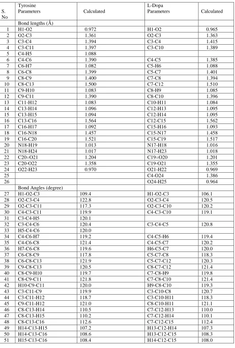
The optimized structural parameters of tyrosine and L-DOPA calculated by DFT, B3LYP method with the 6-311G(d,p) basis set are listed in Table 1 in accordance with the atom numbering scheme given in Fig. 1.2 and Fig. 2.5. For example in tyrosine, the optimized bond lengths of C-C in phenyl ring falls in the range from 1.390Å to 1.400Å, which are in good agreement with those of L-DOPA [1.385Å-1.415Å]. The optimized C8-C13 bond length is 1.500Å for tyrosine which is also in excellent agreement with C7-C12 bond length [1.510Å] for L-DOPA. The optimized C13-C16 bond length is 1.564Å for tyrosine, which is also in very good agreement with C12-C15 bond length [1.562Å] for L-DOPA. The optimized C-N bond length is 1.457Å for tyrosine, which is also in excellent agreement with C-N bond length [1.458Å] for L-DOPA. The optimized C=O bond length is 1.204Å for tyrosine, which is also in very good agreement with C=O bond length [1.201Å] for L-DOPA. The other calculated bond lengths and bond angles also shown an excellent agreement in tyrosine and L-DOPA.
4.2 Vibrational frequencies
Table 1: Optimized geometrical parameters of tyrosine and L-DOPA
S. No
Tyrosine
Parameters Calculated
L-Dopa
Parameters Calculated
Bond lengths (Ǻ)
1 H1-O2 0.972 H1-O2 0.965
2 O2-C3 1.361 O2-C3 1.363
3 C3-C4 1.394 C3-C4 1.415
4 C3-C11 1.397 C3-C10 1.389
5 C4-H5 1.088
6 C4-C6 1.390 C4-C5 1.385
7 C6-H7 1.082 C5-H6 1.088
8 C6-C8 1.399 C5-C7 1.401
9 C8-C9 1.400 C7-C8 1.394
10 C8-C13 1.500 C7-C12 1.510
11 C9-H10 1.083 C8-H9 1.085
12 C9-C11 1.390 C8-C10 1.396
13 C11-H12 1.083 C10-H11 1.084
14 C13-H14 1.096 C12-H13 1.095
15 C13-H15 1.094 C12-H14 1.095
16 C13-C16 1.564 C12-C15 1.562
17 C16-H17 1.092 C15-H16 1.093
18 C16-N18 1.457 C15-N17 1.458
19 C16-C20 1.521 C15-C19 1.517
20 N18-H19 1.013 N17-H18 1.016
21 N18-H24 1.017 N17-H23 1.018
22 C20=O21 1.204 C19=O20 1.201
23 C20-O22 1.358 C19-O21 1.355
24 O22-H23 0.970 O21-H22 0.969
25 C4-O24 1.386
26 O24-H25 0.964
Bond Angles (degree)
27 H1-O2-C3 109.4 H1-O2-C3 106.1
28 O2-C3-C4 122.8 O2-C3-C4 120.5
29 O2-C3-C11 117.3 O2-C3-C10 120.2
30 C4-C3-C11 119.9 C4-C3-C10 119.1
31 C3-C4-H5 120.1
32 C3-C4-C6 120.4 C3-C4-C5 120.8
33 H5-C4-C6 120.0
34 C4-C6-H7 119.2 C4-C5-H6 119.4
35 C4-C6-C8 121.4 C4-C5-C7 120.2
36 H7-C6-C8 119.6 H6-C5-C7 120.0
37 C6-C8-C9 117.8 C5-C7-C8 118.3
38 C6-C8-C13 121.9 C5-C7-C12 120.3
39 C9-C8-C13 120.5 C8-C7-C12 121.4
40 C8-C9-H10 119.7 C7-C8-H9 119.8
41 C8-C9-C11 121.8 C7-C8-C10 121.4
42 H10-C9-C11 120.0 H9-C8-C10 119.3
43 C3-C11-C9 119.9 C3-C10-C8 120.7
44 C3-C11-H12 118.7 C3-C10-H11 118.3
45 C9-C11-H12 121.0 C8-C10-H11 121.1
46 C8-C13-H14 110.5 C7-C12-H13 110.0
47 C8-C13-H15 110.2 C7-C12-H14 110.1
48 C8-C13-C16 112.6 C7-C12-C15 112.4
49 H14-C13-H15 107.2 H13-C12-H14 107.3
50 H14-C13-C16 108.6 H13-C12-C15 108.3
66 C5-C4-O24 124.7
[image:10.595.81.537.331.769.2]67 C4-O24-H25 110.6
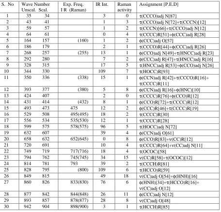
Table 2: Vibrational wave numbers obtained for tyrosine at B3LYP/6-311G(d,p) in cm-1, Experimental frequencies from FT-IR and FT-Raman spectra in cm-1, IR intensities (Km mol-1), Raman scattering activities
(Ǻ4 amu-1) and assignment with P.E.D. percentage in square brackets.
S. No Wave Number Unscal. Scal.
Exp. Freq. I R (Raman)
IR Int. Raman activity
Assignment [P.E.D]
1 35 34 3 0 τ(CCCO)adj N[87]
2 43 41 1 3 τ(CCCO)adj N[72]+τ(CCCN)[12] 3 59 57 1 2 τ(CCCN)[66]+τ(CCCO)adj N[12] 4 64 61 0 4 τ(CCCC)R[51]+φ(CCC)adj R[28] 5 164 157 (160) 1 2 φ(CCC)adj O[57]
6 186 179 2 1 τ(CCCO)R[44]+φ(CCC)adj R[26] 7 268 257 (255) 13 1 φ(CCO)adj N[49]+τ(HNCC)adj R[23] 8 292 280 7 2 φ(CCC)adj R[47]+τ(HNCC)adj R[16] 9 328 315 17 5 τ(HNCC)adj R[53]+φ(CCO)adj N[26]
10 344 330 109 7 τ(HOCC)R[93]
11 350 336 (338) 15 1 φ(CCN)adj R[42]+τ(CCCO)R[16]+
τ(CCCC)R[11]
12 393 377 (380) 5 8 φ(CCN)adj R[16]+φ(HNC)[10] 13 424 407 2 0 τ(CCCC)R[76]+φ(CCO)R[12] 14 431 414 (432) 8 1 φ(CCO)R[72]+τ(CCCC)R[12] 15 493 473 475 12 2 φ(CCC)R[46]+τ(CCCC)R[19] 16 529 508 495(495) 18 2 τ(CCCC)R[30]
17 556 534 535(530) 12 1 τ(CCCC)R[28] 18 599 575 578(575) 96 5 τ(HOCC)adj N[72] 19 632 607 39 4 φ(CCN)adj O[61]
20 658 632 652(645) 0 8 φ(CCO)R[83]+ν(CC)R[12] 21 720 691 10 4 τ(CCCC)R[64]+ν(CC)adj N[11] 22 749 719 717(716) 18 4 τ(OCOC)[58]
23 794 762 745(745) 34 15 ν(CC)R[58]+τ(OCOC)[12] 24 814 781 793 39 2 τ(CCCH)R[81]
25 828 795 (800) 109 6 τ(HCCO)R[59]
31 961 923 942 2 1 τ(HCCC)R[91] 32 1009 969 (945) 40 20 ν(CC)adj N[30] 33 1029 988 985(985) 3 1 φ(CCC)R[85] 34 1094 1050 1048(1047) 85 16 ν(NC)[48]
35 1120 1075 1100(1100) 34 2 φ(HCC)R[34]+ν(CC)R[18]+
φ(HNC)[13]
36 1146 1100 1110(1115) 112 8 φ(HCC)adj R[39]+ν(CC)adj O[15] 37 1194 1146 1157 184 4 ν(CC)R[16]+φ(HNC)[11]
38 1196 1148 13 2 φ(HCC)R[61]+ν(CC)R[13] 39 1206 1158 1178(1180) 41 10 φ(HOC)R[44]+ν(CC)adj O[12] 40 1224 1175 1200(1202) 2 23 ν(CC)adj R[58]+φ(HCC)R[22] 41 1260 1210 1218(1220) 49 3 φ(HNC)[33]+τ(HCCC)adj R[24] 42 1290 1238 1248 101 10 ν(OC)R[68]+φ(HCC)R[13]
43 1295 1243 (1250) 4 19 τ(HCCC)adj R[57]+φ(HCC)adj R[11] 44 1341 1287 1274(1270) 3 16 ν(CC)R[23]+τ(HCCC)adj R[20]+
φ(HCC)R[16]
45 1354 1300 (1288) 39 4 φ(HOC)adj N[45]+ν(CC)adj O[24]+
φ(HCN)[15]
46 1359 1305 8 18 τ(HCCC)adj R[47]
47 1365 1310 1333(1325) 34 2 φ(HCC)R[44]+ν(CC)R[17]+
φ(HOC)R[16]
48 1428 1371 1370(1370) 17 5 φ(HCN)[55]+φ(HNC)[21] 49 1470 1411 1419(1420) 23 1 ν(CC)R[42]+φ(HCC)R[24]+
φ(HOC)R[11]
50 1485 1426 1438(1435) 4 9 φ(HCH)[83]+τ(HCCC)adj R[12] 51 1545 1483 1458 116 2 φ(HCC)R[45]+ν(CC)adj R[17]+
ν(CO)R[13]
52 1631 1566 1518(1520) 17 10 ν(CC)R[64]+φ(HCC)R[12] 53 1655 1589 1593 25 2 φ(HNH)[91]
54 1656 1590 1608(1650) 46 59 ν(CC)R[59]+φ(HCC)R[20] 55 1834 1761 1900 309 9 ν(C=O)[88]
56 3025 2904 25 109 νs(CH2)[98] 57 3064 2941 2925(2930) 3 71 νas(CH2)[95] 58 3083 2960 2960(2970) 20 28 νas(CH2)[96] 59 3145 3019 (3015) 18 81 ν(CH)R[91] 60 3166 3039 3045(3048) 14 46 ν(CH)R[87] 61 3168 3041 8 129 ν(CH)R[87] 62 3198 3070 3205(3060) 9 146 ν(CH)R[93] 63 3495 3355 1 93 νs(NH2)[100] 64 3576 3433 8 41 νas(NH2)[100]
65 3758 3608 63 191 ν(OH)adj N[100]
66 3833 3680 68 139 ν(OH)R[100]
[image:11.595.79.538.80.609.2]Unscal.: Unscaled; Scal.: Scaled; ν: stretching; νs: symmetric stretching; νas: asymmetric stretching; φ: bending; τ: torsion; R: Ring; adj-adjacent.
Table 3: Vibrational wave numbers obtained for L-DOPA at B3LYP/6-311G(d,p) in cm-1, Experimental frequencies from FT-IR and FT-Raman spectra in cm-1, IR intensities (Km mol-1), Raman scattering activities
(Ǻ4 amu-1) and assignment with P.E.D. percentage in square brackets.
S. No Wave Number Unscal. Scal.
Exp. Freq. I R (Raman)
IR Int. Raman activity
Assignment [P.E.D]
15 433 416 68 1 τ(HOCC)R[91]
16 458 440 9 2 τ(HOCC)R[58]+φ(CCC)R[23] 17 467 448 463(465) 11 3 φ(CCC)R[49]+τ(HOCC)R[22] 18 520 499 475 2 9 φ(CCO)R[49]
19 538 516 528 7 2 φ(CC=O)[49]+ν(NC)[14] 20 590 566 555(553) 23 4 τ(HOCC)adj N[44]+φ(CCO)adj
N[29] 21 597 573 50 5 φ(O=CO)[59]
22 621 596 590(592) 73 0 τ(HOCC)adj N[51]+φ(CCO)adj N[11]
23 654 628 615(614) 21 4 φ(CCO)adj N[19]+φ(CC=O)[10] 24 710 682 678(690) 3 4 τ(CCCC)[72]
25 736 707 725(720) 16 8 τ(NCC=O)[38]
26 775 744 738 5 5 ν(CC)adj N[43]+τ(NCC=O)[22] 27 806 774 28 31 ν(CC)R[53]
28 821 788 782(782) 99 7 τ(HCCO)R[71] 29 837 803 809 31 3 τ(HCCO)R[78]
30 854 820 822(814) 113 5 τ(NHCH)[56]+ν(CC)adj N[14] 31 876 841 845(845) 5 4 τ(HCCO)R[29]
32 888 852 868(870) 58 2 τ(HCCC)adj R[28]+τ(NHCH)[21] 33 936 899 5 1 τ(HCCO)R[83]
34 969 930 923(922) 37 4 ν(CC)adj R[57]+φ(CCC)R[14] 35 1015 974 988(986) 31 19 ν(CC)adj N[33]
36 1092 1048 1066(1065) 98 15 ν(NC)[50]+φ(HNC)[13]+φ(HCN)[1 0]
37 1123 1078 18 1 ν(CC)R[19]+φ(HCC)R[18]+
φ(HCC)adj N[12]
38 1133 1088 (1100) 234 8 ν(CC)adj O[28]+φ(HCC)R[14]+
φ(HNC)[11]
39 1164 1117 1122(1124) 19 4 φ(HCC)R[36]+ν(CC)R[17]+
φ(HOC)R[10]
40 1182 1135 1145(1142) 17 7 φ(HOC)R[40]+φ(HCC)adj N[18] 41 1200 1152 1160(1165) 99 9 φ(HCC)R[28]+φ(HOC)adj N[17]+
ν(CC)adj O[12]
47 1341 1287 1285 7 36 τ(HCCC)adj R[54]
48 1350 1296 1300(1300) 69 5 φ(HOC)adj N[46]+ν(CC)adj O[17]+
φ(HCN)[12]
49 1359 1305 16 10 φ(HCC)R[27]+φ(HOC)R[14]+
τ(HCCC)adj R[11]
50 1411 1355 1352(1350) 29 15 ν(CC)R[36]+φ(HOC)R[31] 51 1426 1369 21 5 φ(HCN)[49]+φ(HNC)[25] 52 1474 1415 1405(1407) 95 4 ν(CC)R[52]+φ(HCC)R[16] 53 1491 1431 1460(1445) 9 9 φ(HCH)[88]
54 1558 1496 1500(1500) 136 5 ν(CC)R[53]+φ(HCC)adj N[28] 55 1647 1581 1572 27 6 ν(CC)R[64]
56 1655 1589 1595 25 2 φ(HNH)[92] 57 1661 1595 1605(1610) 25 69 ν(CC)R[68]
58 1830 1757 301 8 ν(C=O)[88]
59 3029 2908 2927 28 121 νs(CH2)[98] 60 3064 2941 (2933) 4 72 νas(CH2)[95] 61 3080 2957 2960(2980) 24 28 νas(CH2)[96]
62 3142 3016 17 66 ν(CH)R[97]
63 3173 3046 (3038) 12 55 ν(CH)R[100] 64 3190 3062 3072(3050) 5 152 ν(CH)R[100] 65 3494 3354 2 95 νs(NH2)[100] 66 3576 3433 8 41 νas(NH2)[100]
67 3758 3608 71 196 ν(OH)adj N[100]
68 3789 3637 105 85 ν(OH)R[100]
69 3848 3694 75 102 ν(OH)R[100]
[image:13.595.82.530.462.607.2]Unscal.: Unscaled; Scal.: Scaled; ν: stretching; νs: symmetric stretching; νas: asymmetric stretching; φ: bending; τ: torsion; R: Ring; adj-adjacent.
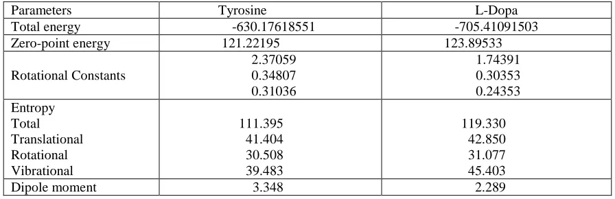
Table 4: Theoretically computed energies (a.u), zero-point vibrational energies (kcal mol-1), rotational constants (GHz), entropies (Cal mol-1 K-1) and dipole moment (D) for Tyrosine and L-Dopa.
Parameters Tyrosine L-Dopa
Total energy -630.17618551 -705.41091503
Zero-point energy 121.22195 123.89533
Rotational Constants 2.37059 0.34807 0.31036 1.74391 0.30353 0.24353 Entropy Total Translational Rotational Vibrational 111.395 41.404 30.508 39.483 119.330 42.850 31.077 45.403 Dipole moment 3.348 2.289
4.3 Vibrational assignment
3100 cm , which is the characteristic region for the ready identification of C-H stretching vibration [39]. Accordingly in the present study for tyrosine, the C-H vibrations are calculated at
3019, 3039 and 3070 cm-1 respectively with significant P.E.D. and are supported by
experimental data. While for L-DOPA, the C-H vibrations are calculated at 3046 and 3062 cm-1
respectively with appropriate P.E.D. and are supported excellently by experimental data.
4.3.3 O-H Vibrations
In a vibrational spectra, the strength of hydrogen bond determines the position of O-H band.
Usually the O-H stretching appears at 3600-3400 cm-1 [40]. In this study tyrosine showed a very
strong absorption peak at 3608 and 3680 cm-1 with P.E.D. [100], which are due to the O-H stretching vibration. While L-DOPA also showed very strong absorption peak at 3608, 3637 and 3694 cm-1 with same P.E.D., which are also due to the O-H stretching vibration. The different bending vibrations of the hydroxyl groups are also identified and they are listed in Table 2 and 3.
4.3.4 NH2 group Vibrations
The scaled NH2 asymmetric stretch is calculated at 3433 cm-1 for tyrosine and also at 3433 cm-1
for L-DOPA with P.E.D.[100]. Whereas NH2 symmetric stretch is calculated at 3355 cm-1 for
tyrosine and at 3354 cm-1 for L-DOPA with P.E.D.[100]. The various bending vibrations of NH2
group for both title compounds are also found to be in good agreement with the experimental data with appropriate P.E.D.
4.3.5 Methylene group Vibrations
The asymmetric CH2 stretching vibrations are generally observed in the region 3100-3000 cm-1,
while the symmetric stretch will appear between 3000-2900 cm-1 [41]. For tyrosine, the
symmetric CH2 stretching vibrations is calculated at 2904 cm-1 and asymmetric CH2 stretching
vibrations are calculated at 2941 and 2960 cm-1 with significant P.E.D. and are also supported by
experimental data. For L-DOPA, the symmetric CH2 stretching vibrations is calculated at 2908
cm-1 and the asymmetric CH2 stretching vibrations are calculated at 2941 and 2957 cm-1 with
appropriate P.E.D. and are also supported by experimental data. The various bending CH2
vibrations are also found to be in excellent agreement with experimental data for both title compounds.
4.3.6 Carbonyl Absorption
4.4 Other Molecular Properties
Several calculated thermodynamic parameters are presented in Table 4. The zero point vibrational energy (ZPVE) of tyrosine is less than the L-DOPA, but the difference seems to be insignificant. The total energies are found less in L-DOPA, in comparison to the tyrosine. The total, translational, rotational and vibrational entropy of tyrosine is less than that of L-DOPA at room temperature, but the difference are only marginal. Values of rotational constants and dipole moment are greater in tyrosine, than that of L-DOPA.
CONCLUSION
Attempts have been made in the present work for the proper frequency assignments for the compound tyrosine and L-DOPA from the FT-IR and FT-Raman spectra. The equilibrium geometries and harmonic frequencies of tyrosine and L-DOPA were determined and analysed at DFT level of theory utilizing 6-311G(d,p) basis set, giving allowance for the lone pairs through diffused functions. The vibrational frequency calculation proved that both structures are stable (no imaginary frequency). The difference between the observed and scaled wave numbers values of most of the fundamentals is very small. Any discrepancy noted between the observed and the calculated frequencies may be due to the fact that the calculations have been actually done on a single molecule in the gaseous state contrary to the experimental values recorded in the presence of intermolecular interactions. The potential energy distribution contribution to each of the observed frequency shows the reliability and accuracy of the normal mode analysis. Therefore, the assignments made at higher levels of theory with only reasonable deviations from the experimental values, seems to be correct.
Acknowledgement
The corresponding author (Neeraj Misra) is grateful to UGC (new Delhi) for providing the financial assistance.
REFERENCES
[1] RJ Wurtman; MC Lewis. Karger, 1991, 32, 94-109. [2] AJ Gelenberg et al. J. Affec. Disord., 1990, 19, 125-132.
[3] W Romanowski; S Grabiec. Acta Physiol. Pol., 1974, 25, 127-134.
[4] HR Lieberman; S Corkin; BJ Spring; RJ Wurtman; JH Growden. Am. J. Clin. Nutr., 1985, 42, 366-370.
[5] LE Banderet; HR Lieberman. Brain Res. Bull., 1989, 22, 759-762.
[6] AJ Gelenberg; CJ Gibson; JD Wojcik. Psychopharmacol. Bull., 1982, 18, 7-18. [7] JS Meyer et al. J. Am. Geriatr Soc., 1977, 7, 289-298.
[8] KE Goodwill; C Sabatier; C Marks; R Raaq; PF Fitzpatrick; RC Stevens. Nat. Struct. Biol.,
1997, 4(7), 578-585.
[9] KE Goodwill; C Sabatier; RC Stevens. Biochem., 1998, 37(39), 13437-13445. [10] JH Waite et al. J. Adhesion, 2005, 81, 1-21.
[11] PB Messersmith et al. Macromol., 2006, 39, 1740-1748.
[12] J Glowinski; RJ Baldessarini. Pharmacol. Rev., 1966, 18, 1201-1238. [13] AH Anton; DF Sayre. J. Pharmacol. Exp. Ther., 1964, 145, 326-336. [14] LJ Poirier; P Singh; TL Sourkes; R Boucher. Brain Res., 1967, 6, 654-666.
[15] J Constantinidis; G Bartholini; R Tissot; A Pletscher. Experientia, 1968, 24(2), 130-131. [16] H Ehringer; O Hornykiewicz. Klin Wochenschr., 1960, 38, 1236-1239.
[25] MH Jamroz. Vibrational Energy Distribution Analysis, VEDA 4 program, Warsaw, 2004. [26] http://www.sigmaaldrich.com/spectra/ftir/FTIR009043.PDF
[27] http://www.sigmaaldrich.com/spectra/rair/RAIR014464.PDF [28] http://www.sigmaaldrich.com/spectra/ftir/FTIR001390.PDF [29] http://www.sigmaaldrich.com/spectra/rair/RAIR001911.PDF [30] MJ Frisch et al. Gaussian, Inc., Wallingford CT, 2004. [31] HB Schlegel. J. Comput. Chem., 1982, 3, 214-218.
[32] G Chang; WC Guida; WC Still. J. Am. Chem. Soc., 1989, 111, 4379-4386. [33] P Hohenberg; W Kohn. Phys. Rev., 1964, B136, 864-871.
[34] AD Becke. J. Chem. Phys., 1993, 98, 5648-5652.
[35] C Lee; W Yang; RG Parr. Phys. Rev., 1988, B37, 785-789.
[36] N Sundaraganesan; H Saleem; S Mohan; M Ramalingam. Spectrochim. Acta, 2005, A61, 377-385.
[37] N Sundaraganesn; S Ilakiamani; H Saleem; PM Wojaechiwsju; D Michalska. Spectrochim.
Acta, 2005, A61, 2995-3001.
[38] PL Fast; J Corchado; ML Sanches; DG Truhlar. J. Phys. Chem., 1999, A103, 3139-3143. [39] V Krishnakumar; RJ Xavier. Ind. J. Pure Appl. Phys., 2003, 41, 597-601.