HIV and Tuberculosis: Overview
Amit Gupta
*, Sushama R Chaphalkar
Department of Immunology, Vidya Pratishthan’s School of Biotechnology (VSBT), India
Copyright © 2015 by authors, all rights reserved. Authors agree that this article remains permanently open access under the terms of the Creative Commons Attribution License 4.0 International License
Abstract
Tuberculosis (air borne disease, caused by Mycobacterium tuberculosis or tubercle bacilli) and HIV (human immunodeficiency virus), enduring human burden, are growing manifold or multiple health problems in the developing world triggering millions of deaths world-over each year. It is crucial to recognize the epidemiology of HIV and tuberculosis involved in transmission and disease progression to comprehend or understand the synergy between the two epidemics.Keywords
HIV, Tuberculosis, Epidemiology, Transmission1. Introduction
Tuberculosis (bacterial, infectious disease) usually affects the lungs; although it can affect almost any component or fragment of our body [1, 2]. The transmission of this disease occurs only when person inhales the droplet nuclei of Mycobacterium tuberculosis and these nuclei moves towards the alveoli of the lungs through nasal passages, upper respiratory tract and bronchi [3, 4]. The main factors responsible for the transmission are environmental factors (space, oxygenating, air pressure, handling of specimen etc); exposure; susceptibility and infectiousness [clinical, protocol/strategy and radiographic (X-ray)]. The currently available vaccine for tuberculosis, Bacilli Calmette-Guerin (BCG), is administered to babies at birth in most countries [5, 6]. BCG vaccine protects young children (age, less than 5 years) against more threatening/hazardous extra pulmonary forms of tuberculosis and thus, is given as a routine vaccination in many countries, as approved by the WHO [7, 8]. However, the efficacy of BCG for protection against pulmonary tuberculosis is suspicious or controversial, based on wide reaching clinical trials [6, 7]. In contrast, BCG vaccination for tuberculosis disease has been shown in some studies to effectively induce a protective immune response against primary infection, but has limited effect on the subsequent course of dormancy and reactivation [7, 8].
Epidemiology
There are approximately 8 million new cases of tuberculosis and nearly 2 million deaths due to this disease each year [1]. According to World Health Assembly (decision making authority of WHO), the meeting which is earlier held in May 2015; Geneva, Switzerland. The members of this committee was agreed and decided to end the global tuberculosis epidemic with in new 20 years (2016 – 2035).
Over the last 20 years, number of tuberculosis cases is still leading in UK, about 9000 people now get tuberculosis each year – around one person in every 7000 of the population [1]. Similar cases of tuberculosis also reported in India, highest burden reported cases in the world, approximately 2 million cases of tuberculosis annually, accounting for roughly one fifth of the global incidence [9]. Generally, tuberculosis can be treated with special antibiotics [10]. Once treatment starts, the treatment has to continue for at least six months. It is crucial or mandatory to complete the whole course/procedure of antibiotics to cure the tuberculosis. If the proper remedy or medication is not taken [11, 12] for this disease, it may lead to death.
Pathogenesis
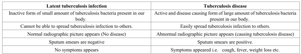
The pathogenesis of tuberculosis is shown in Fig.1. During pathogenesis, the process of latent tuberculosis infection occurs only when extracellular bacteria is ingested by macrophages and also presented to other white blood cells [13]. These cells will provoke or stimulate our immune system and also be able to kill or encapsulate most of the bacteria leading to the formation of granuloma. At this stage, latent tuberculosis infection (LTBI) has been established and diagnosed through various tests such as tuberculin skin test, interferon gamma (IFN-γ) release test etc. if the test is positive i.e. LTBI, it means do not have tuberculosis disease and not be able to spread infection to other people [1, 11, 12, 13]. To understand the phenomenon/concept of LTBI and tuberculosis disease as shown in Table 1.
Antigenic Molecules of Tuberculosis
In Mycobacterium tuberculosis, the most important two immunogenic molecules [(CFP- 10 or Rv3874 and early secretory antigenic target (ESAT)-6 or Rv3875)] are observed and produced abundantly in the culture filtrate [14, 15]. These two molecules are totally dependent on each other and grouped together to form tight dimer and are capable of enhancing both innate as well as adaptive immune system. If there is inactivation of any one component or molecule i.e. ESAT-6/CFP-10 results in dramatically reduced virulence of Mycobacterium tuberculosis. Both these molecules are totally dependent on ESX-1 secretory signal sequences but lack the classical Sec YEG secretory signal sequences [16, 17]. The most interesting feature is that specific mutations in
ESAT-6 did not affect functioning of the ESX-1 secretion system caused a reduction in virulence of mycobacteria but CFP-10 affects the ESX-1 secretion system. It is confirmed that ESAT-6 itself is crucial or important in mediating the virulence functions associated with the ESX-1 secretion system. The key points of immunogenic molecules [16, 17] in order to understand the function i.e.
a) ESAT-6 is likely to interact with toll like receptor i.e. TLR2 resulting in diminish proportion of IL-12p40 secretion in monocytes derived macrophages, apparently benefit to T helper 2 phenotype that helps intracellular persistence and survival of Mycobacterium tuberculosis.
b) Induce apoptosis of macrophages (triggering caspase expression) in case of ESAT-6.
c) ESAT-6 and CFP-10 (alone or in association) inhibits lipolysaccharide (B cell mitogen) increased NF-kappa B dependent gene expression by suppressing the production of reactive oxygen species.
d) ESAT-6 and CFP-10 (alone or in combination) also showed interaction with number of host proteins i.e. laminin on the basolateral surface of pneumocytes leading to rupturing of these cells that aid in the dissemination/dispersal of Mycobacterium tuberculosis in the human lung.
[image:2.595.60.548.656.745.2]The crucial role played by ESAT-6 in the virulence of mycobacteria could be due to its interaction with some host cellular factors, the recognition of which is likely to provide better understanding of the role played by ESAT-6 in modulation of host immune responses.
Figure 1. Pathogenesis of Tuberculosis (Mycobacterium tuberculosis)
Table 1. Comparative difference between Latent tuberculosis infection and Tuberculosis disease
Latent tuberculosis infection Tuberculosis disease
Inactive form of small amount of tuberculosis bacteria present in our
body. Active and disease causing form of large amount of tuberculosis bacteria present in our body.
Cannot be able to spread tuberculosis infection to others. Easily spread tuberculosis infection to others.
Normal radiographic picture appears (No disease) Abnormal radiographic picture appears (causing tuberculosis disease)
Sputum smears are negative Sputum smears are positive.
HIV (Human Immunodeficiency Virus)
Figure 2. Structure of HIV
HIV (Family- Retroviridae; subfamily- Orthoretrovirinae; Genus- Lentivirus) causes AIDS (acquired immunodeficiency syndrome) [18, 19]. The structure and life cycle of HIV [20] as shown in Fig.2 and 3. HIV is divided into 2 subtypes i.e. HIV-1 (provirus, 9.8kb length) and HIV-2 [21, 22]. Out of these, HIV-1 is more virulent as well as infective; this subtype is also called as Lymphadenopathy associated virus (LAV) and Human T lymphotrophic virus (HTLV-III) whereas HIV-2 is very limited and is normally confined to West Africa. In addition, genes in HIV-1 are present in the central region of proviral DNA encodes 9 proteins and these proteins further divided into three classes [23, 24] i.e. structural genes (gag, pol and env); regulatory genes (tat and rev) and accessory proteins (vpr, vpu, vif and nef). Currently, there are approximately or appraise 1.1 million people in the USA and 34 million people globally who are living with HIV infection. In 2011, 1.7 million people expired from AIDS-related causes.
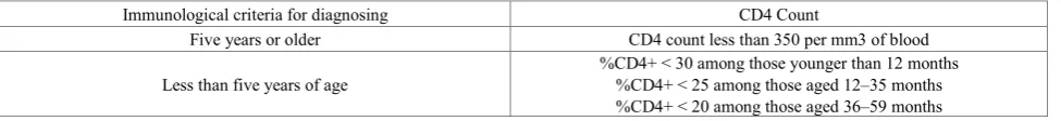
Determination of CD4 Population for Diagnosing HIV Infection
The immunological criteria for diagnosing HIV infection in children or adults in terms of CD4 population as appeared in Table 2. CD4 T cells [25] are the most dominant type of cells in our immune system and it differentiates into T helper 1 and 2. The main function of Th1 and Th2 is to fight against the intracellular and extracellular pathogens. The exact role of CD4 in terms of HIV infection is:
a) If CD4 count is low, viral load (between 100,000 – 1 million) is high- HIV positive
b) If CD4 count is high, viral load (below 10,000) is low- HIV negative
c) If CD4 count is high, viral load (below 50) is low- HIV negative (undetectable viral load)
d) If CD4 count is dropped to 350 or low,- start treatment; if the viral load is low after treatment and CD4 count increased- HIV negative.
Several cohort studies and clinical trials have shown that the CD4 count is the strongest predictor of subsequent disease progression and survival [26]. The use of the CD4 count as an independent and reliable marker for treatment outcome is attractive from various aspects.
a) On the basis of CD4 counts, whether to initiate or start the antiretroviral therapy and opportunistic prophylaxis–all HIV-positive patients in high-income countries and an increasing number of patients in low-income countries have a baseline CD4 count at entry into care.
b) CD4 count (marker of T cells), the cost of this marker has become more reasonable as well as affordable, including in developing countries.
Figure 3. Life cycle of HIV (Human immunodeficiency virus)
Table 2. Criteria for diagnosing HIV infection in terms of CD4 population
Immunological criteria for diagnosing CD4 Count
Five years or older CD4 count less than 350 per mm3 of blood
Less than five years of age %CD4+ < 30 among those younger than 12 months %CD4+ < 25 among those aged 12–35 months
[image:3.595.64.552.684.740.2]Deadly Symbiosis of HIV and Tuberculosis
Although tuberculosis is a remediable and treatable disease, it has catastrophic consequences on human health, second only to the human immunodeficiency virus (HIV) in the number of deaths caused by an infectious disease worldwide. In tandem, HIV infection and tuberculosis create a deadly symbiotic relationship and formed a deadly alliance (when paired each makes the other more lethal). So, there is an urgent need for immediate initiation or requirement of antiretroviral therapy (ART, universal recommendation) for any one co-infected with HIV and Tuberculosis. Till now, there are estimated to be over one million people worldwide who have tuberculosis and HIV co-infection. The overload of disease through HIV/ tuberculosis co-infection is exceptionally high in sub-Saharan Africa, and the dual outbreak of tuberculosis and HIV are of extending concern in Asia -Pacific region, which subscribes more than a half of all tuberculosis cases worldwide, traditionally reports low tuberculosis /HIV co-infection rates. However, routine procedure for testing tuberculosis patients for HIV infection is not invariably implemented and the estimated prevalence of HIV in new tuberculosis cases increased to 6.3 % in 2013. Meanwhile, levels of multi-drug resistant tuberculosis, in Africa and elsewhere are drastically increasing. Tuberculosis is a major cause of death among people living with HIV/AIDS, whose diminished immune systems make them particularly vulnerable to the devastating effects of tuberculosis. The current challenge is to find ways of preventing both tuberculosis and HIV, and to improve diagnosis and management of co-infection.
Tuberculosis is the dominant source of death among persons with HIV infection i.e. in 2013; there were 9 million new cases of tuberculosis, of which 1.1 million were already infected with HIV. Clearly, an improved and effective vaccine is desperately needed for these deadly pathogens [3, 4, 5]. Most of the developing countries including India, the estimated risk rate of developing tuberculosis is higher in case of HIV patients (>25 – 30 times) in comparison with tuberculosis infected alone [2,4]. The most common symptoms appeared in case of HIV- tuberculosis at the time of seroconversion (production of antibodies to HIV) are fever, skin rashes, aching muscles and joints, swollen lymph nodes etc [1,2]. To control the disease burden of HIV- tuberculosis, it involves number of drugs i.e. nucleosides (Truvada and kivexa pills) and non-nucleosides (protease inhibitors or integrase inhibitors e.g. cabotegravir). Recently, there is an urgent need of more effective drug, vaccine and improved diagnostic kit for tuberculosis associated HIV especially important in India, where 40 % of the adult population is infected with Mycobacterium tuberculosis and approximately 60 % HIV-infected persons in India will develop tuberculosis disease during their life-time
.
HIV not only rises in the number of tuberculosis cases, but also converts the clinical course of tuberculosis disease. In addition, this disease is more likely to be disseminated and more difficult to diagnose as the immunosuppression continues. HIV infected tuberculosis patients can also suffer
from other HIV-related diseases [16, 21]. National tuberculosis programmes in the high HIV burden countries are reporting increasing case fatality rates of up to 25 % in the smear-positive and 40-50 % in smear-negative pulmonary tuberculosis patients. In 2000, there were globally an estimated 350,000 deaths from HIV-related tuberculosis.
2. Conclusions
Tuberculosis and HIV epidemic has been a challenge to control and creates lot of problems related to our immune system. There is an urgent need to strengthening shared/collaborative efforts in order to control the HIV and tuberculosis infection. In order to achieve elimination by 2050 e.g. most of the European countries would have to cut down the cases of HIV and tuberculosis infection at least twice as fast. In most low-incidence countries tuberculosis rates are stable or going down very slowly and the majority of patients of this disease are of foreign origin. Most of the countries with high incidence overall face higher rates of re-infection and relapses and report many more MDR tuberculosis cases. The main goal is to eliminate the tuberculosis and HIV infection depends on a more efficient use of current tools and interventions, to be complemented by new and more effective ones.
REFERENCES
[1] Lawn, SD and Zumla AI. Tuberculosis. Lancet 2011; 378 (9785): 57–72.
[2] Volmink J and Garner P. Directly observed therapy for treating tuberculosis. Cochrane Database Syst Rev 2007; 4: CD003343.
[3] Lambert M et al. Recurrence in tuberculosis: relapse or reinfection? Lancet Infect Dis 2003; 3 (5): 282 – 287. [4] Wang, JY, Lee LN, Lai HC, Hsu HL, Liaw YS, Hsueh PR and
Yang PC. Prediction of the tuberculosis reinfection proportion from the local incidence. The Journal of infectious diseases 2007; 196 (2): 281 – 288.
[5] Bonah C. The experimental stable of the BCG vaccine: safety, efficacy, proof, and standards, 1921–1933. Stud Hist Philos Biol Biomed Sci 2005; 36 (4): 696–721.
[6] Behr M. BCG - different strains, different vaccines? The Lancet Infectious Diseases, 2002; 2(2): 86 – 92.
[7] Kernodle D. Decrease in the effectiveness of Bacille Calmette-Guerin Vaccine against Pulmonary Tuberculosis. Clin Infect Dis 2010; 51 (2): 177 - 184.
[8] Dixon G. Pulmonary tuberculosis in childhood. BMJ 1931; 714 (1): 694-697.
[10] Zumla A et al. Tuberculosis. New Eng J of Medicine 2013; 368:745.
[11] Cruz-Knight W et al. Tuberculosis: An overview. Primary Care Clinics Office Practice 2013; 40:743.
[12] Wong EB et al. Rising to the challenge: new therapies for tuberculosis. Trends in Microbiology. 2013; 21:493.
[13] Sutherland I. Recent studies in the epidemiology of tuberculosis, based on the risk of being infected with tubercle bacilli. Adv Tuberc Res 1976; 19: 1–63.
[14] Hsu T, Hingley-Wilson SM, Chen B, Chen M, Dai AZ, et al. The primary mechanism of attenuation of bacillus Calmette-Guerin is a loss of secreted lytic function required for invasion of lung interstitial tissue. Proc Natl Acad Sci USA 2003; 100: 12420–12425.
[15] Brodin P, Eiglmeier K, Marmiesse M, Billault A, Garnier T, et al. (2002) Bacterial artificial chromosome-based comparative genomic analysis identifies Mycobacterium microti as a natural ESAT-6 deletion mutant. Infect Immun 2002; 70: 5568–5578.
[16] Lewis KN, Liao R, Guinn KM, Hickey MJ, Smith S, et al. (2003) Deletion of RD1 from Mycobacterium tuberculosis mimics bacille Calmette-Guerin attenuation. J Infect Dis 187: 117–123.
[17] Pym AS, Brodin P, Brosch R, Huerre M, Cole ST (2002) Loss of RD1 contributed to the attenuation of the live tuberculosis vaccines Mycobacterium bovis BCG and Mycobacterium microti. Mol Microbiol 46: 709–717.
[18] Sepkowitz KA. AIDS—the first 20 years. N Engl J Med 2001; 344 (23): 1764–72.
[19] Sax PE and Baden LR. When to start antiretroviral therapy—ready when you are? The New England Journal of Medicine 2009; 360 (18): 1897–9.
[20] Capon DJ, Ward RH. The CD4-gp120 interaction and AIDS pathogenesis. Annu Rev Immunol 1991; 9: 649-678. [21] Dougan S et al. Diagnoses of HIV-1 and HIV-2 in England,
Wales and Northern Ireland associated with west Africa. Sexually Transmitted Infections 2005; 81:338-341.
[22] Parry JV et al. Towards error-free HIV diagnosis: guidelines on laboratory practice. Comm Dis Pub Health 2003; 6:334-350.
[23] Gallo R, Wong-Staal F, Montagnier L, et al. HIV/HTLV gene nomenclature. Nature 1988; 333:504.
[24] Muesing MA, Smith DH, Cabradilla CD, et al. Nucleic acid structure and expression of the human AIDS/lymphadenopathy retrovirus. Nature 1985; 313:450-458.
[25] Miceli MC, Parnes JR. Role of CD4 and CD8 in T cell activation and differentiation. Advances in Immunology 1993; 53: 59–122.