Single Stage Management of Concomitant Gall Stones
and Common Bile Duct Stones in the Laparoscopic Era
Sandeep Rajta, R.S.Jhobta, A.K.Malhotra, D.K.Verma, A.K.Kaundal, Gopal Singh
Department of Surgery, I.G.M.C. Shimla, Himachal Pradesh University, India
Abstract- Objective: To determine the efficacy, safety and outcome of the single stage management of gall stones and common bile duct stones in the laparoscopic era.
Material and method: From 1st June, 2014 to 31st May, 2015, we treated 20 patients with cholelithiasis and choledocholithiasis with laparoscopic cholecystectomy and laparoscopic common bile duct exploration. Laparoscopic common bile duct exploration was done via choledochotomy in all patients. The male/female ratio was 6/14, with a mean age 53.50 years (range 20 to 70 years). Preoperative evaluation was done through medical history, biochemical tests, ultrasonography and magnetic resonance cholangiopanceatography(MRCP). Intraoperative findings, postoperative complications and hospital stay were analyzed.
Results: One stage stone clearance was successful in all patients. The mean time taken for procedure was 111.75 minutes and the average gas (CO2) consumed was 92.3 litres. Postoperative complications were observed in 2 (10%) patients. In 2 (10%) patients completion of the procedure was achieved by conversion to open choledocholithotomy. Mean hospital stay was 6.75 days. T-tube was inserted in 4(20%) patients. Post operative hospital stay was shorter in the primary closure group than in the T-tube group.
Conclusions: Laparoscopic management of comcomitant gall stones and common bile duct stones has definitely shown decreased post operative discomfort, decreased requirement of post operative analgesia, better cosmesis and early return to work and less morbidity.
Index Terms- alkaline phosphatase (ALP), common bile duct (CBD), endoscopic retrograde cholangiopancreatography (ERCP), magnatic resonance cholangiopancreatography (MRCP)
I. INTRODUCTION
he incidence of choledocholithiasis in patients with cholelithiasis is reported at 5%-10%, with 4%-5% incidence of unsuspected choledocholithiasis when routine cholangiography is performed.1,2,3,4
The management of concomitant gall bladder and common bile duct stones has evolved significantly over the past 20-30 years. In the era of open surgery, open common bile duct exploration would be performed if any common bile duct stones were identified at cholangiography. Following the introduction and rapid uptake of endoscopic retrograde cholangiopancreatography, open common bile duct exploration
was reserved for patients who failed endoscopic retrograde cholangiopancreatography.
During the era of open cholecystectomy the management of choledocholithiasis was relatively straight forward but with the advent of laparoscopic cholecystectomy the treatment of common bile duct (CBD) stones, whether recognized preoperatively or per-operatively remains controversial. Although the treatment of common bile duct stones has changed during the last decade it still seems to be controversial. Currently there are three methods of treatment for choledocholithiasis. The first method is one stage open cholecystectomy with common bile duct exploration. The second one is a two stage procedure of endoscopic retrograde cholangiopancreatography followed by laparoscopic cholecystectomy. The third way is one stage laparoscopic cholecystectomy with common bile duct exploration.
Following the introduction of laparoscopic cholecystectomy5 there has been a gradual increase in laparoscopic common bile duct exploration which has been shown by a few enthusiasts to be as effective as common bile duct clearance and associated with reduced hospital stay compared to preoperative endoscopic retrograde cholangiopancreatography followed by laparoscopic cholecystectomy.6,7,8,9
However laparoscopic common bile duct exploration, either using the transcystic route or via a choledochotomy, does involve more advanced laparoscopic skills, often including flexible choledochoscope and as a result the default procedures in many hospitals remain to be endoscopic retrograde cholangiopancreatography either before or after laparoscopic cholecystectomy. Such is the reliance upon endoscopic retrograde cholangiopancreatography that in some centres surgery for common bile duct stones is now considered a lost art.10
Endoscopic retrograde cholangiopancreatography places the patient at risk of the complications of sphincterotomy including pancreatitis, perforation and bleeding.11,12,13,14 The morbidity of endoscopic retrograde cholangiopancreato-graphy with endoscopic sphincterotomy has been described as around 10% and the mortality around 1-2%.15
Laparoscopic common bile duct exploration can be the option for choledocholithiasis, as it is possible to solve the problem in a single procedure. It also has the advantage of leaving the sphincter of Oddi anatomically intact and avoids the morbidity associated with laparotomy. This study presents our initial experience of laparoscopic common bile duct exploration over a period of twelve months in Indira Gandhi Medical College, Shimla.
II. MATERIAL AND METHODS
This study was conducted in the department of surgery at I.G.M.C., Shimla from 1st June, 2014 to 31st May, 2015 and included patients with cholelithiasis and common bile duct stones. The study protocol was approved by the Ethics Committee of the same institution. Patient consent for laparoscopic surgery and research was obtained before the procedure was started.
III. STUDY POPULATION
20 patients were selected with symptomatic ultrasonography proved common bile duct stones and cholelithiasis. Various parameters were studied pre-operatively, intra-operatively and post-operatively.
IV. INCLUSION CRITERIA
The following patients were included in the study 1. All age groups
1) H/O biliary colic pain and obstructive jaundice.
2) Patient presenting at least four weeks after the acute symptoms.
3) CBD stones detected by ultrasonography and/or MRCP(Magnetic
ResonanceCholangiopancreaticography) with CBD diameter ≥ 8mm
4) Intrahepatic duct dilation as determined by ultrasonography and MRCP
5) Alkaline phosphatase or gamma glutamyle transferase level more than 1.5 times over the limit of normal at the first reference
6) Big and multiple CBD stones without any anatomical anomalies on MRCP.
7) ASA grade I-III.
V. EXCLUSION CRITERIA
The following patients were excluded from the study
1) Clinical, radiological or biochemical evidence of cholangitis and pancreatitis
2) Evidence of cirrhosis, intrahepatic gall bladder, liver mass, abscess andneoplasm
3) Gall bladder empyema and perforation 4) Pregnancy
5) Recurrent choledocholithiasis
6) Previous upper gastrointestinal surgery
7) Patients with contraindications to general anesthesia.. 8) ASA grade IV-V.
9) Complete history was taken and patients were assessed for any other co-morbid condition. Patients were explained about the procedure and informed consent were taken before any procedure.
PREOPERATIVE EVALUATION
The patients were evaluated pre-operatively on the following parameters :
Age, Gender, Height, Weight, BMI (body mass index) Routine blood investigations: CHG, FBS, RFT, Serum electrolytes.
Study-specific investigations: LFT, ultrasound abdomen and M.R.C.P.
VI. THE TECHNIQUE OF LAPAROSCOPIC CHOLESYCSTECTOMY WITH COMMON BILE
DUCT EXPLORATION
Overnight the patients were kept nil orally prior to surgery. Same premedication and anesthetic protocol was followed for all patients.
All patients were operated on under general anaesthesia in a reverse Trendelenburg position.
Pneumoperitoneum was established with CO2 using veress needle and insufflator.
The intra abdominal pressure of 12-13mm of Hg was kept during surgery.
The first 10mm trocar was introduced by Hasson’s technique below the umbilicus for insufflation of carbon dioxide and for insertion of a 30 degree angled laparoscope. Other trocars were placed under direct vision, the second 10mm trocar was introduced in the epigastric region left to the falciform ligament , the third 5mm trocar in the right anterior axillary line in right hypochondrium, the fourth 5mm trocar in the midaxillary line and the last fifth 5mm trocar below the coastal margin, 1-3cm medial to the midclavicular line. Midclavicular trocar was used for cholangioscopy of common bile duct.
After dissection of the calot triangle, liga clips were applied on the cystic duct to prevent spillage of stones into the common bile duct. The anterior surface of the supraduodenal common bile duct was dissected carefully. Common bile duct exploration was performed with an incision at the distal part of common hepatic duct just before the junction with the cystic duct. The longitudinal incision of total length 6-7mm was started on the anterior surface of the common hepatic duct(2-3mm) towards the common bile duct (4-5mm) using the cold knife. Incision was extended to 10-15mm depending upon the size of the stones. Choledochoscopy was performed using a flexible choledochoscope (Karl Storz make) with instrument channel of 7Fr, working length 35cm and outer diameter 15.5Fr.
Stones were extracted after flushing with saline with a dormia basket, fogarty catheter, atraumatic forcep or a milking technique was used. Single 2-8mm stones were removed with forceps through trocar in epigastric port. Large and multiple stones were placed in a bag made of latex glove and removed through trocar in epigastric port. After all stones were extracted, a check cholangioscopy of hepatic ducts and common bile duct was performed to ensure clearance of the biliary system.
There were two types of common bile duct closure after choledochotomy: primary closure or T-tube drainage.
to all patients as and when required. The duration of surgery was counted from port incision to skin closure. The carbon dioxide volume was measured from putting of veress needle to the completion of the procedure. In case of T-tube drainage a check cholangiography was performed on 7th post operative day. In case of no residual stones, T-tube was removed on 10th post operative day. In case of residual stones, distal obstruction or continuous bile leak patients underwent endoscopic retrograde cholangiopancreato- graphy with sphincterotomy. Patients with primary closure of the common bile duct were discharged on post operative day 3-7.
The following post operative complications were analyzed: a) Bile leak
b) Wound infection
c) Paralytic ileus/intestinal obstruction d) Duration of hospital stay
e) Acute pancreatitis f) Retained stones
VII. RESULTS
Twenty (20) selected cases of cholelithiasis and choledocholithiasis admitted in Department of Surgery, Indira Gandhi Medical College, Shimla w.e.f. 1st June, 2014 to 31st May, 2015 were evaluated in this study.
Age & Sex
[image:3.612.53.564.300.383.2]The age of patients in the present study ranged from 20 years to 70 years. The mean age of the patients was 53.50 years. Out of 20 cases, 6 (30%) were male and 14(70%) were female (Table-1).
Table-1 Age Group and Sex
Age group
20-30 31-40 41-50 51- 60 61 -70 Total
No %age No. %age No. %age No. %age No. %age No. %age
Male 2 10% 0 0% 2 10% 1 5% 1 5% 6 30%
Female 0 0% 1 5% 3 15% 5 25% 5 25% 14 70%
Total 2 10% 1 5% 5 25% 6 30% 6 30% 20 100%
BMI
BMI of each case under this study calculated preoperatively varied widely from 17.30 kg/m2 to 29.99 kg/m2. Mean BMI was 21.33kg/m². Three (15%) patients had BMI between 17.30kg/m² to 18.50kg/m², with mean BMI 17.97 kg/m2 and were underweight. Fourteen (70%) patients had BMI between 18.51kg/m² to 24.99kg/m² with mean BMI 22.03 kg/m2 and were having normal weight. Three patients (15%) had BMI ranging
25.00kg/m² to 29.99kg/m² with mean BMI of 26.27 kg/m2 and were overweight.
Mean operating time was 110 minutes in underweight group, whereas it was less in normal weight group i.e. 106.78 minutes. This paradox probably was due to less number of patients (3) in underweight group as compared to normal weight group (14). Mean operative time was 136.66 minutes in overweight group which was more than patients in normal weight group (Table 2).
Table 2
BMI and Mean Operative Time
Weight of Patients (BMI) No. of Patients Mean Operative Time (min)
Under weight (17.30 -18.50 kg/m2) 3 110
Normal weight(18.51-24.99 kg/m 2) 14 106.7857
Over weight(25.00-29.99) kg/m2) 3 136.6667
Operative Complications
None of our patients under study had any per-operative complications like pneumo-omentum, subcutaneous emphysema, pneumothorax, bleeding from abdominal wall, GIT perforation, solid visceral injury, major vascular injury which have been otherwise reported in literature. No post-operative complications like port site infection, abscess or deep vein thrombosis were seen in any patient.
Conversion to Open Common Bile Duct Exploration
common bile duct was grossly dilated with impacted large stone which made the grasping and removal difficult.
Drainage Tube
A drainage tube (14F Ryle’s tube) was kept in Morrison’s space in all (100%) of these cases. This tube was kept for minimum of 3 days and maximum of 15days. Mean duration of drainage in all (20) patients was 4.6 days (Table 3).
Table 3
Duration of Drainage Tube (In Days)
Days No of Patients
0 – 5 18
6 - 10 1
11 – 15 1
T-tube
Keher’s T-tube(14F) was inserted in 2(10%) out of 20 patients under study.
Gas (CO2) Consumed
[image:4.612.308.581.57.96.2]On an average 92.3 litres of gas (CO2) per case was used in this series. The consumption ranged from 60 litres to 120 litres in this series. (Table 4)
Table 4
CO2 Consumed (On Average in Every Four Cases)
1 2 3 4 5
Mean CO2 consumed (litres) 97 109.5 85 83.5 86.5
Time Taken for Operation
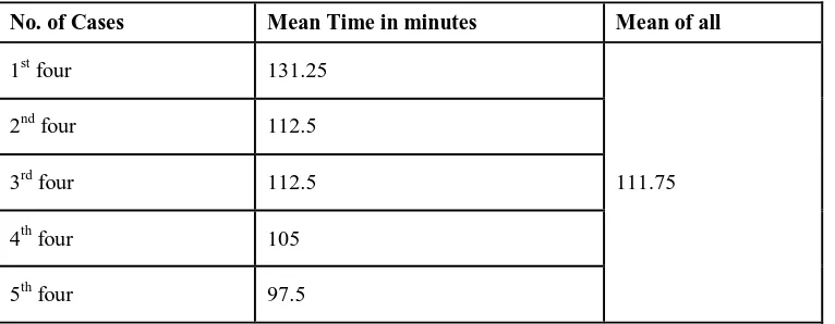
The time taken for completion of the procedure was counted from insertion of Hassan trocar up to the closure of last port. Maximum time taken in the present study was 150 minutes and minimum time taken was 90 minutes. The mean time taken for
the completion of laparoscopic common bile duct exploration was 111.75 minutes. Mean time taken for first 4 cases was 131.25 minutes. (Table 5)
[image:4.612.81.530.254.300.2]
Table-5
Mean Time (Minutes) For Each Four Cases (Duration of Surgery)
No. of Cases Mean Time in minutes Mean of all
1st four 131.25
111.75
2nd four 112.5
3rd four 112.5
4th four 105
5th four 97.5
Postoperative Complications
Out of 20 patients 2 had bile leak (one had bile leak through the abdominal drain and other had persistent bile leakage through the T-tube). Bile coming out of t-tube was due to stricture in lower common bile duct which was diagnosed on t-tube cholangiogram done on 7th postoperative day, for which endoscopic retrograde pancreatocholangiography with sphincterotomy with common bile duct stanting was done on 9th postoperative day. After this bile leak through t-tube gradually decreased over 3-4 days and t-tube was removed. In another case
[image:4.612.118.501.414.563.2]Table-6
Postoperative Complications
S. No. Complication No. of cases
1 Sepsis 0
2 Bile leak 2
3 Paralytic ileus 0
4 Acute pancreatitis 0
5 Retained stones 0
Postoperative Hospital Stay
The mean hospital stay of the patients in the present series was 6.75 days, ranging from 4 days to more than 8 days (including converted cases). One patient had to be hospitalized
[image:5.612.90.527.297.366.2]for 15 days as a result of prolonged postoperative drainage (Table 7).
Table-7 Hospital Stay in Days
No. of days 3-4 days 5-6 days 7-8 days >8 days Total
No. of patients 3 11 3 3 20
%age of patients 15% 55% 15% 15% 100%
Stone Size
All cases under study were having a single or multiple stones in common bile duct. The smallest stone was 0.5cm. and the largest stone was 2.5 cms. in size. The average stone size was
1.342cms.The stone was removed in all cases within single operation. (Table 8).
Table 8
Stone Size and Average Size (In cms.)
Stone Size No. of Patients Average Stone Size
<1.0 7 0.73
1-1.49 7 1.14
1.5-2.49 4 2
2.5 - Above 2 2.5
Total 20 1.31
VIII. FOLLOW UP USG AND LFT
All the patients were called for follow up after four weeks postoperatively in the outpatient department (OPD). All patients had follow-up USG and LFT to evaluate liver function, common bile duct status and residual stone. None of the patient had retained common bile duct stones and LFTs were within normal limits.
DISCUSSION
The age of patients in the present study ranged from 20 years to 70 years. The mean age of the patients was 53.50 years. Chung-Ngai Tang et al16 studied 27 patients who underwent
laparoscopic exploration of common bile duct during the period from 1995-1999. 14 female and 13 male patients were recruited with mean age 59.6 years (range 27-81 years). Eryk Naumowicz et al17 studied 35 patients (27 female and 8 male) who underwent laparoscopic common bile duct exploration, with mean age 58.3 years (range 23-81 years).
[image:5.612.136.479.453.575.2]Variation in time taken for surgery in different cases in the present series can be ascribed to the various factors like initial teething troubles, modern technique, non-availability of trained regular supporting staff familiar with the technique, though all were familiar with the basics of the laparoscopy and of course the learning curve of the procedure. It was the initial stage of the procedure in this institution and none of the operating team members had any “hands on” experience.
On an average 92.3 litres of gas (CO2) per case was used in this series, ranging from 60 to 120 liters. No such reports are available in the literature regarding the amount of CO2 consumed. In the present series 2(10%) of our cases were converted into open choledocholithotomy. However, there is wide variation in open conversion rate in literature. M Jameel et al20 observed open conversion rate of 7%. Alexis Sanchez et al21 did a study on 16 patients who underwent laparoscopic common bile duct exploration and observed conversion rate of 12.5%. One of the patients under study had pericholecystic adhesions and was converted to open procedure due to distorted anatomy. One patient was converted to open due to impacted stone at lower end of common bile duct. These instances of conversion to an open procedure should not be considered as a failure but only completion, because in each instance this course was chosen by the operating surgeon as safest for the patient.Although it is considered to be a sterile procedure, there is certainly chance of infection. In the present series none of the patients had wound (port /incision site) infection, abscess formation, prolonged ileus or deep vein thrombosis as reported in literature. Out of 20 patients, 2 had bile leak post-operatively through t-tube and ryle’s tube drain. Bile coming out of t-tube was due to stricture in lower common bile duct which was diagnosed on t-tube cholangiogram done on 7th postoperative day, for which endoscopic retrograde pancreatocholangiography with sphincterotomy with common bile duct stenting was done. In another case bile leak through drain or into the abdominal cavity was due to disruption of sutures at choledochotomy site for which laparotomy was done on 7thpostoperative day. In a study done by Chung-Ngai et al16 bile leak was present in 14.8% of patients, wound infection 11.1%, wound bleeding 3.7% and mortality (secondary to bile leak and collection) was 3.7%.There is wide variation in hospital stay as observed in literature. In our series mean hospital stay was 6.75 days, ranging from 4 to 20 days (including converted cases). Eryk Naumowicz et al17 reported mean hospital stay of 7.1 days (range 4 to 16). S Aroori et al18 reported mean hospital stay of 3 days (range 1to 6 days). One of the patients had to be hospitalized for 20 days as a result of prolonged postoperative drainage.
The patients in the present study were hospitalized for one extra day than required, as this being an initial experience and we were more watchful of post-operative complications in the patients, who came from far flung and distant hilly areas. The length of hospital stay was shorter in the primary group than in the T-tube group. All cases in the present series underwent post-operative USG abdomen after a period of six weeks. None of the patient had residual stones. From our initial experience of this small series, it can be safely deduced that greatest benefit of laparoscopic common bile duct exploration comes from rapid return of activity that it permits. Most of the patients were discharged from the hospital without activity restrictions and
could return to work as soon as they felt normal. This resulted in to an overall cosmetic procedure for the patient and cost effective for the patient as well as for the society in the long run.
This procedure will have positive economic impact since most of the patients undergoing laparoscopic common bile duct exploration can be discharged from the hospital tolerating oral solids on the third or fourth postoperative day. The potential for reducing the cost of treatment is evident. The decreased hospitalization presently may result in lower hospital costs, but the savings will be currently offset by high cost of the equipment and non availability of expertise. With time these costs will diminish as the equipment becomes rapidly available and less expensive and more and more people are trained in the procedure.
IX. CONCLUSIONS
This procedure has definitely shown decreased post-operative discomfort, decreased requirement of post-post-operative analgesia, better cosmesis, decreased post-operative stay and early return to work and less morbidity. Laparoscopic choledochotomy with primary closure is as effective and safe as T-tube drainage approach. Single stage procedure(LC+LCBDE) has less post operative complications then two stage procedure(ERCP/EST) . In single stage (LC+LCBDE) procedure, anatomy of Ampulla of Vater is not distorted.
REFERENCES
[1] Bagnato VJ, McGee GE, Hatten LE. Justification for routine cholangiography during laparoscopic cholecystectomy. Surg Laparosc Endosc 1991; 1(2): 89-93.
[2] Flowers JL, Zucker KA, Graham SM. Laparoscopic cholangiography. Results and indications. Ann Surg 1992; 215(3): 209-16.
[3] Sackier JM, Berci G, Phillips E, Caroll B, Shapiro S, Paz-Partlow M. The role of cholangiography in laparoscopic cholecystectomy. Arch Surg 1991; 126(8): 1021-26.
[4] Scott TP, Zucker KA, Bailey RW. Laparoscopic cholecystectomy. A review of 12,397 patients. Surg Laparosc Endosc 1992; 2(3): 191-8.
[5] L. K. Nathanson. “Gallstones,” in Hepatobiliary and Pancreatic Surgery. A Companion to Specialist Surgical Practice, chapter 10, pp. 175–196, Saunders Elsevier, Philadelphia, Pa, USA, 4th edition, 2009.
[6] B. V. M. Dasari, C. J. Tan, K. S. Gurusamy “Surgical versus endoscopic treatment of bile duct stones,” Cochrane Database Systematic Reviews, no. 12, Article ID CD003327, 2013.
[7] J. Lu, Y. Cheng, X.Z. Xiong, Y.X. Lin, S.J. Wu, N.S. Cheng. “Two-stage vs single-stage management for concomitant gallstones and common bile duct stones.” World Journal of Gastroenterology, vol. 18, no. 24, pp. 3156– 3166, 2012.
[8] L. K. Nathanson, N. A. O’Rourke, I. J. Martin . Postoperative ERCP versus laparoscopic choledochotomy for clearance of selected bile duct calculi. A randomized trial. Annals of Surgery, vol. 242, no. 2, pp. 188–192, 2005. [9] V. K. Bansal, M. C. Misra, K. Rajan . Single-stage laparoscopic common
bile duct exploration and cholecystectomy versus two-stage endoscopic stone extraction followed by laparoscopic cholecystectomy for patients with concomitant gallbladder stones and common bile duct stones. A randomized controlled trial. Surgical Endoscopy and Other Interventional Techniques, vol. 28, no. 3, pp. 875–885, 2014.
[11] Ponsky JL, Scheeres DE, Simon I. Endoscopic retrograde cholangioscopy. An adjunct to endoscopic exploration of the common bile duct. Am Surg 1990; 56(4): 235-7.
[12] Freeman ML. Complications of endoscopic biliary sphincterotomy. A review. Endoscopy 1997; 29(4): 288-97.
[13] Bergman JJ, van der Mey S, Rauws EA. Longterm follow-up after endoscopic sphincterotomy for bile duct stones in patients younger than 60 years of age. Gastrointest Endosc 1996; 44(6): 643-9.
[14] Neoptolemos JP, London N, Bailey I . The role of clinical and biochemical criteria and endoscopic retrograde cholangiopancreatography in the urgent diagnosis of common bile duct stones in acute pancreatitis. Surgery 1986; 100(4): 732-42.
[15] Cotton PB, Lehman G, Vennes. Endoscopic sphincterotomy complications and their management: an attempt at consensus. Gastrointest Endosc 1991; 37(3): 383-93.
[16] Chung-Ngai Tang,Wing-Tai Siu, Chun-Hanchan, Michael Ka-Wah Li. Laparoscopic exploration of common bile duct. A solution to difficult choledocholithiasis. Ann.Coll.Surg. H.K. 2001;5:104-9.
[17] Eryk Naumowicz1 , Jacek Białecki1 , Krzysztof Kołomecki. Results of treatment of patients with gallstone disease and ductal calculi by
single-stage laparoscopic cholecystectomy and bile duct exploration. Videosurgery Miniinv 2014; 9 (2): 179–189 .
[18] S Aroori, J C Bell.The laparoscopic management of common bile duct stones. Our initial experience . The Ulster Medical Journal, 2002;1(71):22-25.
[19] Dag Arvidsson, Ulf Berggren, Ulf Haglund. Laparoscopic common bile duct exploration. Eur J Surg 1998;164:369-75.
[20] M Jameel, B Darmas, A L Baker . Trend towards primary closure following laparoscopic exploration of the common bile duct. Ann R Coll Surg Engl 2008; 90: 29–35 .
[21] Alexis Sanchez, Omaira Rodriguez, Omar Bellorín, Renata Sa´nchez, Gustavo Benítez . Laparoscopic Common Bile Duct Exploration in Patients With Gallstones and Choledocholithiasis . JSLS (2010)14:246 –250.
AUTHORS
First Author – Sandeep Rajta, IGMC , Shimla (HP), India E.mail: drsandy07@gmail.com