Osteoporosis affects most commonly postmenopausal women, placing them at a significant risk for fractures. It is well known that osteoporotic fractures frequently occur at the skeletal sites including the spine, hip, and distal radius. Thus, evolving a strategy for the prevention of these osteoporotic fractures would appear to be important in postmenopausal women with osteoporosis.
Up to now, numerous preclinical studies on postmenopausal osteoporosis have shown the influence of an ovariectomy (OVX) on the skeleton in animals and the effect of intervention on the skeleton in OVX animals. It has been suggested that OVX nonhuman primates are a good model for postmenopausal osteoporosis because their trabecular and cortical bone remodeling processes, monthly menstrual cycles, and reproductive hormone patterns are similar to those of humans.1-5In particular, Jerome et al.5have shown that OVX cynomolgus monkeys are an excellent model for studying the basic mechanism of postmenopausal osteoporosis and for the development of suitable therapeutic regimens. Peak bone mass is
Influence of Ovariectomy on Bone Turnover and
Trabecular Bone Mass in Mature Cynomolgus Monkeys
Jun Iwamoto,
1Azusa Seki,
2Masao Matsuura,
3Yoshihiro Sato,
4Tsuyoshi Takeda,
1Hideo Matsumoto,
1and James K. Yeh
51 Institute for Integrated Sports Medicine, Keio University School of Medicine, Tokyo; 2Hamri Co., Ltd., Tokyo; 3 Safety Research Institute for Chemical
Compounds Co., Ltd., Hokkaido; 4 Department of Neurology, Mitate Hospital, Fukuoka, Japan; 5 Metabolism Laboratory, Department of Medicine,
Winthrop-University Hospital, NY, USA.
Purpose:To examine the influence of ovariectomy (OVX) on bone turnover and trabecular bone mass at the 3 clinically important skeletal sites in mature cynomolgus monkeys. Materials and Methods: Six female cynomolgus monkeys, aged 17-21 years, were randomized into 2 groups by the stratified weight: the OVX and sham-operation groups (n = 3 in each group). The experimental period was 16 months. Lumbar bone mineral density (BMD) in vivoand serum and urinary bone turnover markers were longitudinally measured, and peripheral quantitative computed tomographic and bone histomorphometric analyses were performed on trabecular bone of the lumbar vertebra, femoral neck, and distal radius at the end of the experiment. Results:OVX induced in a reduction in lumbar BMD compared with the sham controls and the baseline, as a result of increased serum levels of bone-specific alkaline phosphatase and urinary levels of cross-lined N- and C-terminal telopeptides of type I collagen. Furthermore, OVX induced reductions in trabecular volumetric BMD and trabecular bone mass compared with the sham controls, with increased bone formation rate at the lumbar vertebra, femoral neck, and distal radius. Conclusion:The results indicated that OVX in mature cynomolgus monkeys (17-21 years of age) increased bone turnover and induced trabecular bone loss at the three skeletal sites compared with the sham controls. Thus, mature cynomolgus monkeys could be utilized for preclinical studies to examine the effects of interventions on bone turnover and trabecular bone mass at the 3 clinically important skeletal sites.
Key Words :Ovariectomy, monkey, bone turnover, trabecular bone, femoral neck, lumbar spine, radius
Received: May 22, 2008 Revised: June 27, 2008 Accepted: June 27, 2008
Corresponding author: Dr. Jun Iwamoto, Institute for Integrated Sports Medicine, Keio University School of Medicine, 35 Shinanomachi, Shinjuku-ku, Tokyo 160-8582, Japan.
Tel: 81-3-3353-1211, Fax: 81-3-3352-9467 E-mail: jiwamoto@sc.itc.keio.ac.jp
© Copyright:
Yonsei University College of Medicine 2009
reached in female cynomolgus monkeys at 9 years of age,6 2-3 years after growth plate closure, and animals older than this age would be an ideal for studies designed to model skeletally mature women.7The lumbar spine has been most frequently studied in cynomolgus monkeys by measuring bone mineral density (BMD) using dual energy X-ray absorptiometry (DXA).8However, little data exist on the influence of OVX on the 3 clinically important skeletal sites such as the lumbar vertebra, femoral neck, and distal radius in mature cynomolgus monkeys.
Because bone loss at the skeletal site rich in trabecular bone after OVX appears to be more pronounced than that at the skeletal site rich in cortical bone in cynomolgus mon-keys,9a statistically significant loss of trabecular bone mass could be detected at the above described 3 sites even with a smaller sample size. From the point of research and develop-ment (R&D) view, it is important to establish the influence of OVX on the clinically important skeletal sites in mature monkeys. The purpose of the present study was to examine the influence of OVX on bone turnover and trabecular bone mass at the 3 clinically important skeletal sites in mature cynomolgus monkeys.
Treatment of animals
The study was carried out at the Animal Facility of the Simian Conservation Breeding & Research Center (SICONBREC), Inc., Philippines, and the animals were maintained according to the National Institutes of Health (NIH) Guidelines for Care and Use of Laboratory Animals. All the animal proto-cols were approved by the Laboratory Animal Care Com-mittee of SICONBREC Inc., Philippines.
Six wild-caught female cynomolgus monkeys which had been used for breeding at SICONBREC (Macaca Fasci-cularis, conventional grade) were housed individually in bracket cages (500 mm in width; 500 mm in depth; 750 mm in height) and used for the study after quarantine and acclimation. Because nonhuman primates have menstrual cycles until near the end of their maximum life span,10all of them had regular menstruation. According to dentition, their estimated ages ranged from 17 to 21 years. The animals were housed under local vivarium conditions (temperature 26 ±4˚C, humidity 75 ±25% and 12 h on/off light cycle), and were fed a 100 g Standard Monkey Breeder Pellets containing 0.93% calcium and 0.77% phosphorus (Repub-lic Flour Mills Corp., Makati City, Philippines) once a day and given in-house pumped water ad libitum.
After allowing 1-month’s adaptation to the new environ-ment, the monkeys were randomized into 2 groups of 3 monkeys each by the stratified weight method: the OVX group and the sham-operation (Sham) group. After
intramus-cular anesthesia with a mixed solution of ketamine hydro-chloride and xylazine (Troy Laboratories PTY Limited, NSW, Australia) at the dose of 10 mg/kg each, bilateral OVX and a sham-operation were performed for the OVX and Sham groups, respectively. The body weight of the monkeys was monitored weekly and the experimental period was 16 months (64 weeks).
The following parameters were mainly evaluated in the present study; serum and urinary bone turnover markers, lumbar BMD and bone mineral content (BMC) in vivoby DXA, trabecular volumetric BMD (vBMD) of the lumbar vertebra, femoral neck, and distal radius by peripheral quan-titative computed tomography (pQCT), trabecular bone mass and dynamics of the lumbar vertebra, femoral neck, and distal radius by static and dynamic bone histomorphometry, and bone strength of the lumbar vertebra and femoral neck.
Biochemical analysis of serum and urine
Serum and urine samples were collected in the morning after fasting from every animal at the baseline and 16, 32, 48, 53, and 64 weeks after the start of the experiment. In particular, urine excreted for 24 hours was collected using metabolic cages. Serum and urine samples were stored at -70˚C, and then used for the measurements of the following bioche-mical markers.
Serum and urinary levels of calcium and phosphorus were measured by the o-cresolphthalein complexone (OCPC) and Fiske-Subbarow methods, respectively using an auto analyzer 7060E (Hitachi Lt., Tokyo, Japan). Serum levels of alkaline phosphatase (ALP) and creatinine and urinary levels of creatinine were measured by the enzyme method using the Auto analyzer 7060E. Serum levels of bone-specific ALP were measured with an enzyme-linked immunosorbent assay (ELISA) method using Osteolinks BAP (Sumitomo Biomedical Co., Ltd., Osaka, Japan). Serum and urinary levels of cross-linked N-terminal telopeptides of type I collagen (NTX) were measured with an ELISA using Osteomark NTX for serum and urine, respectively (Mochida Pharmaceutical Co., Ltd., Tokyo, Japan). Urinary levels of cross-linked C-terminal telopeptides of type I collagen (CTX) were measured with an ELISA using Furelisa β crosslaps (Fujirebio Inc., Tokyo, Japan). Serum levels of intact parathyroid hormone (iPTH) were measured with an ELISA method using I-PTH ELISA (Diagnostic Systems Laboratories Inc., Texas, USA).
Lumbar BMD and BMC measurement in vivoby DXA DXA scanning of the lumbar spine was performed in vivo at the baseline and 16, 32, 53, and 62 weeks after the start of the experiment. The procedure used for scanning was based on the method previously described.9After anesthesia with an intramuscular injection of a mixed solution of ketamine hydrochloride and xylazine, the lumbar spine (L1-L7) was
scanned by DXA using a DPX-alpha (Lunar Ltd., Madison, WI, USA), with the animals supine on a Styrofoam board together with tap water 10 cm deep. The instrument was set up in a pediatric anteroposterior spine mode (setting: 8 cm scan width, 6 cm/s scan speed, and 0.6 mm×0.6 mm pixel size). The BMD and BMC of the lumbar spine (L4-L6) were determined.
Preparation of bone specimens and measurement of uterus, liver and kidney weight
All the monkeys were labeled with 4 mg/kg of calcein (Dojin Chemical Laboratories Co., Ltd., Kumamoto, Japan) injected intravenously 22 days and 9 days before they were sacrificed. At 64 weeks after the start of the experiment, the animals were anesthetized by intramuscular injection of a mixed solution of ketamine hydrochloride and xylazine, and sacri-ficed by exsanguination. The uterus, liver, and bilateral kidneys were excised from every animal, and their wet weight was measured. The 3rd, 4th and 5th lumbar spines, bilateral femurs, tibiae and forearm bones were also excised from every animal. Immediately, the length of the left femur and tibia was measured with dial calipers. The 4th lumbar vertebra, left femur, and right forearm bone were stored at -20˚C, and then processed for pQCT analysis of trabecular bone of the lumbar vertebra, femoral neck, and distal radius. The 5th lumbar vertebra was stored at -20˚Cand then pro-cessed for biomechanical testing of the lumbar vertebra. The left femur was also processed for biomechanical testing of the femoral neck immediately after pQCT analysis. The 3rd lumbar vertebra, right femur, and left forearm bone were fixed in 70% cold ethanol, and then processed for bone histomorphometric analysis of trabecular bone of the lumbar vertebra, femoral neck, and distal radius.
Bone histomorphometric analysis of trabecular bone of the lumbar vertebra, femoral neck, and distal radius The bones fixed in 70% cold ethanol were cut using an Isomet saw (Buehler, Lake Bluff, IL, USA) to obtain speci-mens of the lumbar vertebra, femoral neck, and distal radius. The bone specimens were stained with Villanueva Osteochrome Bone Stain (Polyscience, Warrington, PA, USA) for 5 days. The specimens were then dehydrated sequentially in ascending concentrations of ethanol (70%, 95%, and 100%) and xylene and then embedded in methyl methacrylate (EM Science, Gibbstown, NJ, USA) at 4˚Cby the method of Erben.11 The frontal-sections of the femoral neck, the sagittal sections of the center of the lumbar vertebra, and the frontal sections of the distal radius were cut into
5-µmslices using a microtome (Leica RM2155; Leica Inc., Nussloch, Germany), transferred onto chromium-gelatin-coated slides, dried overnight under pressure at 42˚C, and coverslipped with Eukitt mounting medium (Calibrated Instruments, Hawthorne, NY, USA) for static and dynamic
histomorphometric analysis of trabecular bone.
A digitizing morphometric system was used to measure bone histomorphometric parameters. The system consisted of an epifluorescence microscope (Olympus BH-2), a color video camera, and a digitizing pad (Numonics 2206) coupled to an IBM computer, and a morphometry program (Osteo-Metrics, Atlanta, GA, USA). The measured parameters for trabecular bone included total tissue volume (TV), bone volume (BV), bone surface (BS), eroded surface (ES), single-and double-labeled surfaces (sLS single-and dLS, respectively), and interlabel width. These data were used to calculate percent trabecular bone volume (BV/TV), trabecular number (TbN), trabecular thickness (Tb Th), trabecular separation (Tb Sp), ES/BS, mineralizing surface (MS)/BS [(sLS/2+dLS)/BS], mineral apposition rate (MAR), bone formation rate (BFR)/BS, and BFR/BV in accordance with the standard nomenclature proposed by Parfitt et al.12In the present study, the region of trabecular bone measured was 1.55-5.95 mm distal from the center of growth plate, which consists of secondary spongiosa.
pQCT analysis of trabecular bone of the lumbar vertebra, femoral neck, and distal radius
The lumbar vertebra, femoral neck, and distal radius were scanned with pQCT (XCT Research SA+; Stratec Medizin-technic GmbH, Pforzheim, Germany) in 50% ethanol/ saline. The bones were placed inside a glass tube and scanned at a slice thickness of 0.77 mm and voxel size of 0.4 mm. The middle parts of the lumbar vertebra and femoral neck were scanned. For the distal radius, the skeletal site distant from the distal joint by a length corresponding to 4% of the bone length was scanned. The scan line was adjusted using the scout view of the pQCT system. For analysis, a threshold of 395 mg/cm3at contour mode 2 was used to separate the bone areas from the marrow regions. To separate the cortical areas from the trabecular areas, we used a constant threshold of 690 mg/cm3. Total and trabecular vBMD and area were measured.
Biomechanical testing of the lumbar vertebra and femoral neck
bath at 37˚CEach specimen was submerged in the saline bath for about 3 minutes before the testing, to allow temper-ature equilibration. Load-displacement curves were record-ed, and the parameters analyzed were maximum load, stiffness, and breaking energy.
Statistical analysis
The data were expressed as means ± standard deviation in tables and means ± standard error in figures. Data com-parisons between the 2 groups were performed with an unpaired t-test. Longitudinal changes in lumbar BMD and BMC and biochemical markers were examined with an one-way analysis of variance (ANOVA) with repeated measurements. Differences in longitudinal changes in these parameters between the 2 groups were examined with a two-way ANOVA with repeated measurements. All the statistical analyses were performed using the Stat View J-5.0 program (SAS Institute Inc., NC, USA) on a Macintosh computer. A significance level of p< 0.05 was used for all the comparisons.
Baseline characteristics of the animals
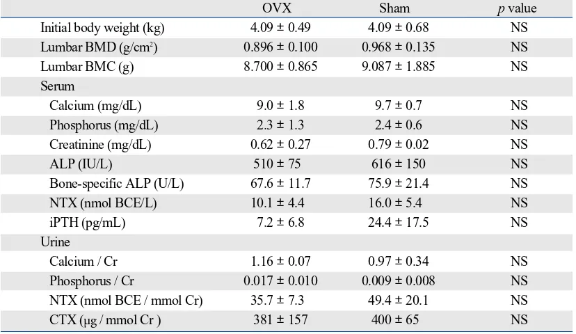
Table 1 shows the baseline characteristics of the animals. There were no significant differences in initial body weight, lumbar BMD and BMC, serum and urinary levels of cal-cium, phosphorus, and creatinine, and serum and urinary
levels of bone turnover markers between the 2 groups.
[image:4.595.316.526.207.359.2]Changes in body weight
Fig. 1 shows longitudinal changes in body weight. Body weight significantly increased in the Sham group, but did not significantly change in the OVX group (one-way ANOVA with repeated measurements, Table 2). However, there was no significant difference in changes in body weight between the 2 groups (two-way ANOVA with repeated measure-ments, Table 2).
[image:4.595.98.512.457.697.2]RESULTS
Table 1.Baseline Characteristics of the Animals
OVX Sham pvalue
Initial body weight (kg) 4.09 ±0.49 4.09 ±0.68 NS
Lumbar BMD (g/cm2) 0.896 ±0.100 0.968 ±0.135 NS
Lumbar BMC (g) 8.700 ±0.865 9.087 ±1.885 NS
Serum
Calcium (mg/dL) 9.0 ±1.8 9.7 ±0.7 NS
Phosphorus (mg/dL) 2.3 ±1.3 2.4 ±0.6 NS
Creatinine (mg/dL) 0.62 ±0.27 0.79 ±0.02 NS
ALP (IU/L) 510 ±75 616
±
150 NSBone-specific ALP (U/L) 67.6 ±11.7 75.9 ±21.4 NS
NTX (nmol BCE/L) 10.1 ±4.4 16.0 ±5.4 NS
iPTH (pg/mL) 7.2 ±6.8 24.4 ±17.5 NS
Urine
Calcium / Cr 1.16 ±0.07 0.97 ±0.34 NS
Phosphorus / Cr 0.017 ±0.010 0.009 ±0.008 NS
NTX (nmol BCE / mmol Cr) 35.7 ±7.3 49.4 ±20.1 NS
CTX (µg / mmol Cr ) 381 ±157 400 ±65 NS
OVX, ovariectomy; BMD, bone mineral density; BMC, bone mineral content; ALP, alkaline phosphatase; NTX, cross-linked N-terminal telopeptides of type I collagen; iPTH, intact parathyroid hormone; CTX, cross-linked C-terminal telopeptides of type I collagen; Cr, creatinine; NS, not significant.
Data are expressed as means ±standard deviation. Unpaired t-test was used to compare the data between the two groups.
Changes in lumbar BMD and BMC
Fig. 2 shows longitudinal changes in the lumbar BMD and BMC. Lumbar BMD significantly decreased in the OVX group, but did not significantly change in the Sham group (one-way ANOVA with repeated measurements, Table 2). Lumbar BMC significantly increased in the Sham group, but did not significantly change in the OVX group (one-way ANOVA with repeated measurements, Table 2). There was a significant difference in changes in the lumbar BMD and BMC between the 2 groups (two-way ANOVA with repeated measurements, Table 2). Thus, OVX induced not only relative osteopenia but also absolute osteopenia, as indicated by a reduction in lumbar BMD compared with Sham controls and the baseline, respectively.
Changes in serum and urine biochemical markers Fig. 3 shows longitudinal changes in the serum and urinary levels of bone turnover markers. Serum and urinary NTX levels significantly increased, while urinary CTX levels did not significantly change in both groups (one-way ANOVA with repeated measurements, Table 2). Bone-specific ALP levels significantly increased in the OVX group, but signi-ficantly decreased in the Sham group (one-way ANOVA with repeated measurements, Table 2). There were signifi-cant differences in changes in urinary NTX and CTX levels and serum bone-specific ALP levels, but not in those in serum NTX levels, between the 2 groups (two-way ANOVA with repeated measurements, Table 2).
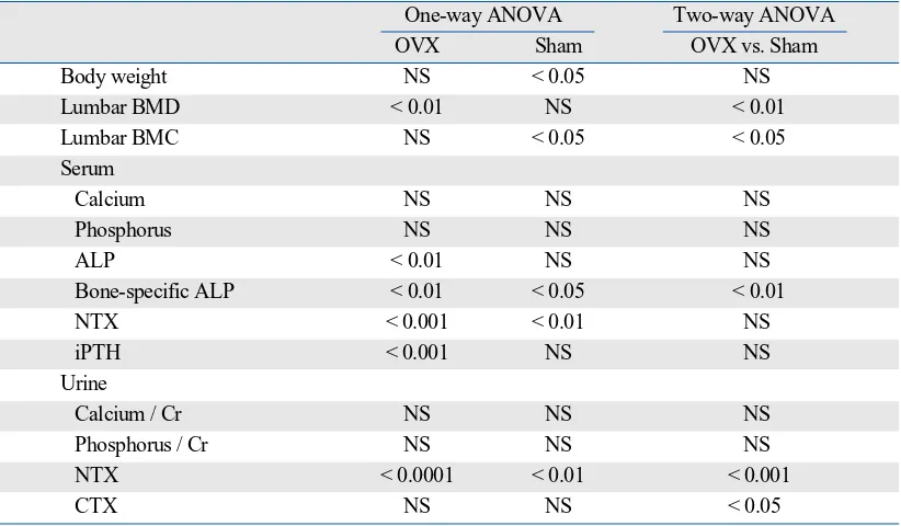
[image:5.595.86.497.74.314.2]Fig. 4 shows longitudinal changes in the serum levels of Table 2.One-way and Two-way ANOVA with Repeated Measurements
One-way ANOVA Two-way ANOVA
OVX Sham OVX vs. Sham
Body weight NS < 0.05 NS
Lumbar BMD < 0.01 NS < 0.01
Lumbar BMC NS < 0.05 < 0.05
Serum
Calcium NS NS NS
Phosphorus NS NS NS
ALP < 0.01 NS NS
Bone-specific ALP < 0.01 < 0.05 < 0.01
NTX < 0.001 < 0.01 NS
iPTH < 0.001 NS NS
Urine
Calcium / Cr NS NS NS
Phosphorus / Cr NS NS NS
NTX < 0.0001 < 0.01 < 0.001
CTX NS NS < 0.05
ANOVA, analysis of variance; OVX, ovariectomy; BMD, bone mineral density; BMC, bone mineral content; ALP, alkaline phosphatase; NTX, cross-linked N-terminal telopeptides of type I collagen; iPTH, intact parathyroid hormone; CTX, cross-linked C-terminal telopeptides of type I collagen; NS, not significant.
62
0 16 32 53 (wks)
Sham
OVX OVX
BMD
.75 .80 .85 .90 .95 1.00 1.05 1.10 1.15 (g/cm2)
62
0 16 32 53 (wks)
Sham BMC
[image:5.595.60.521.359.503.2]7.5 8.0 8.5 9.0 9.5 10.0 10.5 11.0 (g)
64
0 16 32 48 (wks)
Sham OVX OVX Serum NTX 5 10 15 20 25 30 35 40 45 50 (nmol BCE/L)
53 0 16 32 53 64(wks)
Sham Urinary NTX 20 40 60 80 100 120 140 160 180 200
(nmol BCE/mmol Cr)
48
64
0 16 32 48 (wks)
Sham OVX OVX Urinary CTX 100 200 300 400 500 600 700 800
53 0 16 32 53 64(wks)
[image:6.595.72.537.63.323.2]Sham Bone-specific ALP 40 50 60 70 80 90 100 110 120 130 (U/L) 48 (µg/mmol Cr)
Fig. 3.Changes in serum and urinary levels of bone turnover markers. Data are expressed as means ±standard error. Serum and urinary NTX levels significantly increased and urinary CTX levels did not in both groups {one-way analysis of variance (ANOVA) with repeated measurements, Table 2}. Bone-specific ALP levels significantly increased in the OVX group, but significantly decreased in the Sham group (one-way ANOVA with repeated measurements, Table 2). There were significant differences in changes of urinary NTX and CTX levels and serum bone-specific ALP levels, but not in those in serum NTX levels, between the 2 groups (two-way ANOVA with repeated measurements, Table 2). OVX, ovariectomy; NTX, cross-linked N-terminal telopeptides of type I collagen; CTX, cross-linked C-terminal telopeptides of type I collagen; Cr, creatinine; ALP, alkaline phosphatase.
64
0 16 32 48 (wks)
Sham OVX OVX Calcium 6.0 6.5 7.0 7.5 8.0 8.5 9.0 9.5 10.0 10.5 11.0 11.5 (mg/dL)
53 0 16 32 53 64 (wks)
Sham Phosphorus 1.0 1.5 2.0 2.5 3.0 3.5 4.0 4.5 (mg/dL) 48 64
0 16 32 48 (wks)
Sham OVX OVX ALP 300 400 500 600 700 800 900 1,000 1,100
53 0 16 32 53 64 (wks)
Sham iPTH 0 20 40 60 80 100 120 140 160 180 (pg/mL) 48 (IU/L)
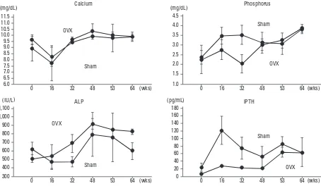
[image:6.595.79.533.416.680.2]calcium, phosphorus, ALP, and iPTH. Serum calcium and phosphorus levels did not significantly change in both groups (one-way ANOVA with repeated measurements, Table 2). Serum ALP and iPTH levels significantly increas-ed in the OVX group, but did not significantly change in the Sham group (one-way ANOVA with repeated measure-ments, Table 2). There were no significant differences in changes in serum calcium, phosphorus, ALP, and iPTH levels between the 2 groups (two-way ANOVA with repeat-ed measurements, Table 2).
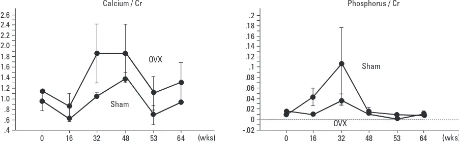
Fig. 5 shows longitudinal changes in the urinary levels of calcium and phosphorus. Urinary calcium/Cr and phos-phorus/Cr did not significantly change in both groups (one-way ANOVA with repeated measurements, Table 2). There were no significant differences in changes in urinary calcium/ Cr and phosphorus/Cr between the 2 groups (two-way ANOVA with repeated measurements, Table 2).
[image:7.595.64.515.395.537.2]Final body weight, femoral and tibial length, and uterus, liver, and kidney weight at necropsy
Table 3 shows final body weight, femoral and tibial length, and uterus, liver, and kidney weight at necropsy. There were no significant differences in final body weight, femoral and tibial length, or liver and kidney weight. However, the uterus weight was significantly lower in the OVX group than in
the Sham group, suggesting successful surgery in the OVX group.
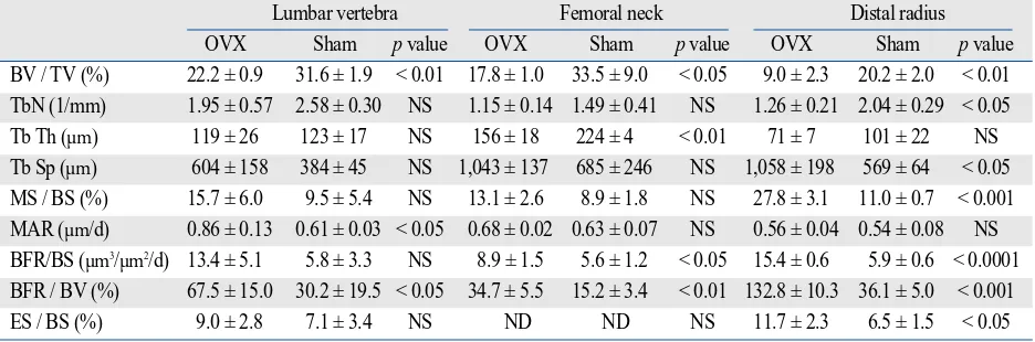
Bone histomorphometric analysis of trabecular bone of the lumbar vertebra, femoral neck, and distal radius Table 4 shows the results of static and dynamic bone histomorphometric analysis of trabecular bone of the lumbar vertebra, femoral neck, and distal radius. BV/TV of the lumbar vertebra, femoral neck, and distal radius was significantly lower in the OVX group than in the Sham group, which was associated with increased BFR/BV. How-ever, the response of trabecular bone structural parameters and bone formation and erosion parameters differed among the skeletal sites.
pQCT analysis of trabecular bone of the lumbar vertebra, femoral neck, and distal radius
Table 5 shows the results of pQCT analysis of trabecular bone of the lumbar vertebra, femoral neck, and distal radius. Trabecular vBMD of the lumbar vertebra, femoral neck, and distal radius was lower in the OVX group than in the Sham group. In particular, the total area of the femoral neck was greater in the OVX group than in the Sham group, suggesting cortical expansion in the femoral neck caused by OVX.
64
0 16 32 53 (wks)
Sham
OVX
OVX Calcium / Cr
.4 .6 .8 1.0 1.2 1.4 1.6 1.8 2.0 2.2 2.4 2.6
48 0 16 32 53 64 (wks)
Sham Phosphorus / Cr
-.02 0 .02 .04 .06 .08 .1 .12 .14 .16 .18 .2
48
[image:7.595.88.497.604.715.2]Fig. 5.Changes in urinary levels of calcium and phosphorus. Data are expressed as means ±standard error. Urinary calcium/Cr and phosphorus/Cr did not significantly change in the both groups {one-way analysis of variance (ANOVA) with repeated measurements, Table 2}. There were no significant differences in changes in urinary calcium/Cr and phosphorus/Cr between the 2 groups (two-way ANOVA with repeated measurements, Table 2). OVX, ovariectomy; Cr, creatinine.
Table 3.Final Body Weight, Femoral and Tibial Length, and Uterus, Liver, and Kidney Weight at Necropsy
OVX Sham pvalue
Final body weight (kg) 3.95 ±0.45 4.33 ±1.02 NS
Femoral length (cm) 13.9 ±0.3 14.1 ±0.8 NS
Tibial length (cm) 12.6 ±0.2 12.7 ±0.9 NS
Uterus weight (g) 1.77 ±0.41 9.15 ±1.84 < 0.01
Liver weight (g) 91.9 ±9.9 86.3 ±6.6 NS
Right kidney weight (g) 11.1 ±1.3 9.4 ±1.4 NS
Left kidney weight (g) 11.3 ±1.5 9.2 ±2.1 NS
OVX, ovariectomy NS, not significant.
Biomechanical analysis of the lumbar vertebra and femoral neck
Table 6 shows the results of biomechanical testing of the lumbar vertebra and femoral neck. There were no signifi-cant differences in the maximum load, stiffness, and breaking energy of the lumbar vertebra and femoral neck between the 2 groups. The lack of any significant differences in the parameters might be attributable to the low statistical power due to the small sample size.
The present study clearly demonstrated that OVX in mature cynomolgus monkeys, aged 17-21 years, increased bone turnover markers such as serum bone-specific ALP and
urinary NTX and CTX, and induced trabecular bone loss associated with increased BFR/BV compared with Sham controls at the lumbar vertebra, femoral neck, and distal radius. OVX induced not only relative osteopenia but also absolute osteopenia as indicated by a reduction in lumbar BMD compared with Sham controls and the baseline, res-pectively, confirming that mature cynomolgus monkeys could be utilized for preclinical studies of postmenopausal osteoporosis to examine the effects of interventions on bone turnover and trabecular bone mass at the 3 clinically impor-tant skeletal sites. Thus, we support the concept that nonhu-man primates are a good model for postmenopausal osteo-porosis based on densitometric, histomorphometric and serum bone marker analysis.7
Concern has been expressed that the OVX monkey model does not produce consistent, statistically significant
[image:8.595.71.539.75.230.2]DISCUSSION
Table 4.Bone Histomorphometric Analysis of Trabecular Bone of the Lumbar Vertebra, Femoral Neck, and Distal Radius
Lumbar vertebra Femoral neck Distal radius
OVX Sham pvalue OVX Sham pvalue OVX Sham pvalue
BV / TV (%) 22.2 ± 0.9 31.6 ± 1.9 < 0.01 17.8 ± 1.0 33.5 ± 9.0 < 0.05 9.0 ± 2.3 20.2 ± 2.0 < 0.01 TbN (1/mm) 1.95 ± 0.57 2.58 ± 0.30 NS 1.15 ± 0.14 1.49 ± 0.41 NS 1.26 ± 0.21 2.04 ± 0.29 < 0.05 Tb Th (µm) 119 ± 26 123 ± 17 NS 156 ± 18 224 ± 4 < 0.01 71 ± 7 101 ± 22 NS Tb Sp (µm) 604 ± 158 384 ± 45 NS 1,043 ± 137 685 ± 246 NS 1,058 ± 198 569 ± 64 < 0.05 MS / BS (%) 15.7 ± 6.0 9.5 ± 5.4 NS 13.1 ± 2.6 8.9 ± 1.8 NS 27.8 ± 3.1 11.0 ± 0.7 < 0.001 MAR (µm/d) 0.86 ± 0.13 0.61 ± 0.03 < 0.05 0.68 ± 0.02 0.63 ± 0.07 NS 0.56 ± 0.04 0.54 ± 0.08 NS BFR/BS (µm3/µm2/d) 13.4 ± 5.1 5.8 ± 3.3 NS 8.9 ± 1.5 5.6 ± 1.2 < 0.05 15.4 ± 0.6 5.9 ± 0.6 < 0.0001 BFR / BV (%) 67.5 ± 15.0 30.2 ± 19.5 < 0.05 34.7 ± 5.5 15.2 ± 3.4 < 0.01 132.8 ± 10.3 36.1 ± 5.0 < 0.001 ES / BS (%) 9.0 ± 2.8 7.1 ± 3.4 NS ND ND NS 11.7 ± 2.3 6.5 ± 1.5 < 0.05 OVX, ovariectomy; BV, bone volume; TV, total tissue volume; TbN, trabecular number; Tb Th, trabecular thickness; Tb Sp, trabecular separation; MS, mineralizing surface; BS, bone surface; MAR, mineral apposition rate; BFR, bone formation rate; ES, eroded surface; ND, not detected; NS, not significant.
[image:8.595.71.540.298.379.2]Data are expressed as means ±standard deviation. Unpaired t-test was used to compare the data between the two groups.
Table 5.pQCT Analysis of Trabecular Bone of the Lumbar Vertebra, Femoral Neck, and Distal Radius
Lumbar vertebra Femoral neck Distal radius
OVX Sham pvalue OVX Sham pvalue OVX Sham pvalue
Total vBMD (mg/cm3) 438 ± 72 577 ± 56 < 0.05 718 ± 7 918 ± 97 < 0.05 470 ± 27 515 ± 17 NS Trabecular vBMD (mg/cm3) 259 ± 60 384 ± 45 < 0.05 287 ± 71 587 ± 170 < 0.05 123 ± 23 216 ± 24 < 0.01 Total area (mm2) 106.9 ± 8.5 114.9 ± 14.4 NS 71.2 ± 2.4 63.6 ± 2.9 < 0.05 61.1 ± 0.1 66.1 ± 7.5 NS Trabecular area (mm2) 47.9 ± 4.3 51.8 ± 6.4 NS 21.4 ± 0.7 19.1 ± 0.8 < 0.05 27.5 ± 0.1 29.8 ± 3.4 NS pQCT, peripheral quantitative computed tomography; OVX, ovariectomy; vBMD, volumetric bone mineral density; NS, not significant.
[image:8.595.70.541.429.497.2]Data are expressed as means ±standard deviation. Unpaired t-test was used to compare the data between the two groups.
Table 6.Bone Strength of the Lumbar Vertebra and Femoral Neck
Lumbar vertebra Femoral neck
OVX Sham pvalue OVX Sham pvalue
Maximum load (N) 548 ± 169 836 ± 303 NS 1,463 ± 116 1,681 ± 649 NS Stiffness (N/m) 13,091 ± 1,463 13,730 ± 997 NS 9,260 ± 3,299 9,952 ± 3,506 NS Breaking energy (N m 10-3) 7,257 ± 2,140 10,181 ± 3,835 NS 20,193 ± 6,171 19,871 ± 3,384 NS OVX, ovariectomy; NS, not significant.
osteopenia and bone fragility, and frequently exhibits a “failure to gain” bone, rather than the clear bone loss expected of a model for postmenopausal osteoporosis.8 Relative osteopenia compared with intact animals is observ-ed in younger OVX cynomolgus monkeys who were fobserv-ed a normal calcium diet, whereas absolute osteopenia develops in OVX cynomolgus monkeys at age > 9 years and conco-mitantly fed a low calcium diet.13In the present study, however, OVX in cynomolgus monkeys, aged 17-21 years, induced absolute osteopenia under the condition of a normal calcium diet (0.93%), as indicated by a reduction in lumbar BMD from the baseline. Thus, not only the dietary calcium content but also ages of the animals might be important to develop absolute osteopenia in OVX cynomolgus monkeys. Urinary levels of NTX and CTX and serum levels of bone-specific ALP levels increased after OVX in the present study. Previous studies have shown that OVX in cynomo-lgus monkeys increased urinary levels of deoxypyridinoline and serum levels of osteocalcin, ALP, bone-specific ALP, and tartrate-resistant acid phosphatase.5,9,14-16However, recent studies demonstrated OVX-induced elevations in urinary levels of CTX in addition to the above markers in cynomolgus monkeys.17,18Serum and urinary levels of bone turnover markers were shown to increase within a few months, peak at 6-12 months, and remain elevated for 18 months or longer.8All of these results indicate that OVX rapidly increases the serum and urinary levels of bone turnover markers and maintains this state for a couple of years in cynomolgus monkeys, consistent with our results (Fig. 3). However, the present study confirmed changes in newer bone markers including urinary NTX and CTX in OVX cynomolgus monkeys.
In the present study, trabecular vBMD and trabecular bone mass (BV/TV) of the lumbar vertebra, femoral neck, and distal radius were similarly lower in the OVX group than in the Sham group. Trabecular bone loss compared with Sham controls at the 3 skeletal sites was clearly associat-ed with an increasassociat-ed bone formation rate (BFR/BV). This result is consistent with those of the study conducted by Jerome et al.5,16They reported that vertebral trabecular bone loss was associated with dramatically increased bone forma-tion rates, primarily caused by higher activaforma-tion frequency of basic multicellular units of bone in cynomolgus monkeys (4-14 years of age),5and that OVX increased the bone formation rates in trabecular bone obtained by iliac biopsy in cynomolgus monkeys (5-8 years of age).16Thus, increased bone formation rates appear to be observed at multiple trabecular bone sites with bone histomorphometry.8
Because of the small sample size, we could not detect any significant changes after OVX in bone strength of the lumbar vertebra and femoral neck, as observed in cynomol-gus monkeys, aged 11-15 years.14Furthermore, we could not find any significant changes after OVX in cortical bone
mass and strength of the femoral and tibial diaphysis, evaluated by pQCT analysis and biomechanical testing (three-point bending test), respectively (data not shown). No significant changes in cortical bone mass as well as cortical porosity were detected at the femoral neck and distal radius, even though we performed histomorphometry of cortical bone (data not shown). Jerome et al.13 recommend-ed a group size of 25 for studies in cynomolgus monkeys. Thus, further studies are needed to clarify the influence of OVX on bone strength and cortical bone mass in mature cynomolgus monkeys. However, the statistically significant changes in bone turnover and trabecular bone mass at the 3 clinically important skeletal sites are worth reporting in the present study, as well as absolute osteopenia as indicated by a longitudinal decrease in lumbar BMD from the baseline even with a small sample size in mature cynomolgus monkeys.
In conclusion, the present study clearly demonstrated that OVX in mature cynomolgus monkeys, aged 17-21 years, increased bone turnover markers and induced trabecular bone loss compared with Sham controls at the lumbar vertebra, femoral neck, and distal radius. OVX also induced absolute osteopenia as indicated by a reduction in lumbar BMD from the baseline. Thus, the results strongly indicate that mature cynomolgus monkeys could be utilized for pre-clinical studies of postmenopausal osteoporosis to examine the effects of interventions on bone turnover and trabecular bone mass at the three clinically important skeletal sites.
We thank Dr. Takashi Kanda (Nippon Chemiphar Co., Ltd., Tokyo, Japan) for the serum and urine biochemical analyses, Drs. Kiichi Nonaka and Kaoru Shindo-Yamamoto (Elk Co., Ltd., Tokyo, Japan) for the pQCT analysis, Dr. Tsuyoshi Ishii (Maruto Co., Ltd., Tokyo, Japan) for the biomecha-nical testing, and Dr. Toshihiro Takahashi (Nippon Chemiphar Co., Ltd., Tokyo, Japan) for the preparation of the manuscript.
1. Balena R, Toolan BC, Shea M, Markatos A, Myers ER, Lee SC, et al. The effects of 2-year treatment with the aminobisphosphonate alendronate on bone metabolism, bone histomorphometry, and bone strength in ovariectomized nonhuman primates. J Clin Invest 1993;92:2577-86.
2. Pope NS, Gould KG, Anderson DC, Mann DR. Effects of age and sex on bone density in the rhesus monkey. Bone 1989;10:109-12. 3. Johansson ED, Neill JD, Knobil E. Periovulatory progesterone
concentration in the peripheral plasma of the rhesus monkey with a methodologic note on the detection of ovulation. Endocrinology 1968;82:143-8.
4. Neill JD, Johansson ED, Knobil E. Patterns of circulating proge-sterone concentrations during the fertile menstrual cycle and the remainder of gestation in the rhesus monkey. Endocrinology 1969; 84:45-8.
5. Jerome CP, Carlson CS, Register TC, Bain FT, Jayo MJ, Weaver DS, et al. Bone functional changes in intact, ovariectomized, and ovariectomized, hormone-supplemented adult cynomolgus monkeys (Macaca fascicularis) evaluated by serum markers and dynamic histomorphometry. J Bone Miner Res 1994;9:527-40.
6. Jayo MJ, Jerome CP, Lees CJ, Rankin SE, Weaver DS. Bone mass in female cynomolgus macaques: a cross-sectional and longitu-dinal study by age. Calcif Tissue Int 1994;54:231-6.
7. Jerome CP, Carlson CS, Jayo MJ, Register TC, Weaver DS, Lees CJ, et al. Histomorphometric and mineral density fractionation studies of lumbar vertebrae of intact and ovariecto-mized (OVX) monkeys. Bone Miner 1994;26:275-8.
8. Jerome CP, Peterson PE. Nonhuman primate models in skeletal research. Bone 2001;29:1-6.
9. Itoh F, Kojima M, Furihata-Komatsu H, Aoyagi S, Kusama H, Komatsu H, et al. Reductions in bone mass, structure, and strength in axial and appendicular skeletons associated with increased turnover after ovariectomy in mature cynomolgus monkeys and preventive effects of clodronate. J Bone Miner Res 2002;17:534-43. 10. Pavelka MS, Fedigan LM. Reproductive termination in femdle
Japanese monkeys: A comparative life history perspective. Am J Phys Anthropol 1999;109:455-64.
11. Erben RG. Embedding of bone samples in methylmethacrylate: an improved method suitable for bone histomorphometry, histoche-mistry, and immunohistochemistry. J Histochem Cytochem 1997;
45:307-13.
12. Parfitt AM, Drezner MK, Glorieux FH, Kanis JA, Malluche H, Meunier PJ, et al. Bone histomorphometry: standardization of nomenclature, symbols, and units. Report of the ASBMR Histo-morphometry Nomenclature Committee. J Bone Miner Res 1987; 2:595-610.
13. Jerome CP, Lees CJ, Weaver DS. Development of osteopenia in ovariectomized cynomolgus monkeys (Macaca fascicularis). Bone 1995;17:403S-8S.
14. Jerome CP, Turner CH, Lees CJ. Decreased bone mass and strength in ovariectomized cynomolgus monkeys (Macaca fascicularis). Calcif Tissue Int 1997;60:265-70.
15. Hotchkiss CE, Stavisky R, Nowak J, Brommage R, Lees CJ, Kaplan J. Levormeloxifene prevents increased bone turnover and vertebral bone loss following ovariectomy in cynomolgus monkeys. Bone 2001;29:7-15.
16. Jerome CP, Power RA, Obasanjo IO, Register TC, Guidry M, Carlson CS, et al. The androgenic anabolic steroid nandrolone decanoate prevents osteopenia and inhibits bone turnover in ovariectomized cynomolgus monkeys. Bone 1997;20:355-64. 17. Lees CJ, Register TC, Turner CH, Wang T, Stancill M, Jerome
CP. Effects of raloxifene on bone density, biomarkers, and histo-morphometric and biomechanical measures in ovariectomized cynomolgus monkeys. Menopause 2002;9:320-8.