Endobronchial Ultrasound-Guided Transbronchial
Needle Aspiration for the Diagnosis of Central
Lung Parenchymal Lesions
Akash Verma,
1,2Kyeongman Jeon,
1Won-Jung Koh,
1Gee Young Suh,
1Man Pyo Chung,
1Hojoong Kim,
1O Jung Kwon,
1and Sang-Won Um
11Division of Pulmonary and Critical Care Medicine, Department of Medicine, Samsung Medical Center,
Sungkyunkwan University School of Medicine, Seoul, Korea.
2Department of Pulmonary and Critical Care Medicine, Changi General Hospital, Singapore.
Received: July 20, 2012 Revised: August 23, 2012 Accepted: August 24, 2012
Corresponding author: Dr. Sang-Won Um, Division of Pulmonary and Critical Care Medicine, Department of Medicine, Samsung Medical Center, Sungkyunkwan University School of Medicine,
50 Irwon-dong, Gangnam-gu, Seoul 135-710, Korea.
Tel: 82-2-3410-3429, Fax: 82-2-3410-3849 E-mail: sangwonum@skku.edu
∙ The authors have no financial conflicts of interest.
© Copyright:
Yonsei University College of Medicine 2013
This is an Open Access article distributed under the terms of the Creative Commons Attribution Non-Commercial License (http://creativecommons.org/ licenses/by-nc/3.0) which permits unrestricted non-commercial use, distribution, and reproduction in any medium, provided the original work is properly cited.
Purpose: The purpose of this study was to evaluate the usefulness of convex
probe endobronchial ultrasound-guided transbronchial needle aspiration (EBUS-TBNA) for detecting malignancy in parenchymal pulmonary lesions located adja-cent to the adja-central airways. Materials and Methods: We retrospectively reviewed the diagnostic performance of EBUS-TBNA in consecutive patients with high clinical suspicion of a centrally located primary lung cancer who had undergone EBUS-TBNA at the Samsung Medical Center between May 2009 and June 2011.
Results: Thirty-seven patients underwent EBUS-TBNA for intrapulmonary
le-sions adjacent to the central airways. Seven lele-sions were located adjacent to the trachea and 30 lesions were located adjacent to the bronchi. Cytologic and histo-logic samples obtained via EBUS-TBNA were diagnostic in 32 of 37 (86.4%) of patients. The final diagnosis was lung cancer in 30 patients (7 small cell lung can-cer, 23 non-small cell lung cancer), lymphoma in one and malignant fibrous histio-cytoma in one patient. The diagnostic sensitivity of EBUS-TBNA in detecting ma-lignancy and detecting both mama-lignancy and benignity was 91.4% and 86.5%, respectively. Two patients experienced minor complications. Conclusion: EBUS-TBNA is an effective and safe method for tissue diagnosis of parenchymal lesions that lie centrally close to the airways. EBUS-TBNA should be considered the pro-cedure of choice for patients with centrally located lesions without endobronchial involvement.
Key Words: Bronchoscopy, endobronchial ultrasound, transbronchial needle as-piration, lung cancer, parenchymal lesion
INTRODUCTION
Lung cancer is the leading cause of cancer related deaths worldwide, with an esti-mated one to two million deaths each year.1 Lung cancer causes more deaths than
co-UC260F-OL8, Olympus Ltd., Tokyo, Japan) connected to an ultrasound unit (EU-C60 Olympus Ltd., Tokyo, Japan). The procedures were performed under local anesthesia (ligno-caine) and moderate sedation (midazolam). For paratracheal lesions, the scope was positioned endotracheally. For peri-bronchial lesions the scope was positioned in the respective bronchi in order to visualize the lung lesion. TBNA was per-formed using a 22-gauge needle (NA-2015X-4022, Olym-pus Ltd., Tokyo, Japan). Two aspirates were performed with 15 passes (moving needle back & forth in the lesion) per as-pirate. The core tissue was expelled onto piece of paper for histological examination and the needle was flushed with sa-line onto glass slides for cytological examination. The aspi-rate was smeared onto glass slides, air dried, fixed immedi-ately with 95% alcohol, and stained with Hematoxylin and Eosin (HE). Histological cores were fixed with 10% neutral buffered formalin and stained with HE. Rapid on-site cyto-logical examination was not available. A post-procedure chest X-ray was routinely performed to exclude any proce-dure-related complications.
Definitions of diagnosis for study patient
Cyto-pathological specimens from EBUS-TBNA were cate-gorized as malignant (presence of malignant cells), benign (normal lung tissue without malignant cells), or inadequate (no or scanty cellular component, blood only, mucus, or be-nign bronchial cells only). Malignant and bebe-nign samples diagnosed by EBUS-TBNA were defined as adequate ples. Tumor-positive findings from the EBUS-TBNA sam-ples were not surgically validated. The diagnostic standard for benign disease was pathological or microbiological con-firmation of a specific benign disease, surgical concon-firmation of lesions showing no malignant cells, or spontaneous re-gression of lung parenchymal lesion after EBUS-TBNA during follow-up of more than 6 months. Malignant disease was defined as confirmation of malignant cells or tissues from EBUS-TBNA, CT-guided transthoracic needle aspira-tion or surgical lung biopsy. Cases which did not satisfy the criteria of benign or malignant disease were classified as in-determinate cases.
If the pathology from the EBUS-TBNA samples resulted in a diagnosis of malignancy, corroborative evidence of ma-lignancy was looked for in the form of surgical lung biopsy or biopsy results of tissue obtained from some other site sus-pected of malignant involvement. The results of the concur-rent EBUS-TBNA of mediastinal lymph nodes were ex-cluded from analysis.
lon, pancreas and prostate.1 If lung cancer is suspected, a
histological diagnosis, in conjunction with accurate staging, is needed in order to guide therapy and prognosis.2,3
The conventional methods for establishing diagnosis of intraparenchymal lesions are transthoracic needle aspiration (TTNA) or transbronchial needle aspiration/biopsy by bronchoscopy with or without fluoroscopy. Other newer techniques such as computed tomography (CT) guided bronchoscopy,4 virtual bronchoscopy,5,6 electromagnetic
navigation bronchoscopy7,8 and endobronchial ultrasound
guide-sheath9,10 have been developed to improve diagnostic
yield, but have not become mainstream modalities and are only available at limited centers.
Convex probe endobronchial ultrasound-guided trans-bronchial needle aspiration (EBUS-TBNA) has been shown to be useful for staging of the mediastinum with high sensitivity, and when combined with endoscopic ultra-sound, accessing lymph node stations inaccessible even by mediastinoscopy, enabling near complete mediastinal eval-uation.11-14
Although the utility of EBUS-TBNA to diagnose paren-chymal lesions in cases of non-diagnostic conventional bron-choscopy is being recognized and emerging, there are limited reports about its application for this purpose.15-17 The purpose
of this study was to evaluate the usefulness of EBUS-TBNA for detecting malignancy in the parenchymal lesions locat-ed adjacent to the central airways.
MATERIALS AND METHODS
Study design and patients
We retrospectively reviewed the diagnostic performance of EBUS-TBNA in consecutive patients with high clinical sus-picion of a centrally located primary lung cancer who had undergone EBUS-TBNA at the Samsung Medical Center be-tween May 2009 and June 2011. The centrally located lung lesions were defined as an intrapulmonary nodule or mass lo-cated adjacent to the tracheobronchial tree as visualized on chest CT scan. This study was approved by the Institutional Review Board of the Samsung Medical Center, which waived the requirements for informed consent of the individual pa-tients, given the retrospective nature of the study.
EBUS-TBNA procedure
were performed in relation to diagnosis of malignancy and diagnosis of all disease (benignity and malignancy). Pa-tients who had an indeterminate final diagnosis were con-sidered a false negative result of EBUS-TBNA. The sensi-tivity, specificity, and diagnostic accuracy for detecting malignancy were calculated using the standard definitions.
RESULTS
Characteristics of study patients
Thirty-seven patients underwent EBUS-TBNA for intrapul-monary lesions adjacent to the central airways. There were 25 (68%) males and the median age was 63 years (40-81). The size of pulmonary lesions on CT varied from 8 to 82 mm in short axis with a median of 27.5 mm (22 lesions were less than 3 cm and 15 were larger than 3 cm). Positron emission tomography (PET) scan was done in 34 (92%) patients and increased FDG uptake was seen in the paren-chymal mass lesion in all patients. However, median stan-dard uptake value (SUVmax) was only available in 29 (78%)
patients as 8 patients had PET scan done outside our institu-tion. Median SUVmax was 12 (3.3-47.3) (Table 1). Fifteen
(40.5%) patients had endobronchial abnormality. Twelve (32%) had mucosal or submucosal abnormality such as exophytic tumor or hyperaemia, and 3 (8%) had extrinsic compression. Twenty-two (59%) had no endobronchial ab-normality (Table 1).
EBUS-TBNA procedures
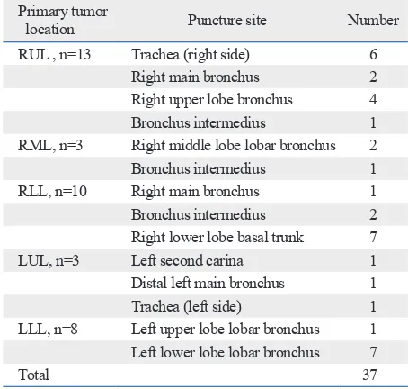
Locations of primary tumors sampled by EBUS-TBNA and puncture sites are shown in Table 2. Right upper lobe lesions were targeted predominantly through the trachea, right main stem bronchus, or bronchus intermedius. Left upper lobe masses were sampled through the left upper lobar bronchus at its orifice, the distal left main bronchus and the left side of the trachea. Both lower lobar masses were targeted through the distal main bronchus, bronchus intermedius or the basal trunk. Two patients experienced complications. One devel-oped pneumothorax requiring chest tube insertion and an-other patient developed moderate bleeding which resolved spontaneously. Both patients recovered from their complica-tions uneventfully. A representative case of EBUS-TBNA for lung parenchymal lesion is described in Fig. 1.
Other diagnostic modalities
Twenty-two out of 37 (59.4%) patients had undergone
mu-Data analysis
[image:3.595.57.282.135.406.2]All data are presented as a number (%) or median (range) unless otherwise stated. Diagnostic performance analyses
Table 1. Baseline Characteristics of the Study Patients
Variables median (range) Number (%) or
Number of patients 37
Age (yrs) 63 (40-81)
Male gender 25 (68)
Type of disease
First presentation with suspected
lung cancer 32 (86.4)
Prior lung cancer with surgical
resection and suspected recurrence 5 (13.5) Performance of PET/CT scan before
EBUS-TBNA 34 (92)
Number of patients with increased
FDG uptake in lung lesion 34 (100) Maximum standardized uptake
value (n=30) 12 (3.3-47.3)
Short axis diameter of the lesion (mm) 27.5 (8-82) Types of endobronchial lesions
No endobronchial abnormality 22 (59)
Endobronchial abnormality 15 (40.5) Extrinsic compression only 3 (8) Mucosal and submucosal abnormality 12 (32) EBUS-TBNA, endobronchial ultrasound-guided transbronchial needle; PET, positron emission tomography; CT, computed tomography; FDG, fluorode-oxyglucose.
Table 2. Location of Lung Parenchymal Lesions and Punc-ture Sites of EBUS-TBNA
Primary tumor
location Puncture site Number
RUL , n=13 Trachea (right side) 6
Right main bronchus 2
Right upper lobe bronchus 4
Bronchus intermedius 1
RML, n=3 Right middle lobe lobar bronchus 2
Bronchus intermedius 1
RLL, n=10 Right main bronchus 1
Bronchus intermedius 2
Right lower lobe basal trunk 7
LUL, n=3 Left second carina 1
Distal left main bronchus 1
Trachea (left side) 1
LLL, n=8 Left upper lobe lobar bronchus 1
Left lower lobe lobar bronchus 7
Total 37
[image:3.595.55.282.480.696.2]cosal and transbronchial biopsies by conventional bron-choscopy. Out of these, 19 patients had it done at a median (range) interval of 4.5 (1-7) days prior to EBUS-TBNA and 3 patients had it done in the same sitting as EBUS-TBNA. The results of conventional bronchoscopic biopsy were non-diagnostic in 17 patients (15 patients in the prior to EBUS-TBNA group and in 2 patients in the same sitting group) with an overall non-diagnostic rate of 17/22 (77.3%). Out of 5 cases that had positive bronchoscopic biopsy, one had a slit like narrowing of the distal left main bronchus with a submu-cosal lesion, one had a submusubmu-cosal lesion in the left upper lobe, two had mucosal abnormality in the right upper lobe and one had a left lower lobe mucosal lesion. EBUS-TBNA was performed on these patients, despite positive yield, to as-certain the cell type when the cell type was not clearly dis-cernible (doubtful) based on conventional bronchoscopy, or to obtain additional tissue for mutation analysis (for patients who had bronchoscopy done outside our institute). Seven-teen cases were judged to be non-accessible by bronchoscop-ic biopsies based on chest CT scans and bronchoscopbronchoscop-ic find-ings. Sputum was evaluated in 21 patients and was positive in one (2.7%). TTNA was done in 7 patients and was posi-tive in one (2.7%) of these patients (Table 3).
Out of the 32 cases of malignancy detected by EBUS-TBNA, corroborative evidence of malignancy was found in the cerebrospinal fluid in 1 case, brain biopsy in 1 case, lo-bectomy in 1 case, tonsillectomy in 1 case, colon polypec-tomy in 1 case, tonsil and duodenal biopsy along with bone marrow biopsy in 1 case, pleural fluid in 1 case, thyroid fine-needle aspiration in 1 case and supraclavicular lymph node aspiration in 4 cases (Table 4).
Final diagnosis of study patients
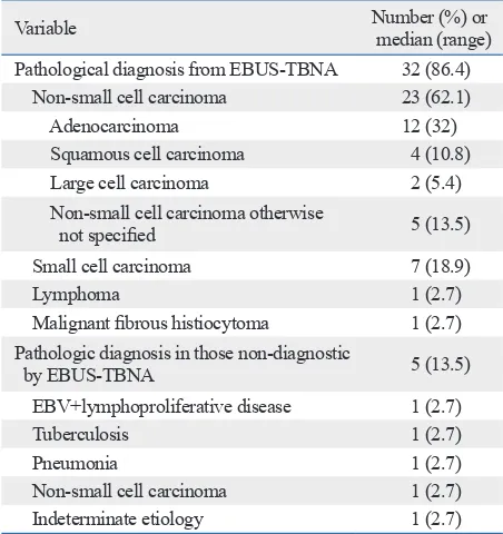
Pathologic diagnosis was achieved in 32 patients (86.4%) by EBUS-TBNA (Table 5, Fig. 2). The diagnoses
[image:4.595.72.540.68.195.2]estab-Fig. 1. A representative case of EBUS-TBNA for lung parenchymal lesions. (A) CT scan of a 43-year-old male with lung cancer showing a mass in the right upper lobe adjacent to the right lateral wall of the trachea. (B) Endobronchial ultrasound image of the same lesion with the ultrasound probe placed next to the right lateral wall of the trachea. EBUS-TBNA, endobronchial ultrasound-guided transbronchial needle aspiration.
Table 3. Diagnostic Modalities Used for 37 Study Patients
Modalities Positivity for malignancy,
number (%)
EBUS-TBNA (n=37) 32 (86.4)
Conventional bronchoscopic biopsy (n=22) 5 (13.5) Transbronchial lung biopsy (n=12) 0 (0)
Mucosal biopsy (n=10) 5 (13.5)
Timing of conventional bronchoscopy (n=22) 5 (13.5) Prior to EBUS-TBNA session (n=19) 4 (10.8) At the same time of EBUS-TBNA session
(n=3) 1 (2.7)
Bronchoalveolar lavage (n=17) 0 (0)
Transthoracic needle aspiration (n=7) 1 (2.7)
Pleural fluid cytology (n=3) 1 (2.7)
Supraclavicular lymph node aspiration (n=6) 4 (10.8) Thyroid aspiration cytology (n=1) 1 (2.7)
Sputum cytology (n=21) 1 (2.7)
EBUS-TBNA, endobronchial ultrasound-guided transbronchial needle aspi-ration.
Table 4. Sites & Modalities Providing Corroborative Evi-dence of Malignancy (n=37)
Modalities Positivity for malignancy,
number (%)
Cerebrospinal fluid (n=1) 1 (2.7)
Brain biopsy (n=1) 1 (2.7)
Lobectomy (n=1) 1 (2.7)
Tonsillectomy (n=1) 1 (2.7)
Colon polypectomy (n=1) 1 (2.7)
Tonsil, duodenal and bone marrow biopsy
(n=1) 1 (2.7)
Transthoracic needle aspiration (n=7) 1 (2.7)
Pleural fluid cytology (n=3) 1 (2.7)
Supraclavicular lymph node aspiration (n=6) 4 (10.8)
Thyroid aspiration cytology (n=1) 1 (2.7)
Sputum cytology (n=21) 1 (2.7)
[image:4.595.313.539.252.474.2] [image:4.595.314.539.535.730.2]lished were lung cancer in 30 patients [7 small cell lung cancer (SCLC), 23 non-small cell lung cancer (NSCLC)], lymphoma in one and malignant fibrous histiocytoma in one (Table 5, Fig. 2). EBUS-TBNA was non-diagnostic in 5 patients. Out of these, one patient was diagnosed with Ep-stein-Barr virus (EBV) related lymphoproliferative disorder by lobectomy, one was diagnosed with tuberculosis based on positive culture of bronchoalveolar lavage fluid, one was diagnosed with benign disease due to interval radiological resolution, one was diagnosed with non-small cell lung car-cinoma on bronchoscopic biopsy and one did not undergo any confirmatory test due to refusal. Accordingly, diagno-ses of malignant disease, benign disease and indeterminate cases were confirmed in 34, 2, and 1 patient, respectively. Among 23 patients with a final diagnosis of NSCLC, 16 had advanced disease (14 with stage IV disease and 2 with stage IIIB disease), five had stage IIIA disease, one had stage IIB disease and one had stage IB disease. Among pa-tients with a final diagnosis of SCLC, five had extensive disease and two had limited disease. Overall, three (8%) patients underwent surgery with curative intent. The diag-nosis in these patients was EBV related lymphoprolifera-tive disease (n=1), stage IB NSCLC (n=1) and malignant fibrous histiocytoma (n=1).
Diagnostic performances of EBUS-TBNA
[image:5.595.59.528.67.248.2]The sensitivity (95% confidence interval), specificity (95% confidence interval) and diagnostic accuracy of EBUS-TBNA for the diagnosis of malignancy was 91.4% (89.2- 100), 100% (15.0-85.0) and 91.9%, respectively. The diag-nostic sensitivity (95% confidence interval) of EBUS-TBNA in detecting both malignancy and benignity (all cases) was
Table 5. The Results of EBUS-TBNA Procedures in 37 Study Patients
Variable median (range)Number (%) or
Pathological diagnosis from EBUS-TBNA 32 (86.4)
Non-small cell carcinoma 23 (62.1)
Adenocarcinoma 12 (32)
Squamous cell carcinoma 4 (10.8)
Large cell carcinoma 2 (5.4)
Non-small cell carcinoma otherwise
not specified 5 (13.5)
Small cell carcinoma 7 (18.9)
Lymphoma 1 (2.7)
Malignant fibrous histiocytoma 1 (2.7) Pathologic diagnosis in those non-diagnostic
by EBUS-TBNA 5 (13.5)
EBV+lymphoproliferative disease 1 (2.7)
Tuberculosis 1 (2.7)
Pneumonia 1 (2.7)
Non-small cell carcinoma 1 (2.7)
Indeterminate etiology 1 (2.7)
EBUS-TBNA, endobronchial ultrasound-guided transbronchial needle aspi-ration; EBV, Epstein-Barr virus.
Table 6. Diagnostic Performances of EBUS-TBNA Sensitivity Specificity Accuracy Diagnosis of
malignancy* 32/35 (91.4) 2/2 (100) 34/37 (91.9) Diagnosis of
benignity and
malignancy* 32/37 (86.5) NA 32/37 (86.5)
NA, not applicable; EBUS-TBNA, endobronchial ultrasound-guided trans-bronchial needle aspiration.
[image:5.595.56.282.307.547.2]*One case that was negative by EBUS-TBNA and did not undergo any confirmation by any method was included as a false negative result of EBUS-TBNA.
Fig. 2. Diagnostic algorithm of study patients. EBUS-TBNA, endobronchial ultrasound-guided transbronchial needle aspiration; BAL, bronchoalveolar la-vage; EBV, Ebstein-Barr virus.
Non-small cell carcinoma (n=23)
Malignant fibrous histiocytoma (n=1)
Lymphoma (n=1)
Small cell carcinoma (n=7)
Tuberculosis (BAL fluid culture positive) (n=1)
Pneumonia (spontaneous radiological resolution) (n=1)
Indeterminate (patient refused evaluation) (n=1)
EBV related lymphoproliferative disorder (lobectomy) (n=1)
Non-small cell carcinoma (bronchoscopic biopsy) (n=1) No diagnosis obtained by EBUS-TBNA
(n=5) Patients undergoing EBUS-TBNA of
lung parenchymal lesions (n=37)
[image:5.595.56.283.597.671.2]eral lesions. In cases of central lesions, needles must tra-verse a significant amount of lung tissue along its transtho-racic route due to the distance between the chest wall and the lesion, and thus, pose a substantial risk of pneumotho-rax and bleeding.22,23
All of these issues can be overcome by using EBUS- TBNA, as lesions can be localized and visualized well. Sur-rounding structures and vessels can be identified by Dop-pler mode to ensure safety, and the real time nature of the procedure allows for continuous visualization of the needle throughout the lesion sampling. Additionally, specific areas within the lesion (non-necrotic) can be targeted for punc-ture to increase the yield if necessary.
Previously, Tournoy, et al.15 reported a sensitivity of 82%
and negative predictive value of 23% for EBUS-TBNA in diagnosing centrally located parenchymal lesions, similar in location to our series. They defined central lesions as those with a medial border lying within the inner third of the hemi thorax. In another study, Nakajima, et al.16
demon-strated a sensitivity, specificity and accuracy of 94.1%, 100% and 94.3% respectively in similar group of patients. Our findings were similar to these results and help strengthen the evidence in favor of this application of EBUS-TBNA. In our study, we adopted a very conservative approach to calculate the diagnostic sensitivity of EBUS-TBNA. We considered one patient who had a negative result of EBUS-TBNA and did not undergo any confirmation by any meth-od as a false negative result of EBUS-TBNA. The diagnos-tic sensitivity of EBUS-TBNA in detecting malignancy and detecting both malignancy and benignity (all cases) was 91.4% and 86.5%, respectively, in this study.
The present study clearly has limitations. First, due to its retrospective nature, “gold standard” surgical lung biopsy confirmation was not done in all patients. This was because of the unresectability of these lesions either due to advanced lung cancer, small cell lung cancer or prior lobectomy or pneumonectomy. It was judged unethical to subject these patients to a more invasive procedure when the diagnosis had been successfully established by EBUS-TBNA, and additional corroborative evidence was available in 31% of patients. Secondly, the low negative predictive value dem-onstrated in our study might be the result of the relatively high prevalence of lung cancer. Thirdly, the diagnostic yield of this technique for benign lesion remains to be estab-lished. A prospective and a larger study will help to over-come these issues. Another technical limitation arose due to the larger external diameter of the convex probe EBUS, 86.5% (71.2-95.4) (Table 6).
DISCUSSION
The current study demonstrates that in addition to the well es-tablished usefulness of lymph node aspiration for mediastinal staging, EBUS-TBNA is a useful technique for establishing a diagnosis of parenchymal lesions located adjacent to the tra-cheobronchial tree. EBUS-TBNA provided the diagnosis of malignancy in 86.4% of cases. The sensitivity to diagnose malignancy was 91.4% and the specificity was 100%.
The diagnostic yield of EBUS-TBNA in this study was higher than that of conventional bronchoscopy reported for centrally located parenchymal lesions in the literature.18
Where the yield of bronchial lung biopsy is up to 95% in endobronchially visible lesions, it drops to 60-75% for pa-renchymal lesions ≥2 cm in diameter even with fluorosco-py.19 With TBNA, the yield has been reported to be 69.3%
in parenchymal lesions with a mean diameter of 3.5 cm (range 0.8-8 cm) with fluoroscopy.20 However, in cases of
submucosal or peribronchial lesions, biopsy forceps are un-able to penetrate the wall of the tracheobronchial tree in or-der to reach the peribronchial lesion, and blindly perform-ing bronchoscopic needle aspiration is risky, especially when there are no mucosal changes to guide the site of puncture.
Forceps/brush biopsy of parenchymal lesions with the help of radial probe EBUS with a guide sheath and fluoros-copy has been shown to have a diagnostic yield of 72.8%, 77% and 90% in lesions less than 2 cm, 2-3 cm and larger than 3 cm in size, respectively.10 Without fluoroscopy, the
yield is even lower, especially in lesions that are less than 3 cm in size.9 Furthermore, this technique is more suitable for
peripheral lesions than for central lesions, and it is not a real time procedure.
The diagnostic yield with newer techniques such as elec-tromagnetic navigation bronchoscopy has been shown to be 59% when used alone, and 88% when used in conjunction with EBUS, in a randomized controlled trial.21 However,
EBUS guided biopsy is more suitable for peripherally lo-cated lesions than central lesions, and is not real time in na-ture as the CT images used to guide the navigation are ob-tained much earlier than the actual time of performing the procedure.
periph-Harada M, et al. Diagnostic value of endobronchial ultrasonogra-phy with a guide sheath for peripheral pulmonary lesions without X-ray fluoroscopy. Chest 2007;131:1788-93.
10. Kurimoto N, Miyazawa T, Okimasa S, Maeda A, Oiwa H, Miyazu Y, et al. Endobronchial ultrasonography using a guide sheath in-creases the ability to diagnose peripheral pulmonary lesions endo-scopically. Chest 2004;126:959-65.
11. Herth FJ, Eberhardt R, Vilmann P, Krasnik M, Ernst A. Real-time endobronchial ultrasound guided transbronchial needle aspiration for sampling mediastinal lymph nodes. Thorax 2006;61:795-8. 12. Herth FJ, Ernst A, Eberhardt R, Vilmann P, Dienemann H,
Krnik M. Endobronchial ultrasound-guided transbronchial needle as-piration of lymph nodes in the radiologically normal mediastinum. Eur Respir J 2006;28:910-4.
13. Yasufuku K, Nakajima T, Motoori K, Sekine Y, Shibuya K, Hiro-shima K, et al. Comparison of endobronchial ultrasound, positron emission tomography, and CT for lymph node staging of lung cancer. Chest 2006;130:710-8.
14. Yasufuku K, Chiyo M, Koh E, Moriya Y, Iyoda A, Sekine Y, et al. Endobronchial ultrasound guided transbronchial needle aspiration for staging of lung cancer. Lung Cancer 2005;50:347-54. 15. Tournoy KG, Rintoul RC, van Meerbeeck JP, Carroll NR, Praet M,
Buttery RC, et al. EBUS-TBNA for the diagnosis of central paren-chymal lung lesions not visible at routine bronchoscopy. Lung Cancer 2009;63:45-9.
16. Nakajima T, Yasufuku K, Fujiwara T, Chiyo M, Sekine Y, Shibuya K, et al. Endobronchial ultrasound-guided transbronchial needle aspiration for the diagnosis of intrapulmonary lesions. J Thorac Oncol 2008;3:985-8.
17. Lee JE, Kim HY, Lim KY, Lee SH, Lee GK, Lee HS, et al. Endo-bronchial ultrasound-guided transEndo-bronchial needle aspiration in the diagnosis of lung cancer. Lung Cancer 2010;70:51-6. 18. Rivera MP, Mehta AC; American College of Chest Physicians.
Initial diagnosis of lung cancer: ACCP evidence-based clinical practice guidelines (2nd edition). Chest 2007;132(3 Suppl):131S-48S.
19. Gould MK, Fletcher J, Iannettoni MD, Lynch WR, Midthun DE, Naidich DP, et al. Evaluation of patients with pulmonary nodules: when is it lung cancer?: ACCP evidence-based clinical practice guidelines (2nd edition). Chest 2007;132(3 Suppl):108S-30S. 20. Gasparini S, Ferretti M, Secchi EB, Baldelli S, Zuccatosta L,
Gu-sella P. Integration of transbronchial and percutaneous approach in the diagnosis of peripheral pulmonary nodules or masses. Experi-ence with 1,027 consecutive cases. Chest 1995;108:131-7. 21. Eberhardt R, Anantham D, Ernst A, Feller-Kopman D, Herth F.
Multimodality bronchoscopic diagnosis of peripheral lung lesions: a randomized controlled trial. Am J Respir Crit Care Med 2007; 176:36-41.
22. Geraghty PR, Kee ST, McFarlane G, Razavi MK, Sze DY, Dake MD. CT-guided transthoracic needle aspiration biopsy of pulmo-nary nodules: needle size and pneumothorax rate. Radiology 2003;229:475-81.
23. Cox JE, Chiles C, McManus CM, Aquino SL, Choplin RH. Trans-thoracic needle aspiration biopsy: variables that affect risk of pneumothorax. Radiology 1999;212:165-8.
which prevented its use for parenchymal lesions located ad-jacent to the more distal bronchial tree. Here is where radial probe EBUS will be more useful.
In conclusion, EBUS-TBNA seems to be an effective and safe method for tissue diagnosis of parenchymal lesions that lie centrally close to the airways and esophagus. EBUS-TB-NA should be considered the procedure of choice for pa-tients with such centrally located paratracheobronchial le-sions without endobronchial involvement.
ACKNOWLEDGEMENTS
This work was supported by the Samsung Biomedical Re-search Institute (C-B0-312-1).
REFERENCES
1. Alberg AJ, Ford JG, Samet JM; American College of Chest Physi-cians. Epidemiology of lung cancer: ACCP evidence-based clini-cal practice guidelines (2nd edition). Chest 2007;132(3 Suppl): 29S-55S.
2. Schwartz AM, Henson DE; American College of Chest Physi-cians. Diagnostic surgical pathology in lung cancer: ACCP evi-dence-based clinical practice guidelines (2nd edition). Chest 2007;132(3 Suppl):78S-93S.
3. Wahidi MM, Govert JA, Goudar RK, Gould MK, McCrory DC; American College of Chest Physicians. Evidence for the treatment of patients with pulmonary nodules: when is it lung cancer?: ACCP evidence-based clinical practice guidelines (2nd edition). Chest 2007;132(3 Suppl):94S-107S.
4. Shinagawa N, Yamazaki K, Onodera Y, Miyasaka K, Kikuchi E, Dosaka-Akita H, et al. CT-guided transbronchial biopsy using an ultrathin bronchoscope with virtual bronchoscopic navigation. Chest 2004;125:1138-43.
5. Asano F, Matsuno Y, Shinagawa N, Yamazaki K, Suzuki T, Ishida T, et al. A virtual bronchoscopic navigation system for pulmonary peripheral lesions. Chest 2006;130:559-66.
6. Tachihara M, Ishida T, Kanazawa K, Sugawara A, Watanabe K, Uekita K, et al. A virtual bronchoscopic navigation system under X-ray fluoroscopy for transbronchial diagnosis of small peripheral pulmonary lesions. Lung Cancer 2007;57:322-7.
7. Eberhardt R, Anantham D, Herth F, Feller-Kopman D, Ernst A. Electromagnetic navigation diagnostic bronchoscopy in peripheral lung lesions. Chest 2007;131:1800-5.
8. Gildea TR, Mazzone PJ, Karnak D, Meziane M, Mehta AC. Elec-tromagnetic navigation diagnostic bronchoscopy: a prospective study. Am J Respir Crit Care Med 2006;174:982-9.