Citation:
Boos, C and Vincent, E and Mellor, A and O’Hara, JP and Newman, C and Cruttenden, R and Scott, P and Cooke, M and Matu, J and Woods, D (2017) The effect of Sex on Heart Rate Variability at High Altitude. Medicine and Science in Sports and Exercise, 49 (12). ISSN 1530-0315 DOI: https://doi.org/10.1249/MSS.0000000000001384
Link to Leeds Beckett Repository record:
http://eprints.leedsbeckett.ac.uk/3921/
Document Version: Article
The aim of the Leeds Beckett Repository is to provide open access to our research, as required by funder policies and permitted by publishers and copyright law.
The Leeds Beckett repository holds a wide range of publications, each of which has been checked for copyright and the relevant embargo period has been applied by the Research Services team.
We operate on a standard take-down policy. If you are the author or publisher of an output and you would like it removed from the repository, please contact us and we will investigate on a case-by-case basis.
Manuscript (include title page, abstract, text, references)
The effect of Sex on Heart Rate Variability at High Altitude
Christopher John Boos1,2,3, Emma Vincent4 , Adrian Mellor3-5, John O’Hara3, Caroline
Newman4, Richard Cruttenden4, Phylip Scott4, Mark Cooke3, Jamie Matu3, David
Richard Woods4,6,7
1Department of Cardiology, Poole Hospital NHS Foundation trust, Poole, UK
2 Dept of Postgraduate Medical Education, Bournemouth University, Bournemouth, UK
3Research Institute, for Sport, Physical Activity and Leisure, Leeds Beckett University,
Leeds, UK
4Defence Medical Services, Lichfield, UK
5James Cook University Hospital, Middlesbrough, UK
6Northumbria and Newcastle NHS Trusts, Wansbeck General and Royal Victoria
Infirmary, Newcastle, UK
7University of Newcastle, Newcastle upon Tyne, UK
Key words: sex, high altitude, Heart rate variability, hypoxia
Corresponding author: Dr Christopher J Boos, Department of Cardiology, Poole Hospital
NHS Foundation Trust, Longfleet Rd. Poole, Dorset, BH15 2JB
Abstract
There is evidence to suggest that high altitude (HA) exposure leads to a fall in heart rate
variability (HRV) that is linked to the development of acute mountain sickness (AMS).
The effects of sex on changes in HRV at HA and its relationship to AMS are unknown.
Methods
HRV (5-minute single lead ECG) was measured in 63 healthy adults (41 men and 22
women) aged 18-56 years at sea level (SL) and during a HA trek at 3619m, 4600m and
5140m respectively. The main effects of altitude (SL, 3619, 4600 and 5140m) and sex
(men vs women) and their potential interaction were assessed using a Factorial Repeated
Measures ANOVA. Logistic regression analyses were performed to assess the ability of
HRV to predict AMS.
Results
Men and women were of similar age (31.2 ±9.3 vs 31.7±7.5 years), ethnicity, body and
mass index. There was main effect for altitude on heart rate, SDNN (standard deviation
[SD] of normal-to-normal [NN] intervals), RMSSD (Root mean square of successive
differences), NN50 (number of pairs of successive NNs differing by >50 ms), pNN50
(NN50 / total number of NNs), very low frequency (VLF), low frequency (LF), high
frequency (HF) and total power (TP). The most consistent effect on post hoc analysis was
reduction in these HRV measures between 3619 and 5140m at HA. Heart rate was
significantly lower and SDNN, RMSSD, LF, HF and TP were higher in men compared
with women at HA. There was no interaction between sex and altitude for any of the
HRV indices measured. HRV was not predictive of AMS development.
Increasing HA leads to a reduction in HRV. Significant differences between men and
Introduction
The assessment of heart rate variability (HRV) has rapidly evolved from what was
predominantly a research tool to an increasingly appreciated clinical modality (1). Its
most widespread translational use at present is in the assessment of psychological stress,
physical fitness and the prevention of overtraining (1, 26). The improved portability and
reduced cost of HRV-measurement equipment have also played a significant role in this
regard. HRV assessment relies on the detailed assessment of the variations in the time-
intervals between consecutive heart beats which are subject to continuous autonomic
control (26,28). From these data, the changes in the beat-beat intervals over time (time-
domain analyses) can be more robustly quantified from as little as 1-5 minutes of
recording (1,27,29). The beat-to-beat data can be further examined by frequency domain
analysis whereby the generated sinusoidal waveforms of these intervals over time, are
placed into various frequency components, allowing for a more in depth analysis of
autonomic balance (26,28).
An area of recent interest has been in the effects of high altitude (HA) on HRV
(6-8, 20). HA exposure challenges several physiological systems that are heavily reliant
on continuous autonomic control and are likely to influence HRV (7,8,21,22). Acute
hypoxia and HA leads to marked sympathetic activation yet paradoxically there is also
evidence of increased competing parasympathetic activity which contributes to the
reduction in maximal heart rate in proportion to the altitude gained (7,8,19,21).
Hypobaric hypoxia, cold, exercise, stress and fatigue, which are synonymous with HA
exposure, are all known individually to influence HRV (8,16).
There are data to suggest that acute hypoxia and HA exposure leads to a decline
in HRV (8,20) with conflicting data on its potential link to HA-related symptoms and
acute mountain sickness (AMS) (12,13,29). Published studies on HRV at HA have been
terrestrial, rather than simulated HA (6,12,13). Moreover, there has been an under-
representation of women, in current datasets, despite their obvious physical and
potentially important physiological differences compared to men. Resting heart rate tends
to be higher in women than men, yet their stroke volumes and cardiac outputs are lower
and these differences are sustained and even enhanced with hypoxia (5,14). Resting
minute ventilation, which affects HRV, is relatively greater in women under both
normoxia and hypoxia (23). Time-domain measures of HRV are typically higher in
healthy men (<50 years) compared with age-matched women (5,14,24). However, the
power spectral density (PSD) of HRV in females is usually characterized by less total
power (TP), greater or similar high-frequency (HF) and lower low-frequency (LF) power
and LF/HF ratios (14,27,28).
There are some data to suggest that women may be also more vulnerable to both
AMS development and worsening symptom severity compared with men (9,18,25).
Given the possible sexual dimorphism in HRV and AMS incidence/severity coupled with
the reported links between HRV and AMS an investigation of comparative HRV in men
and women and its relationship to AMS development is warranted.
In this study, we aimed to investigate, for the first time, the influence of sex on
time and frequency-domain measures of HRV with increasing terrestrial HA and its
potential link to AMS development.
Methods
Study design and participants
Sixty-three healthy British Military servicemen aged >18years were included. They were
all assessed at near SL (<200m) and again at three further altitudes during progressive
was confirmed following a detailed baseline questionnaire. For inclusion, all subjects
needed to be low altitude dwellers and were required to be deemed medically fit for HA
exposure by their medical practitioners. All participants were required to have
successfully completed their mandatory military Personal Fitness Assessment 1.5 mile
run in accordance with published standards (adjusted to age and sex) prior to inclusion.
This run was undertaken in sports clothing on a flat surface. Subjects with a history of
cardiac arrhythmias were excluded. The subjects were studied consecutively in groups of
8-14 at sea level and at HA with a two-day stagger between successive groups. All
trekking groups followed an identical ascent and exercise recovery profile with similar
morning wake times. Sea level (SL) baseline assessments were performed in the UK
approximately six weeks prior to each departure.
High Altitude Ascent and descent profile
The subjects flew from the UK to Kathmandu (1400m) where they underwent two days
of local acclimatisation (Days 1-3). Thereafter, they travelled by road over two days to
1030m (Darbang). From there they commenced trekking on foot over the ensuing 11
days to an altitude of 5140m (with an overpass of 5360m) before commencing their
decent on foot to Marpha (2719m) and then by road back to Kathmandu. Research
assessments were performed at SL and at static research camps at 3619m, 4600m and
5140m during HA ascent.
Physiological Assessments and Heart Rate Variability
Oxygen saturations (SpO2) were measured using a Nonin Onyx (Nonin Medical Inc,
Plymouth, Minnesota) pulse oximeter with sampling over approximately 15 seconds.
was defined as a LLS of >3 in the presence of headache (11,15). HRV assessments were
undertaken using dedicated battery-operated portable HRV devices which records a
single lead ECG at a sampling rate of 250/second (CheckMyHeart Plus™ Daily Care
Biomedical, Taiwan) as previously described (5). The first of the two surface ECG
electrodes were placed at the right sternal edge at one finger breathe below the
suprasternal notch and the second over the left 5th intercostal space at the mid clavicular
line (i.e. cardiac apex). Measurements were taken on fully rested subjects over a five-
minute period in the early morning post-micturition and prior to breakfast or caffeine (6).
All subjects were studied seated in a warm building at sea level and wearing warm
clothing and in a tent at HA and were advised not to talk during HRV assessment. All
stored recordings were exported via USB hook up for offline data analysis
(CheckMyHeart Plus™ R30 V4, Daily Care Biomedical, Taiwan).
The R waves of the stored ECG were used as the fucidal point to determine the beat to
beat interval with full ECG disclosure. Non-normal-to-normal-(NN) intervals and ectopic
beats were identified using customised software non-linear algorithms and were
highlighted by colour coding within the HRV software to ease their identification. All
ECG data was inspected in six second windows for further identification and manual
editing of potential non NN intervals if necessary. All confirmed non NN intervals due to
ectopy were excluded. The average five-minute heart rate, and the SDNN, RMSSD,
NN50 and pNN50 time domain measures, as previously described, were recorded
(26,28). The SDNN refers to the standard deviation of the NN intervals from the acquired
ECG. The RMSSD (Root mean square of successive differences) is the square root of the
mean of the squares of the successive differences between adjacent NN intervals. The
NN50 describes the number of pairs of successive NNs that differ by >50 ms and the
Frequency-domain analysis was performed using the non-detrend method of fast Fourier
frequency band data collected were the HF power (0.15-0.40 Hz), LF power (LF; 0.04-
0.15 Hz), very low frequency power (VLF; 0.01-0.04 Hz), TP and the LF: HF ratio as
previously defined (21,23). Normalized HRV values of LF (LFnu) and HF (HFnu) were
calculated as a percentage of the total spectral power minus the VLF respectively (28).
Ethics
All participation was voluntary and all subjects underwent detailed written informed
consent. This study was approved by the Ministry of Defence Research and Medical
Ethics Committee (MODREC) and was conducted according to the standards of the
Declaration of Helsinki.
Statistical analysis
Data were analysed using GraphPad InStat version 3.05 and SPSS® statistics
version 22 with all graphical figures presented using GraphPad Prism version 4.00 for
Windows (GraphPad Software, San Diego, CA, USA). Sample size calculations were
performed using a proprietary determined sample size calculator using (GraphPad
StatMate version 2.00 for Windows). Data inspection and the Kolmogorov-Smirnov test
was undertaken to assess normality of all continuous data, which were presented as mean
± standard deviations and as the standard error of the mean (SEM) for figures.
Categorical variables were compared using a Chi-Squared test. Comparison of unpaired
data was performed using an independent t test and a Mann-Whitney test for parametric
and non-parametric data respectively. Correlations were performed using Pearson and
Spearman rank correlation (±95% confidence interval, CI) for parametric and non-
parametric data respectively. A Factorial Repeated Measures ANOVA with Bonferroni
correction (to minimise type I error) was performed to assess the main effect of sex (men
interaction of altitude x sex on HRV scores. Binary logistic regression analyses (enter)
were undertaken to assess potential continuous HRV and other univariate predictors of
AMS development (yes or no) and its coefficient (B). We also undertook an additional
exploratory analysis of the categorical HRV measures of RMSSD <30ms, LF:HF >1.3
and LFnu <20% that have been previously reported to be associated with AMS (12,13) .
Non-parametric data was log (Ln) transformed and normality confirmed for the ANOVA
and logistic regression analyses. A two tailed P value <0.05 was considered statistically
significant for all comparisons.
Sample size calculations
In a recent pilot study in Dhaulagiri, which included 12 subjects, we observed a non-
significant but 11% (-7.9 ms) fall in the RMSSD time domain measure of HRV from
baseline to 3600m, using identical HRV (CheckMyHeart Plus™) devices (6). Hence, by
studying an even greater altitude of >5000m we calculated that a sample size of >18
subjects in each group (men vs women) would have >80% power to detect a significant
change in the RMSSD at HA at a significance level (alpha) of 0.05 (two-tailed). In
another recent study Saleem et al documented that the SDNN was significantly higher in
27 healthy men versus 18 healthy women (24). We estimated that a sample size of at
least 18 women and >30 men studied across four differing altitudes would have sufficient
power to detect a significant sex difference in HRV including SDNN.
Results
HRV data were available on 62 subjects at SL and at 3619 m and on 58 subjects at
4600m and 5140m respectively. The men (31.2±9.3 years) and women (31.7±7.5 years)
expected, the men were on average taller and heavier with higher systolic blood pressures
at baseline, with faster completion times for their mandatory annual 1.5 mile running
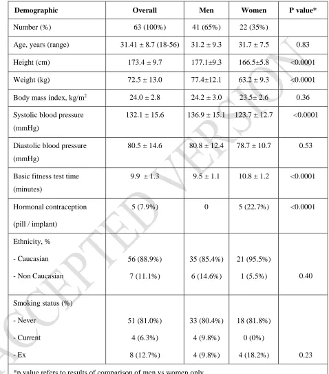
fitness test (P<0.0001) (table 1).
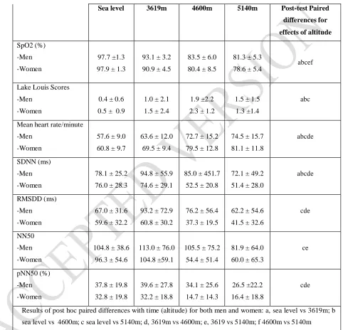
HA exposure led to a significant fall in SpO2 and an increase LLS among
both men and women, compared to baseline with no effect for subject sex (table 2). Heart
rate (five-minute average) increased at HA in both sexes, with women having
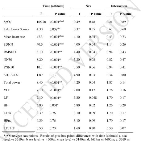
consistently higher rates than men both at SL and at HA (table 2 and 4).
There was a significant main effect for altitude on all time-domain measures of
HRV. On post-hoc analysis this represented a significant reduction in time-domain
measures of HRV most consistently between 3619m to 5140m (table 2 and 4; figure 1).
There was a significant main effect for altitude on LF, HF and TP. This difference was
again most marked on post-hoc analyses between 3619m and 5140m where significant
reductions in LF, HF and TP were observed (table 3 and 4; Figures 2).
Time domain measures of HRV were non-significantly higher in men at SL and
significant differences emerged at HA, where all measures were notably higher in men
(table 2 and 4; figure 1). There was also a main effect for sex among the frequency
domain measures of TP, LF and HF power which were all significantly higher in men at
HA (table 3 and 4, figure 2). There were no interactions between sex (men vs women) x
altitude (SL, 3619m, 4600m and 5140m) on any measures of HRV (table 4) or heart rate.
SpO2 inversely correlated with LLS (r=-0.38; 95% CI -0.50 to -0.24; p<0.0001)
and positively with RMSSD (r=0.16; p=0.02), SDNN (r=0.18; 0.05 to 0.30: p=0.007),
VLF (r=0.17; 0.04 to 0.30; p=0.01), LF (r=0.16; 0.03 to 0.29; p=0.2) and TP (r=0.17;
0.03 to 0.29; P=0.02).
The prevalence of AMS increased at HA from 15.2% at 3619m to 27.3% at
increasing altitude (B 0.80; P <0.0001) and mean heart rate (B 0.03; P=0.04) were the
only univariate predictors of AMS. None of the continuous measured HRV parameters or
the categorical variable of subject sex (men vs women) were predictive of AMS. RMSSD
<30ms, LF:HF >1.3 and LFnu <20% were not predictive of or associated with AMS.
Discussion
This is the largest study to assess the effects of HA on HRV, and to the author’s
knowledge the first study to investigate the influence of sex on HRV at terrestrial HA. In
this study HRV was influenced by HA. Minor sex-related differences in HRV that were
observed at SL were sustained at genuine terrestrial HA. A link between HRV and
symptoms of AMS were not found.
We observed a significant fall in resting SpO2 and an increase in LLS with
increasing HA. There was also a significant main effect for altitude on heart rate (which
increased) and all the evaluated time-domain measured of HRV. The most consistent
change was between 3619 and 5140m, and hence at higher altitude, where there was a
significant fall in SDNN, RMSSD, NN50, PNN50, LF and HF power. These findings
are in keeping with published data that has shown a fall in time-domain measures of
HRV at HA (12,13,29). These changes are in part explained by a number of factors
linked to the HA environment. These include reducing sleep quality, extremes of cold
and heat, physical exhaustion and increasing anxiety which are all known to adversely
affect and reduce time domain measures of HRV (13,19,30).
We also observed a significant main effect for sex on heart rate and time domain
measures of HRV at HA, with men having consistently higher scores and greater
higher time domain measures of HRV in young adult men versus women at SL (3,14,24).
This is the first comparative study at HA. The trend to higher time domain HRV
measures at SL became significant at HA. There was no interaction of altitude on sex on
the time domain HRV parameters. This finding can be partly explained by the sex
differences in heart rate which was consistently lower in the men. Heart rate is well
known inversely correlate with all main time domain measures of HRV (28).
We also observed a significant effect of altitude (SL, 3619m, 4600m and 5140m)
on TP, VLF, LF and HF power. The most consistent finding, on post-hoc analyses was a
reduction in these parameters at the highest altitude of 5140m vs sea level and 3619m.
HA exposure was also associated with a significant main effect of sex with greater TP,
LF and HF power among the men. Results from a very recently published meta-analysis
of comparative HRV measures among men and women at SL, that included more than
60,000 participants, demonstrated that when compared to that seen in women, PSD in
men is generally characterised by lower HF power and greater LF, TP and LF/HF ratios
(14). This is thought to reflect their higher resting sympatho-vagal tone (hence greater LF
and LF/HF ratios) compared with women. Our LF data supports this previous data.
However, contrary to the published data we found that HF power was actually higher
with variable effects on LF/HF power among the men. There are several potential factors
that might explain these results. It is known that LF, HF power and their relative ratios
(LF/HF) can be markedly influenced by a number of factors which include age,
respiratory rate, recording length and heart rate (3,14,23,26,28). Whilst the ages were
similar between the men and the women the greater heart rates in women would have led
to the analysis of a higher number of beat-to-beat intervals, despite an identical recording
period, which could be an important confounder. Secondly, whilst increasing heart rate
and minute ventilation are HA are thought to relate to enhanced sympathetic activation
there is also evidence of elevated parasympathetic neural activity (4,7,21,22). This
maximal heart rate at HA (7). LF power and the LF/HF ratio have been traditionally
thought to represent sympathetic activation and net sympatho-vagal balance respectively
with RMSSD and HF power reflecting parasympathetic nerve activity (28). However,
there is evolving evidence to show that these arbitrary assumptions about the discrete
autonomic effects these HRV measures, may be overly simplistic (2).
Our identified sex-related dissimilarities in the time and frequency domain HRV
measures at HA could also relate to differences in fitness levels. Indeed, the men in our
study had higher time domain measures of HRV and lower 1.5 mile run times. Our
findings could also relate dissimilarities in acclimatisation in men vs women.
Acclimatisation encompasses the cumulative effects of multiple factors such as
hydration, ventilation and enuresis that are known to influence autonomic balance and
HRV (10). HRV, and in particular frequency domain analysis can be significantly
affected by breathing pattern and ventilation, which are markedly affected HA where
hypoxia driven hyperventilation predominates (14,17,18). In our study paced breathing
during HRV assessments were not performed, but the participants were encouraged to
relax and breathe normally. The majority of published studies on HRV at HA have
utilised spontaneous non-paced breathing, hence were keen to utilise a comparative
methodology (7,9,10,24). Our participants were assessed at far higher altitudes and
under greater hypoxia than most of the previous HA HRV studies to date, hence the
potential challenge to paced breathing was likely to have been greater. We anticipated
that at 4600 and 5140m controlled breathing under significant hypoxia and a high
ventilatory drive might paradoxically increase subject anxiety and perceived
breathlessness. By enforcing a similar paced breathing protocol in both men and women
we risked neutralising genuine sex-related differences in HRV related to well-reported
dissimilarities in ventilation between men and women at HA (2,19). Unfortunately, we
This is an obvious limitation as sex-related differences in their spontaneous breathing it
could have provided further insight into the observed differences in HRV identified.
We did not observe a link between AMS and HRV in this current study. There is
limited evidence linking changes in HRV to AMS, raising the prospect of using HRV as
a non-invasive predictor of AMS development (9,10). In a previous study Karinen et al
investigated 36 different healthy climbers ascending from 2400 m to 6300 m altitudes
during five differing expeditions and noted that a lower RMSSD and HF at 2400m was a
marker of AMS at 3000 to 4300m (10). However, contrary to our study, the speed of
ascent varied between their five studied groups. Furthermore, they measured HRV over
two rather than five minutes. In another study, of similar size (n=32), Hang et al noted
that a HF% <20% (nu) or LF/HF ratio >1.3 at lower altitudes was predictive of AMS at
3400m (9). These HRV parameters failed to be either associated or predict AMS in or
study. Willie et al in a prolonged normobaric hypoxia study and our group in another
recent study (ithlete RMSSD-derived HRV score), failed to identify a clear link between
AMS and HRV supporting our data (16, 23).
The potential reasons for the contradictory findings in HRV to predict AMS may
relate to differences in study design, HA environment, ascent/hypoxic profile, HRV
recording time as well as the actual HRV parameters measures. Even the definitions of
AMS that were used differed between these studies. For example, Karinen et al defined
AMS as a LLS of >3 in their study whereas in the study by Willie they a LLS ≥4 was
used to define AMS (10,24). In our study we used the Lake Louise Consensus definition
(1992) for AMS, which is refers to LLS score of >3 in the presence of headache (4,8,12).
It is well known that AMS is a highly complex and heterogeneous condition. Its
causative mechanisms include changes in cerebral arterial blood flow and increased
local autonomic control (18). Whilst HRV reflects overall cardiac autonomic control it is
This study has several additional imitations that should be mentioned. The
subjects were studied in consecutive groups of 8-14 two days apart and not all together in
one batch. This was because of the large sample size for this type of remote field study
and the need to undertake at HRV in the early morning pre-breakfast and caffeine. We
measured five minute HRV which may be more vulnerable to short-term sex and
situational bias than that obtained from longer recordings (22). However, five minute
HRV measurement is well-validated and endorsed by the current HRV Task Force
Guidelines and is more potentially applicable to clinical practice than that of longer
recordings (22). We included a larger proportion of men than women and cannot exclude
the possibility of sample bias, despite their similarities in age, ethnicity, smoking history
and body mass index.
In conclusion our findings indicate that increasing HA was associated with a
reduction in HRV which was most notable at 4600m and above. There were significant
sex related differences in HRV between men and women which were sustained at HA.
There was no interaction between sex and altitude on any of the HRV parameters
measured. These sex-related differences may reflect dissimilarities in their autonomic
balance and acclimatisation to HA. HRV was not predictive of AMS.
Acknowledgments
The authors would like to thank the Surgeon General. We also like to sincerely thank the
subjects for their time and for volunteering to take part in this study.
This projected was funded by a variety of sources and the main funders include the Royal
Centre for Defence Medicine, a grant from the Royal Navy Royal Marines Charity
(RNRMC) and the Poole Hospital Cardiac Research Fund.
Disclosures
Professor Boos has received speaker’s fee and consultation fees from Pfizer, Bristol
Myers Squibb, Astra Zeneca and Boehringer-Ingelheim. Professor Mellor has received
References
1. Bellenger CR, Fuller JT, Thomson RL, Davison K, Robertson EY, Buckley
JD. Monitoring Athletic Training Status Through Autonomic Heart Rate
Regulation: A Systematic Review and Meta-Analysis. Sports Med. 2016;
46(10): 1461-86.
2. Billman GE. The LF/HF ratio does not accurately measure cardiac sympatho-
vagal balance. Front Physiol. 2013; 4(26):1-5.
3. Bonnemeier H, Richardt G, Potratz J, Wiegand UK, Brandes A, Kluge N,
Katus HA. Circadian profile of cardiac autonomic nervous modulation in
healthy subjects: differing effects of aging and sex on heart rate variability. J
Cardiovasc Electrophysiol. 2003; 14(8):791-9.
4. Boos CJ, O'Hara JP, Mellor A, Hodkinson PD, Tsakirides C, Reeve N,
Gallagher L, Green ND, Woods DR. A Four-Way Comparison of Cardiac
Function with Normobaric Normoxia, Normobaric Hypoxia, Hypobaric
Hypoxia and Genuine High Altitude. PLoS One. 2016; 11(4): e0152868.
5. Boos CJ, Mellor A, O'Hara JP, Tsakirides C, Woods DR. The Effects
of Sex on Cardiopulmonary Responses to Acute Normobaric Hypoxia. High
Alt Med Biol. 2016 Jun;17(2):108-15. doi: 10.1089/ham.2015.0114. Epub
2016 Mar 23.
6. Boos CJ, Bakker-Dyos J, Watchorn J, Woods DR, O'Hara JP, Macconnachie
L, Mellor A. A comparison of two methods of heart rate variability
assessment at high altitude. Clin Physiol Funct Imaging. 2016 Jan 14. doi:
10.1111/cpf.12334. [Epub ahead of print].
7. Boushel R, Calbet J-AL, Rådegran G, Søndergaard MS, Wagner PD, Saltin B.
Parasympathetic neural activity accounts for the lowering of exercise heart
8. Buchheit M, Richard R, Doutreleau S, Lonsdorfer-Wolf E, Brandenberger G,
Simon C. Effect of acute hypoxia on heart rate variability at rest and during
exercise. Int J Sports Med. 2004; 25(4): 264-9.
9. Canouï-Poitrine F, Veerabudun K, Larmignat P, Letournel M, Bastuji-Garin
S, Richalet JP. Risk prediction score for severe high altitude illness: a cohort
study. PLoS One. 2014 ;9(7):e100642. doi: 10.1371/journal.pone.0100642.
eCollection 2014.
10. Dishman RK, Nakamura Y, Garcia ME, Thompson RW, Dunn AL, Blair SN.
Heart rate variability, trait anxiety, and perceived stress among physically fit
men and women. Int J Psychophysiol. 2000; 37(2):121-33.
11. Hackett, P.H. & Oelz, O. The Lake Louise consensus on the quantification of
altitude illness. In: Sutton JR, Houston CS & Coates G (eds) Hypoxia and
Mountain Medicine: Queen City Printers, Burlington, VT 1992: 327–330.
12. Huang HH, Tseng CY, Fan JS, Yen DH, Kao WF, Chang SC, Kuo TB, Huang
CI, Lee CH. Alternations of heart rate variability at lower altitude in the
predication of trekkers with acute mountain sickness at high altitude. Clin J
Sport Med. 2010; 20(1): 8-63.
13. Karinen HM, Uusitalo A, Vähä-Ypyä H, Kähönen M, Peltonen JE, Stein PK,
Viik J, Tikkanen HO. Heart rate variability changes at 2400m altitude predicts
acute mountain sickness on further ascent at 3000-4300 m altitudes. Front
Physiol. 2012; 336.
14. Koenig J, Thayer JF. Sex differences in healthy human heart rate variability:
A meta-analysis. Neurosci Biobehav Rev. 2016; 64: 288-310.
15. Lake Louise Consensus on Definition and Quantification of Altitude Illness
In: Sutton JR, Coates G, Houston CS. (eds.) Hypoxia: Mountain Medicine.
16. Liu W, Lian Z, Liu Y. Heart rate variability at different thermal comfort
levels. Eur J Physiol. 2008; 103 (3):61-6.
17. Loeppky JA, Scotto P, Charlton GC, Gates L, Icenogle M, Roach RC.
Ventilation is greater in women than men, but the increase during acute
altitude hypoxia is the same. Respir Physiol. 2001; 125(3): 225-37.
18. MacInnis MJ, Carter EA, Freeman MG, Pandit BP, Siwakoti A, Subedi A,
Timalsina U, Widmer N, Thapa GB, Koehle MS, Rupert JL. A prospective
epidemiological study of acute mountain sickness in Nepalese pilgrims
ascending to high altitude (4380 m). PLoS One. 2013 Oct 9;8(10):e75644.
doi: 10.1371/journal.pone.007564
19. Mazzeo RS, Reeves JT. Adrenergic contribution during acclimatization to
high altitude: perspectives from Pikes Peak. Exerc Sport Sci Rev. 2003; 31(1):
13-8.
20. Mellor A, Bakker-Dyos J, OʼHara J, Woods DR, Holdsworth DA, Boos CJ.
Smartphone-Enabled Heart Rate Variability and Acute Mountain Sickness.
Clin J Sport Med. 2017 Apr 12. doi: 10.1097/JSM.0000000000000427.
21. Paparde A, Plakane L, Circenis K, Aivars JI. Effect of acute systemic hypoxia
on human cutaneous microcirculation and endothelial, sympathetic and
myogenic activity. Microvasc Res. 2015; 102: 1-5.
22. Richalet JP. Physiological and Clinical Implications of Adrenergic Pathways
at High Altitude. Adv Exp Med Biol. 2016; 903: 343-56.
23. Sajkov D, Neill A, Saunders NA, McEvoy RD. Comparison of effects of
sustained isocapnic hypoxia on ventilation in men and women. J Appl Physiol
(1985). 1997; 83(2): 599-607.
24. Saleem S, Hussain MM, Majeed SM, Khan MA. Sex differences of heart rate
25. Santantonio M, Chapplain JM, Tattevin P, Leroy H, Mener E, Gangneux JP,
Michelet C, Revest M. Prevalence of and risk factors for acute mountain
sickness among a cohort of high-altitude travellers who received pre-travel
counselling.Travel Med Infect Dis. 2014;12(5): 534-40.
26. Sassi R, Cerutti S, Lombardi F, Malik M, Huikuri HV, Peng CK, Schmidt G,
Yamamoto Y. Advances in heart rate variability signal analysis: joint position
statement by the e-Cardiology ESC Working Group and the European Heart
Rhythm Association co-endorsed by the Asia Pacific Heart Rhythm Society.
Europace. 2015; 17(9): 1341-53.
27. Sztajzel J, Jung M, Bayes de Luna A. Reproducibility and sex-related
differences of heart rate variability during all-day activity in young men and
women. Ann Noninvasive Electrocardiol. 2008; 13(3): 270-7.
28. Task Force of the European Society of Cardiology and the North American
Society of Pacing and Electrophysiology. Heart rate variability: standards of
measurement, physiological interpretation and clinical use. Circulation 1996;
93: 1043-65.
29. Wille M, Mairer K, Gatterer H, Philippe M, Faulhaber M, Burtscher M.
Changes in cardiac autonomic activity during a passive 8 hour acute exposure
to 5500 m normobaric hypoxia are not related to the development of acute
mountain sickness. Int J Sports Med. 2012; 33(3): 186-91.
30. Woo JM, Kim TS. Sex plays significant role in short-term heart rate
Legends for Figures
Figure 1 Comparative Changes in the RMSSD (mean ± SEM) among men and women at
sea level and increasing high altitude. Post-test differences on repeated measures ANOVA:
[image:24.596.46.520.187.746.2]* versus sea level, ‡ 3619 vs 5140m, †3619 vs 4600m.
Figure 2 Comparative Changes in Low frequency (LnLF) Power (mean ± SEM) among
men and women at sea level and increasing high altitude. Post-test differences on repeated
Table 1
Table 1: Baseline Demographics
Demographic Overall Men Women P value*
Number (%) 63 (100%) 41 (65%) 22 (35%)
Age, years (range) 31.41 ± 8.7 (18-56) 31.2 ± 9.3 31.7 ± 7.5 0.83
Height (cm) 173.4 ± 9.7 177.1±9.3 166.5±5.8 <0.0001
Weight (kg) 72.5 ± 13.0 77.4±12.1 63.2 ± 9.3 <0.0001
Body mass index, kg/m2 24.0 ± 2.8 24.2 ± 3.0 23.5± 2.6 0.36
Systolic blood pressure
(mmHg)
132.1 ± 15.6 136.9 ± 15.1 123.7 ± 12.7 <0.0001
Diastolic blood pressure
(mmHg)
80.5 ± 14.6 80.8 ± 12.4 78.7 ± 10.7 0.53
Basic fitness test time
(minutes)
9.9 ± 1.3 9.5 ± 1.1 10.8 ± 1.2 <0.0001
Hormonal contraception
(pill / implant)
5 (7.9%) 0 5 (22.7%) <0.0001
Ethnicity, %
- Caucasian
- Non Caucasian
56 (88.9%)
7 (11.1%)
35 (85.4%)
6 (14.6%)
21 (95.5%)
1 (5.5%) 0.40
Smoking status (%)
- Never - Current - Ex 51 (81.0%) 4 (6.3%) 8 (12.7%) 33 (80.4%) 4 (9.8%) 4 (9.8%) 18 (81.8%) 0 (0%)
4 (18.2%) 0.23
Table 2
Table 2 Changes in SpO2 and time domain measures of Heart rate Variability at sea level to
increasing high altitude
Sea level 3619m 4600m 5140m Post-test Paired
differences for
effects of altitude
SpO2 (%)
-Men
-Women
97.7 ±1.3
97.9 ± 1.3
93.1 ± 3.2
90.9 ± 4.5
83.5 ± 6.0
80.4 ± 8.5
81.3 ± 5.3
78.6 ± 5.4 abcef
Lake Louis Scores
-Men
-Women
0.4 ± 0.6
0.5 ± 0.9
1.0 ± 2.1
1.5 ± 2.4
1.9 ±2.2
2.3 ± 1.2
1.5 ± 1.5
1.3 ±1.4
abc
Mean heart rate/minute
-Men
-Women
57.6 ± 9.0
60.8 ± 9.7
63.6 ± 12.0
69.5 ± 9.4
72.7 ± 15.2
79.5 ± 12.8
74.5 ± 15.7
81.1 ± 11.8
abcde
SDNN (ms)
-Men
-Women
78.1 ± 25.2
76.0 ± 28.3
94.8 ± 55.9
74.6 ± 29.1
85.0 ± 451.7
52.5 ± 20.8
72.1 ± 49.2
51.4 ± 28.0
abcde
RMSDD (ms)
-Men
-Women
67.0 ± 31.6
59.6 ± 32.2
93.2 ± 72.9
60.8 ± 30.2
76.2 ± 56.4
37.3 ± 19.5
62.2 ± 54.6
41.5 ± 32.6
cde
NN50
-Men
-Women
104.8 ± 38.6
96.3 ± 54.6
113.0 ± 76.0
104.8 ±59.1
105.5 ± 75.2
54.4 ± 51.4
81.9 ± 64.0
60.0 ± 65.3
ce
pNN50 (%)
-Men
-Women
37.8 ± 19.8
32.8 ± 19.8
39.6 ± 27.8
32.2 ± 18.8
34.1 ± 25.6
14.7 ± 14.3
26.5 ±22.2
16.4 ± 18.8
cde
Results of post hoc paired differences with time (altitude) for both men and women: a, sea level vs 3619m; b
Table 3
Table 3 Changes Frequency domain measures of Heart rate Variability at sea level to
increasing high altitude
Sea level 3619m 4600m 5140m Post-test Paired
differences for
effects of altitude
Total power (ms2)
-Men
-Women
7.87 ± 0.7
7.75 ± 0.7
7.95 ± 1.4
7.73 ± 0.8
7.86 ± 1.6
7.05 ± 0.7
7.34 ± 1.5
7.0 ± 1.0
cef
VLF (ms2)
-Men
-Women
6.06 ± 0.7
6.16 ± 0.8
6.11 ± 1.2
5.92 ± 0.9
6.00 ± 1.3
5.44 ± 0.7
5.62 ± 1.3
5.28 ± 1.1
ce
LnLF (ms2)
-Men
-Women
6.80 ± 0.8
6.44 ± 0.7
6.61 ± 1.4
6.67 ± 1.0
6.62 ± 1.7
5.89 ± 0.9
6.19 ± 1.4
5.76 ± 1.0
ce
LnHF (ms2)
-Men
-Women
6.55 ± 0.8
6.23 ± 1.0
6.87 ± 1.7
6.36 ± 0.9
6.50 ± 2.1
5.47 ± 1.0
6.09 ± 1.8
5.43 ± 1.3
e
LF%, nu
-Men
-Women
55.4 ± 15.9
44.5 ± 18.8
44.7 ± 18.0
56.8 ± 15.6
52.7 ± 17.9
58.8 ± 16.4
52.0 ± 18.8
56.8 ± 20.6
NS
HF%, nu
-Men
-Women
44.6 ± 15.9
45.7 ± 18.4
55.4 ± 18.0
43.2 ± 15.6
47.3 ± 17.6
41.2 ± 16.4
48.0 ± 18.8
43.2 ± 20.6
NS
LF / HF
-Men
-Women
1.7 ± 1.3
1.4 ± 1.1
1.0 ± 0.80
1.6 ± 1.0
1.6 ± 1.5
2.1 ± 1.8
1.6 ± 1.5
2.2 ± 2.2
NS
NS, Non-significant; Results of post hoc paired differences with time for men and women
combined: a, sea level vs 3619m; b sea level vs 4600m; c sea level vs 5140m; d, 3619m vs
Table 4
Table 4 Results of Two-way Repeated Measures ANOVA comparing the main effects of
altitude (SL, 3619m, 4600m and 5140m) and sex (men vs women) on measures of heart rate
variability
Time (altitude) Sex Interaction
F P value F P value F P Value
SpO2 165.20 <0.001abcef 0.49 0.48 0.21 0.89
Lake Louis Scores 4.30 0.008abc 0.37 0.55 0.63 0.60
Mean heart rate 47.3 <0.001abcde 4.10 0.04 0.43 0.73
SDNN 40.6 <0.001abcde 4.00 0.04 1.14 0.26
RMSDD 8.10 <0.001cde 4.40 0.04 0.94 0.43
NN50 8.20 <0.001ce 3.20 0.08 0.82 0.47
PNN50 10.7 <0.001cde 3.50 0.06 0.94 0.41
SD1 / SD2 1.89 0.13 4.90 0.03 0.34 0.80
Total power 8.40 <0.001cef 4.20 0.04 1.87 0.14
VLF 7.10 <0.001ce 2.00 0.17 1.76 0.16
LF 7.10 <0.001ce 3.80 0.048 1.70 0.17
HF 5.80 0.001e 5.80 0.02 1.26 0.29
LFnu 0.39 0.76 3.10 0.09 1.70 0.17
HFnu 0.39 0.76 3.10 0.09 1.70 0.17
LF / HF 0.50 0.70 1.60 0.20 3.50 0.07
SpO2, oxygen saturations; Results of post hoc paired differences with time (altitude) a, sea