INTRODUCTION
Pathological gait patterns are generally observed in patients with hemiplegia caused by stroke, cerebral palsy (CP), or trau-matic brain injury.1-3 Hemiplegic patients exhibit heteroge-neous gait impairments in both the affected and unaffected lower extremities, resulting in abnormal kinematic character-istics and temporospatial asymmetry during their gait.4-6 Com-mon pathological gait patterns exhibited by hemiplegic pati-ents are abnormal proximal joint movement, such as excessive anterior tilt of the pelvis, as well as abnormal distal joint move-ment, including foot drop during gait.7-10 In addition, these ab-normal kinematic patterns lead to decreased cadence and walk-ing velocity and an unbalanced stance and swwalk-ing phase,
ev-Effect of Rhythmic Auditory Stimulation
on Hemiplegic Gait Patterns
Yoon-Kyum Shin
1,2, Hyun Ju Chong
3, Soo Ji Kim
4, and Sung-Rae Cho
1,2,51Department and Research Institute of Rehabilitation Medicine, Yonsei University College of Medicine, Seoul;
2Brain Korea 21 PLUS Project for Medical Science, Yonsei University College of Medicine, Seoul;
3Department of Music Therapy, Graduate School and Ewha Music Rehabilitation Center, Ewha Womans University, Seoul;
4Music Therapy Education, Graduate School of Education and Ewha Music Rehabilitation Center, Ewha Womans University, Seoul;
5Rehabilitation Institute of Neuromuscular Disease, Yonsei University College of Medicine, Seoul, Korea.
Purpose: The purpose of our study was to investigate the effect of gait training with rhythmic auditory stimulation (RAS) on both kinematic and temporospatial gait patterns in patients with hemiplegia.
Materials and Methods: Eighteen hemiplegic patients diagnosed with either cerebral palsy or stroke participated in this study.
All participants underwent the 4-week gait training with RAS. The treatment was performed for 30 minutes per each session, three sessions per week. RAS was provided with rhythmic beats using a chord progression on a keyboard. Kinematic and temporospa-tial data were collected and analyzed using a three-dimensional motion analysis system.
Results: Gait training with RAS significantly improved both proximal and distal joint kinematic patterns in hip adduction, knee
flexion, and ankle plantar flexion, enhancing the gait deviation index (GDI) as well as ameliorating temporal asymmetry of the stance and swing phases in patients with hemiplegia. Stroke patients with previous walking experience demonstrated significant kinematic improvement in knee flexion in mid-swing and ankle dorsiflexion in terminal stance. Among stroke patients, subacute patients showed a significantly increased GDI score compared with chronic patients. In addition, household ambulators showed a significant effect on reducing anterior tilt of the pelvis with an enhanced GDI score, while community ambulators significantly increased knee flexion in mid-swing phase and ankle dorsiflexion in terminal stance phase.
Conclusion: Gait training with RAS has beneficial effects on both kinematic and temporospatial patterns in patients with
hemi-plegia, providing not only clinical implications of locomotor rehabilitation with goal-oriented external feedback using RAS but also differential effects according to ambulatory function.
Key Words: Gait, rhythmic auditory stimulation, hemiplegia
Yonsei Med J 2015 Nov;56(6):1703-1713
http://dx.doi.org/10.3349/ymj.2015.56.6.1703 pISSN: 0513-5796 · eISSN: 1976-2437
Received: September 15, 2014 Revised: December 28, 2014 Accepted: February 12, 2015
Co-corresponding authors: Dr. Sung-Rae Cho, Department and Research Insti-tute of Rehabilitation Medicine, Yonsei University College of Medicine, 50-1 Yonsei-ro, Seodaemun-gu, Seoul 03722, Korea.
Tel: 82-2-2228-3715, Fax: 82-2-363-2795, E-mail: [email protected] and Dr. Soo Ji Kim, Music Therapy Education, Graduate School of Education and Ewha Music Rehabilitation Center, Ewha Womans University, 52 Ewhayeodae-gil, Seo-daemun-gu, Seoul 03760, Korea.
Tel: 82-2-3277-6916, Fax: 82-2-3277-6918, E-mail: [email protected]
•The authors have no financial conflicts of interest.
© Copyright: Yonsei University College of Medicine 2015
entually reducing energy efficiency.11-13 Due to these gait defici-encies, patients with hemiplegia experience serious barriers to functional recovery, as locomotive ability is essential for many daily activities.
Several therapeutic interventions, including conventional treadmill training, body weight-supported treadmill training, robot-assisted gait training, and hippotherapy, are components of rehabilitation for patients with hemiplegia.14-17 As a specific gait-training intervention has not yet been deemed superior by a sufficient amount of evidence, the clinical issue of gait impro-vement has continually drawn considerable attention from cli-nicians and researchers.18 Recently, one promising gait train-ing method developed to improve gait impairment is rhythmic auditory stimulation (RAS), which has been applied for a vari-ety of neurological diseases including stroke, CP, Parkinson’s disease, traumatic brain injury, and spinal cord injury.19-29 Gait training with RAS emphasizes rhythmic bilateral movements providing rhythmic cueing using music elements such as a tem-po and beats with chords to ameliorate asymmetry, which is an unrelenting problem in patients with hemiplegia.30 The ba-sic mechanism of gait training with RAS is to regulate repeated movements by auditory-motor synchronization in the central nervous system. An auditory-motor synchronization mecha-nism is organized isochronously by neural substrates and re-flects auditory rhythm and tempo in functional motor output, such as a gait pattern (i.e., velocity, cadence, and stride length in a given period).31 RAS is based on an entrainment model in which rhythmic auditory cues synchronize motor responses into a stable time relationship. In other words, rhythm serves as an anticipatory and continuous time reference on which functional movements are paced or mapped within a stable temporal template. Therefore, entrainment between auditory stimulation and motor responses makes gait pattern regulated and stable in patients with gait deficit.19 Based on the results of previous studies, external auditory cues may rhythmically sti-mulate neural circuits entraining subcortical systems and lead to the optimization of motor commands.19,31-33
Most previous studies regarding gait training with RAS in patients with neurological impairments have demonstrated improvement in temporospatial gait parameters, including cadence, walking velocity, and stride length.19,21,22,30,32,34,35 Thaut, et al.19,32 showed that RAS significantly improved walking veloc-ity, stride length, cadence, and symmetry in acute hemiplegic patients who had suffered from stroke. Suh, et al.35 found that three-week gait training with RAS had a significant effect on the gait parameters of walking velocity, stride length, and ca-dence as well as standing balance in hemiplegic patients fol-lowing stroke. Hashiguchi, et al.36 reported that RAS resulted in significant gait improvement by increasing gait velocity while simultaneously decreasing the gait variability of stride time in subacute hemiplegic patients after stroke.
In patients with CP who have bilateral involvement, recent studies on gait training with RAS revealed that improvements
of both kinematic and temporospatial gait parameters can ben-efit patients with gait impairments.21,22 Kim, et al.21,22 showed that significant improvement in proximal limb movements was observed after immediate and long-term gait training with RAS in patients with CP. Thus, statistical significance in amelio-rating the anterior tilt of the pelvis and hip flexion has been id-entified in patients with bilateral spastic CP.
Throughout these previous studies, gait training with RAS was shown to establish a promising therapeutic purpose in the rehabilitation of gait disturbances. However, there is little evi-dence indicating that RAS treatment for hemiplegic gait pat-terns pertains to three-planar analysis in the sagittal, coronal, and transverse planes of lower extremity joint kinematics, such as those of the pelvis, hip, knee, ankle, and foot. Therefore, it remains an important challenge to refine the effects of gait training with RAS in order to confirm the changes of both kine-matic and temporospatial characteristics in patients with he-miplegia and to compare the kinematic changes according to the etiology of the neurological disorder, onset duration, and ambulatory status.
MATERIALS AND METHODS
Participants
Eighteen individuals with hemiplegia who were diagnosed with either CP or stroke were recruited in this study. The pro-cedure was approved by the Institutional Review Board (4-2012-0483). Informed consent was obtained from all partici-pants after the experimental procedures were sufficiently ex-plained and before the study began. The inclusion criteria for individuals with hemiplegia were as follows: each participant 1) had no discernible hearing deficit, 2) was able to walk inde-pendently for a distance of at least 10 m without the use of a walking aid or supporter, and 3) was able to understand the command to walk following RAS.22
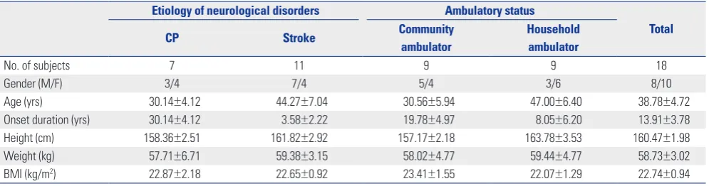
Demographical and clinical characteristics including age, gender, height, weight, body mass index, diagnosis, and am-bulatory status are shown in Table 1. Participants were classi-fied as either community ambulators or household ambulators according to their ambulatory status. Community ambulators were able to independently walk around on level ground, curbs, and uneven terrain outdoors as well as indoors for a minimum of 150 feet; they were also able to manage stairs and challeng-ing community activities, such as makchalleng-ing visits in their neigh-borhood. Conversely, household ambulators were only able to walk indoors for short distances (a maximum of 50 feet) either independently or with orthopedic devices; they also encoun-tered difficulty with stairs and uneven terrain and required as-sistance or walking aids when leaving the house.21,37
Gait training
on the carpet of the same clinic. The RAS treatment was per-formed for 30 minutes per each session, three sessions per week. The intervention procedure consisted of the following established protocol:21,22 1) A participant independently walked barefoot along a 10-m flat walkway three times without RAS at the individual’s preferred walking speed. 2) Walking cadence (steps/min) was calculated based on the gait parameters out-lined in step 1. 3) The identified initial tempo signaled by met-ronome beats (beats per minute) was set to the participant’s cadence obtained in step 2 in each session. 4) RAS was then provided by the music therapists, who played a live rhythmic pattern using a composed four-chord progression with metro-nome beats on a keyboard (PSR-E213, Yamaha Electronics Co., Hamamatsu, Japan). To ensure accuracy and consistency of the rhythmic stimulus, the same therapists performed the pro-cedure for each patient. These specialized therapists provided a regular and stable rhythm. To avoid rhythmic monotonous-ness, several chord progressions were applied. 5) The same chord pattern was repeated to provide a continuous timing cue and a period of 2 minutes to help each participant quickly ad-apt to the RAS. In this step, the music therapist instructed each participant to finger-tap the rhythm for 1 minute to ensure that they could hear the sounds and also confirmed that each partici-pant could walk comfortably by allowing them to adapt to the RAS for 1 minute. 6) Each participant then walked the length of 10 m three to six times with RAS and rested for 1–3 minutes between walks, depending on the endurance level of the pa-tient. This step was repeated 5–8 times in a session. To moni-tor compliance of the participants to the RAS, three music ther-apists evaluated the change in walking speed.21,22 7) The final 1–2 minutes was spent by fading out the rhythmic stimulation to monitor the independent carryover effect, which was qu-antified by calculating cadence. These seven steps were applied in each training session during a period of 4 weeks.
Testing and gait analysis
All participants were pre- and post-tested 2 days before start-ing and 2 days after conductstart-ing the 4-week gait trainstart-ing with RAS in the gait analysis laboratory. Kinematic and
temporospa-tial data from the pelvis, hip, knee, ankle, and foot were col-lected and analyzed for the gait trials without RAS. To ensure that reflective foot markers could be recognized from the infra-red signal of the motion analysis system, participants walked barefoot along a 10-m flat walkway three times at the individu-al’s preferred walking speed without RAS. For kinematic anal-ysis, 15 passively reflective markers were adhered with special-ized tape to the sacrum, both sides of the anterior superior iliac spine, middle thigh, lateral knee, middle shank of the tibia, lat-eral malleolus, heel, and forefoot.22 A three-dimensional mo-tion analysis system with six cameras (Vicon Nexus ver. 1.8.5, Vicon Motion System Ltd., Oxford, UK) was used to record ki-nematics by measuring the degree of joint motion during gait performance. This system comprised six infrared-sensitive cam-eras for locating and tracking the fixed retro-reflective mark-ers through space.
The motion analysis system was calibrated before each gait analysis. Participants were simultaneously videotaped from the front and side, and measurements were recorded in the sa-gittal, coronal, and transverse planes. Kinematic data included the angle of pelvic tilt, pelvic obliquity, pelvic rotation, hip flex-ion and extensflex-ion, hip adductflex-ion and abductflex-ion, hip internal and external rotation, knee flexion and extension, ankle plan-tar and dorsiflexion, and foot internal and external rotation. All kinematic and temporospatial data were processed and plot-ted, and the graphs were visualized using Polygon software ver. 3.5.1 (Oxford Metrics Inc., Oxford, UK), which was inter-worked with a three-dimensional motion analysis system. To perform the statistical analysis, three points from each joint range of motion from continuous raw data in the lower extrem-ity were used: initial contact (the moment when the heel struck the floor), the minimal joint angle, and the maximal joint an-gle during the whole gait cycle.22
Gait deviation index
In order to evaluate the overall gait pathology, a global index of three-dimensional kinematic changes, the gait deviation index (GDI),38 was utilized in the pre- and post-RAS treatment. The GDI is a scaled distance between nine individual kinematic va-Table 1. General Characteristics of Participants
Etiology of neurological disorders Ambulatory status
Total
CP Stroke Community
ambulator
Household ambulator
No. of subjects 7 11 9 9 18
Gender (M/F) 3/4 7/4 5/4 3/6 8/10
Age (yrs) 30.14±4.12 44.27±7.04 30.56±5.94 47.00±6.40 38.78±4.72
Onset duration (yrs) 30.14±4.12 3.58±2.22 19.78±4.97 8.05±6.20 13.91±3.78
Height (cm) 158.36±2.51 161.82±2.92 157.17±2.18 163.78±3.53 160.47±1.98
Weight (kg) 57.71±6.71 59.38±3.15 58.02±4.77 59.44±4.77 58.73±3.02
BMI (kg/m2) 22.87±2.18 22.65±0.92 23.41±1.55 22.07±1.29 22.74±0.94
[image:3.595.57.555.85.216.2]riables of pathological gait (pelvic tilt, pelvic obliquity, pelvic rotation, hip flexion, hip adduction and abduction, hip internal and external rotation, sagittal angles of the knee and ankle, and foot progression in the transverse plane) and the average of ki-nematic variables of the reference normal gait. One stride ob-tained from each pre- and post-treatment gait analysis was se-lected from the hemiplegic side of every participant. All kine-matic variables were extracted and sampled at every 2% inter-val in the whole gait cycle. Each set of 459 data points (9 joint angles×51 points) was computed as gait vectors using the gait feature of a normal gait referred to as orthonormal f-basis. The computing method is explained in detail in another referenced paper.38 The GDI score can be interpreted as follows: a score of 100 indicates a GDI equal to the average of the normal con-trol. In our study, the GDI was utilized to ensure that individu-al pathologicindividu-al gait improved after gait training with RAS com-pared to the normal reference. The GDI score was calculated to take into account the overall effect of kinematic changes of the pelvis, hip, knee, ankle, and foot throughout the gait cycle. In
other words, the GDI incorporated the pelvic tilt, pelvic obliq-uity, pelvic rotation, hip flexion, hip adduction and abduction, hip internal and external rotation, the sagittal angles of the knee and ankle, and the foot progression in the transverse plane.
Temporospatial measures and side-to-side asymmetry
Temporospatial parameters such as cadence (steps/min), walking velocity (m/sec), stride length (m), step length (m), stride time (sec), step time (sec), single limb support (%), dou-ble limb support (%), stance phase (%), and swing phase (%) were calculated using Polygon software ver. 3.5.1 (Oxford Met-rics Inc., Oxford, UK). To evaluate the side-to-side asymmetry between the lower limbs, the absolute difference between the unaffected and affected sides was analyzed using temporo-spatial data.22
Statistical analysis
[image:4.595.43.541.333.704.2]All statistical analyses were performed with SPSS Statistics 20 (IBM Corp., Armonk, NY, USA). Wilcoxon signed-rank tests
Table 2. Changes in Kinematic Patterns of Hemiplegic Limbs after Gait Training with RAS
Joint Plane Joint motion Pre Post
Pelvis Sagittal Anterior tilt at IC 10.07 ±1.67 8.84 ±1.43
Minimal angle of anterior tilt 9.20 ±1.74 8.14 ±1.47
Maximal angle of anterior tilt 15.07 ±2.11 13.72 ±1.63
Coronal Upward/downward tilt at IC 1.37 ±0.59 1.29 ±0.47
Maximal angle of downward tilt -3.33 ±0.73 -3.01 ±0.60
Maximal angle of upward tilt 4.19 ±0.49 4.21 ±0.54
Transverse External/internal rotation at IC -0.36 ±0.90 -0.49 ±0.95
Maximal angle of external rotation -8.68 ±1.48 -8.87 ±1.29
Maximal angle of internal rotation 4.37 ±0.95 2.83 ±0.94
Hip Sagittal Flexion at IC 34.89 ±2.16 34.27 ±2.20
Minimal flexion at push-off 3.38 ±2.51 1.89 ±1.87
Maximal flexion in terminal swing 39.01 ±2.48 39.00 ±2.25
Coronal Abduction/adduction at IC 1.05 ±0.81 1.93 ±0.73
Maximal abduction in mid-swing -4.43 ±1.22 -3.30 ±1.12
Maximal adduction in mid-stance 7.21 ±1.00 8.75 ±0.82*
Transverse External/internal rotation at IC -6.27 ±2.50 -6.81 ±2.11
Maximal external rotation in mid-swing -16.32 ±2.11 -17.64 ±1.64
Maximal internal rotation in terminal stance 12.67 ±1.75 11.75 ±2.14
Knee Sagittal Flexion at initial contact 21.33 ±2.32 21.84 ±1.63
Minimal flexion in terminal stance 13.41 ±2.42 13.70 ±2.05
Maximal flexion in mid-swing 56.27 ±3.66 60.18 ±3.06*
Ankle Sagittal PF/DF at IC -5.10 ±1.38 -3.10 ±1.50*
Maximal PF at push-off -9.04 ±1.73 -6.91 ±1.74*
Maximal DF in terminal stance 15.32 ±1.97 17.09 ±1.57*
Foot Transverse External/internal rotation at IC -8.23 ±2.59 -9.53 ±2.03
Maximal external rotation in mid-stance -16.62 ±2.80 -17.74 ±2.22
Maximal internal rotation at push-off -1.57 ±2.91 -4.07 ±2.24
GDI 83.89 ±2.72 87.29 ±2.65*
RAS, rhythmic auditory stimulation; IC, initial contact; PF, plantar flexion; DF, dorsiflexionl; GDI, gait deviation index; SE, standard error. Values are mean±SE.
were used to evaluate intra-subject pairwise comparisons be-tween gait parameters: kinematics and temporospatial data and the GDI score in the pre- and post-RAS treatment. Mann-Whitney U tests were also used to compare the inter-group de-signs of general characteristics and gait parameters. All statis-tical significance levels were set at p<0.05.
RESULTS
General characteristics of participants
Among total 18 hemiplegic patients, seven subjects diagnosed with CP and eleven subjects diagnosed with stroke participated in this study. Nine subjects with community ambulatory func-tion and nine subjects with household ambulatory funcfunc-tion participated. All participants showed mild spasticity of the lower extremities with a grade of 1 to 1+ on the Modified Ash-worth Scale. The onset duration of stroke was 3.58±2.22 years. Detailed information on gender, age, height, weight, and body mass index is shown in Table 1. Considering that CP is a de-velopmental disorder that affects the initial acquisition of gait while stroke patients have previous walking experience, these two neurologic disorders represented in the study population were separately analyzed. There were no significant differences in the parameters of general characteristics between CP and stroke (Table 1). In addition, there were no significant differ-ences in the general parameters between community ambula-tors and household ambulaambula-tors (Table 1).
Changes in kinematic patterns in patients with hemiplegia
When kinematic characteristics of the hemiplegic side were evaluated before and after RAS treatment, pelvic anterior tilt in the sagittal plane largely tended to decrease after gait training with RAS. Hip adduction significantly increased in the mid-stance phase after the RAS treatment (p=0.039 by Wilcoxon
signed-rank test) (Table 2). Maximal knee flexion increased in the mid-swing phase (p=0.022), and ankle plantar flexion de-creased at initial contact (p=0.025) and push off (p=0.006), while ankle dorsiflexion increased in the terminal stance (p=0.031). When a comprehensive measure of overall gait pathology, GDI, was calculated, the GDI score was found to significantly im-prove in patients with hemiplegia after RAS treatment (83.89± 2.72 to 87.29±2.65, p=0.043) (Table 2).
Changes in temporospatial parameters in patients with hemiplegia
When temporospatial parameters on the hemiplegic side were evaluated both before and after RAS treatment, there were no significant changes in the statistical analysis after gait training with RAS (Table 3). However, when the side-to-side differenc-es between the unaffected and the hemiplegic side of the tem-porospatial parameters were evaluated, the side-to-side differ-ence of the stance phase (6.95±0.99% to 4.62±1.15%; p=0.006 by Wilcoxon signed-rank test) and the swing phase (12.12± 5.06% to 4.62±1.15%; p=0.006) significantly decreased after the RAS treatment (Table 3), suggesting that gait training with RAS could improve the side-to-side symmetry in patients with he-miplegia.
Comparison of kinematic patterns according to the etiology of neurologic disorders
[image:5.595.56.558.550.706.2]We additionally analyzed the kinematic data of CP and stroke separately. Stroke patients showed significant kinematic im-provement in maximal knee flexion in mid-swing phase (48.88± 4.31 to 55.31±3.90; p=0.021 by Wilcoxon signed-rank test) and in maximal ankle dorsiflexion in terminal stance (13.79±1.27 to 16.15±1.42; p=0.026) (Table 4). Patients with CP, on the oth-er hand, did not show kinematic improvement aftoth-er gait train-ing with RAS (Table 4). This result suggests that the previous walking experience in stroke patients who have a history of typical gait may have an effect on their response to RAS.
Table 3. Changes in Temporospatial Parameters of Hemiplegic Limbs after Gait Training with RAS
Temporospatial data Temporospatial data Side-to-side difference
Pre Post Pre Post
Cadence (steps/min) 90.00 ±5.66 97.49 ±6.74 3.63 ±0.62 3.26 ±0.79
Walking speed (m/s) 0.66 ±0.08 0.66 ±0.08 0.02 ±0.00 0.02 ±0.01
Stride length (m) 0.83 ±0.06 0.80 ±0.07 0.02 ±0.00 0.03 ±0.01
Step length (m) 0.42 ±0.03 0.43 ±0.03 0.06 ±0.01 0.05 ±0.01
Stride time (s) 1.45 ±0.12 1.33 ±0.08 0.06 ±0.01 0.05 ±0.01
Step time (s) 0.76 ±0.08 0.68 ±0.06 0.13 ±0.03 1.02 ±0.92
Single support (s) 0.39 ±0.02 0.40 ±0.01 0.11 ±0.02 0.07 ±0.02
Double support (s) 0.59 ±0.11 0.50 ±0.06 0.06 ±0.01 0.04 ±0.01
Stance phase (%) 65.42 ±1.28 65.99 ±1.24 6.95 ±0.99 4.62 ±1.15*
Swing phase (%) 34.58 ±1.28 34.01 ±1.24 12.12 ±5.06 4.62 ±1.15*
RAS, rhythmic auditory stimulation; SE, standard error. Values are mean±SE.
Comparison of kinematic patterns according to onset duration in stroke patients
Based on longitudinal studies suggesting that motor recovery reaches a plateau until the first 6 months post-stroke,39 we addi-tionally analyzed the effects of RAS on kinematic patterns in both subacute and chronic stroke patients separately. Although chronic stroke patients did not show an increase in GDI score, these patients did show improvement in hip external rotation (0.26±3.57 to -3.98±3.13; p=0.028), maximal ankle dorsiflexion at terminal stance (14.08±1.98 to 16.85±1.72; p=0.028), and foot external rotation (-6.23±2.47 to -9.69±1.44; p=0.028) at initial contact after gait training with RAS (Table 5). On the other hand, subacute stroke patients showed a significant increase in GDI score (80.88±3.82 to 88.99±5.00; p=0.043 by Wilcoxon signed-rank test) with improvement in maximal knee flexion in mid-swing phase (45.15±3.59 to 56.42±4.74; p=0.043) (Table 5). This result suggests that subacute stroke patients are more likely to respond to RAS than chronic patients.
Comparison of kinematic patterns according to ambulatory status
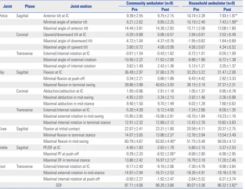
In the analysis of kinematic characteristics, household ambu-lators showed that pelvic anterior tilt at initial contact (10.74± 2.28 to 7.93±1.97; p=0.038 by Wilcoxon signed-rank test) and the minimal angle of anterior tilt (10.19±2.40 to 7.43±1.99; p= 0.038) were ameliorated after gait training with RAS. On the other hand, community ambulators were found to have signifi-cant increases in maximal knee flexion in the mid-swing phase (60.79±4.67 to 63.82±4.40; p=0.021) and ankle dorsiflexion in the terminal stance (13.86±2.42 to 16.97±2.12; p=0.008), where-as ankle plantar flexion at push-off decrewhere-ased (-9.39±2.20 to -6.92±2.06; p=0.021) (Table 6). In the analysis of GDI score, household ambulators, and not community ambulators, show-ed significant gait improvement (80.07±3.36 to 86.32±3.82;
p=0.008 by Wilcoxon singed-rank test) (Table 6). The kinematic
changes of the proximal pelvic joint and distal ankle joint at pre- and post-RAS treatment showed differential patterns ac-Table 4. Comparison of Kinematic Patterns after Gait Training with RAS According to the Etiology of Neurologic Disorders
Joint Plane Joint motion in the gait event CP (n=7) Stroke (n=11)
Pre Post Pre Post
Pelvis Sagittal Anterior tilt at IC 10.99 ±3.34 9.72 ±3.33 9.48 ±1.83 8.28 ±1.17
Minimal angle of anterior tilt 10.27 ±3.46 9.23 ±3.42 8.52 ±1.94 7.45 ±1.18
Maximal angle of anterior tilt 17.38 ±4.90 15.62 ±3.94 13.60 ±1.64 12.51 ±1.06
Coronal Upward/downward tilt at IC 0.09 ±0.95 0.88 ±0.59 2.19 ±0.66 1.55 ±0.69
Maximal angle of downward tilt -6.12 ±0.87 -4.36 ±0.88 -1.56 ±0.61 -2.15 ±0.71
Maximal angle of upward tilt 2.89 ±0.54 4.05 ±1.11 5.02 ±0.61 4.31 ±0.58
Transverse External/internal rotation at IC -0.09 ±2.03 0.24 ±2.08 -0.54 ±0.80 -0.96 ±0.89
Maximal angle of external rotation -12.93 ±2.76 -13.04 ±2.27 -5.97 ±1.15 -6.22 ±0.91
Maximal angle of internal rotation 2.54 ±1.99 2.15 ±1.88 5.54 ±0.81 3.27 ±1.03
Hip Sagittal Flexion at IC 40.58 ±2.93 38.98 ±3.91 31.27 ±2.52 31.28 ±2.32
Minimal flexion at push-off 8.48 ±4.74 5.45 ±3.31 0.14 ±2.50 -0.37 ±2.06
Maximal flexion in terminal swing 43.48 ±4.38 41.06 ±4.50 36.16 ±2.78 37.69 ±2.42
Coronal Abduction/adduction at IC 2.02 ±1.32 3.32 ±1.32 0.43 ±1.03 1.04 ±0.78
Maximal abduction in mid-swing -3.33 ±2.14 -1.31 ±2.24 -5.14 ±1.50 -4.56 ±1.09
Maximal adduction in mid-stance 8.57 ±1.55 9.90 ±1.65 6.35 ±1.29 8.02 ±0.83
Transverse External/internal rotation at IC -9.33 ±4.27 -8.22 ±4.55 -4.33 ±3.07 -5.91 ±2.06
Maximal external rotation in mid-swing -16.21 ±3.16 -15.29 ±1.92 -16.39 ±2.93 -19.14 ±2.34 Maximal internal rotation in terminal stance 10.59 ±2.64 11.89 ±2.68 13.99 ±2.33 11.66 ±3.16
Knee Sagittal Flexion at initial contact 27.52 ±3.80 26.82 ±2.40 17.39 ±2.33 18.67 ±1.62
Minimal flexion in terminal stance 20.58 ±4.11 19.10 ±3.17 8.85 ±2.13 10.26 ±2.19
Maximal flexion in mid-swing 67.88 ±3.48 67.84 ±3.49 48.88 ±4.31 55.31 ±3.90*
Ankle Sagittal PF/DF at IC -3.26 ±1.68 -0.98 ±2.05 -6.27 ±1.97 -4.44 ±2.05
Maximal PF at push-off -8.69 ±2.84 -5.60 ±2.90* -9.26 ±2.29 -7.74 ±2.25
Maximal DF in terminal stance 17.73 ±4.75 18.56 ±3.47 13.79 ±1.27 16.15 ±1.42*
Foot Transverse External/internal rotation at IC -7.53 ±4.79 -7.59 ±3.28 -8.68 ±3.15 -10.76 ±2.64
Maximal external rotation in mid-stance -14.93 ±4.44 -15.57 ±3.91 -17.69 ±3.74 -19.13 ±2.73
Maximal internal rotation at push-off 0.49 ±4.21 0.10 ±3.25 -2.87 ±4.04 -6.72 ±2.85
GDI 80.16 ±5.05 83.64 ±5.01 86.27 ±3.06 89.61 ±2.91
RAS, rhythmic auditory stimulation; CP, cerebral palsy; IC, initial contact; PF, plantar flexion; DF, dorsiflexion; GDI, gait deviation index; SE, standard error. Values are mean±SE.
[image:6.595.44.539.86.470.2]cording to the ambulatory status (Fig. 1).
DISCUSSION
Hemiplegic gait pattern, which is observed in patients with stroke or CP, is characterized by laborious and imbalanced limb movement, as an asymmetrical kinematic and temporo-spatial pattern occurs during locomotion. In addition, patients with hemiplegia have difficulties maintaining shock absorp-tion and weight acceptance in the stance phase as well as ac-celerating forward propulsion with adequate limb excursion in the swing phase.40
CP is a developmental disorder that affects the initial acqui-sition of gait, while stroke patients do have a history of typical gait. Previous walking experience in stroke patients may affect their response to RAS, as evidenced by significant kinematic improvement in maximal knee flexion in mid-swing and
maxi-mal ankle dorsiflexion in terminal stance. Nevertheless, when kinematic and temporospatial data were evaluated to compare inter-group design using the Mann-Whitney U test, there were no significant differences between CP and stroke patients. Therefore, all data from CP and stroke participants were inte-grated with hemiplegic patients to present the effect of gait training with RAS in this study. Overall, gait training with RAS significantly improved the GDI score, demonstrating the ben-efits of RAS treatment on the overall kinematic gait patterns in patients with hemiplegia. Additionally, subacute stroke pa-tients were shown to have significant increases in GDI score, suggesting that subacute patients are more likely to respond to RAS than chronic patients.
[image:7.595.59.555.88.470.2]In the analysis of proximal joint motion, excessive anterior tilt of the pelvis was significantly reduced in household ambu-lators. In concordance with previous studies of CP patients with spastic diplegia,21,22 this finding supports the hypothesis that gait training with RAS also alleviates excessive anterior tilt Table 5. Comparison of Kinematic Patterns after Gait Training with RAS According to Onset Duration in Stroke Patients
Joint Plane Joint motion in the gait event Subacute patients (n=5) Chronic patients (n=6)
Pre Post Pre Post
Pelvis Sagittal Anterior tilt at IC 11.08 ±3.57 9.37 ±2.35 8.14 ±1.74 7.38 ±1.00
Minimal angle of anterior tilt 10.54 ±3.80 8.49 ±2.51 6.83 ±1.71 6.58 ±0.78
Maximal angle of anterior tilt 14.99 ±3.50 11.77 ±2.15 12.45 ±1.03 13.14 ±0.91
Coronal Upward/downward tilt at IC 2.92 ±1.01 3.06 ±0.79 1.58 ±0.87 0.29 ±0.79*
Maximal angle of downward tilt -1.36 ±1.15 -1.39 ±1.21 -1.72 ±0.67 -2.78 ±0.83
Maximal angle of upward tilt 4.97 ±0.73 4.55 ±0.45 5.05 ±1.01 4.12 ±1.05
Transverse External/internal rotation at IC -0.13 ±1.33 -0.38 ±1.26 -0.88 ±1.06 -1.44 ±1.33
Maximal angle of external rotation -3.56 ±1.23 -4.01 ±1.24 -7.98 ±1.45 -8.06 ±0.74
Maximal angle of internal rotation 5.93 ±1.40 2.29 ±1.21 5.21 ±1.03 4.08 ±1.62
Hip Sagittal Flexion at IC 29.45 ±4.08 31.84 ±3.24 32.79 ±3.33 30.81 ±3.56
Minimal flexion at push-off 3.56 ±5.07 1.44 ±3.75 -2.71 ±1.47 -1.89 ±2.28
Maximal flexion in terminal swing 34.68 ±4.22 37.44 ±2.97 37.39 ±3.97 37.90 ±3.96
Coronal Abduction/adduction at IC -0.07 ±1.68 1.16 ±0.95 0.86 ±1.40 0.93 ±1.27
Maximal abduction in mid-swing -5.82 ±1.97 -4.25 ±0.97 -4.56 ±2.36 -4.82 ±1.92
Maximal adduction in mid-stance 4.53 ±1.62 8.15 ±0.80 7.86 ±1.84 7.91 ±1.45
Transverse External/internal rotation at IC -9.83 ±4.36 -8.23 ±2.51 0.26 ±3.57 -3.98 ±3.13*
Maximal external rotation in mid-swing -20.06 ±2.23 -22.86 ±1.15 -13.34 ±4.92 -16.03 ±3.87 Maximal internal rotation in terminal stance 11.17 ±4.28 10.31 ±6.09 16.34 ±2.29 12.78 ±3.36
Knee Sagittal Flexion at initial contact 14.94 ±4.39 17.60 ±2.78 19.43 ±2.29 19.56 ±2.04
Minimal flexion in terminal stance 9.02 ±3.82 11.22 ±3.60 8.71 ±2.63 9.47 ±2.95
Maximal flexion in mid-swing 45.15 ±3.59 56.42 ±4.74* 51.98 ±7.43 54.38 ±6.35
Ankle Sagittal PF/DF at IC -6.87 ±3.45 -5.47 ±2.63 -5.77 ±2.48 -3.59 ±3.25
Maximal PF at push-off -9.85 ±4.00 -9.69 ±2.83 -8.76 ±2.91 -6.12 ±3.48
Maximal DF in terminal stance 13.45 ±1.84 15.31 ±2.53 14.08 ±1.89 16.85 ±1.72*
Foot Transverse External/internal rotation at IC -11.61 ±6.43 -12.04 ±5.87 -6.23 ±2.47 -9.69 ±1.44*
Maximal external rotation in mid-stance -20.32 ±7.21 -19.71 ±6.00 -15.50 ±3.82 -18.63 ±1.77 Maximal internal rotation at push-off -7.04 ±8.15 -7.85 ±6.15 0.60 ±3.19 -5.77 ±2.02*
GDI 80.88 ±3.82 88.99 ±5.00* 90.76 ±3.97 90.13 ±3.79
RAS, rhythmic auditory stimulation; IC, initial contact; PF, plantar flexion; DF, dorsiflexion; GDI, gait deviation index; SE, standard error. Values are mean±SE.
of the pelvis in hemiplegic patients with stroke or CP who have household ambulatory function. Abnormal pelvic control is a major problem that can lead to a distorted gait pattern due to the linkage of distal joint movement.41 Hip adduction in the mid-stance phase was also ameliorated in patients with hemi-plegia after gait training with RAS. Hemiplegic patients com-monly show less hip adduction than healthy controls in the stance phase. As this coronal kinematic deviation reflects poor stability in both the stance and swing phase during gait,42 RAS treatment eventually improves walking stability.
In the analysis of distal joint motion, knee flexion in the mid-swing phase increased in patients with hemiplegia, particu-larly in community ambulators. This finding suggests that in-creased knee flexion may reduce toe dragging and compens-ative pelvic hiking, inducing relcompens-atively adequate limb propul-sion during the swing phase of gait.43 Ankle dorsiflexion in the terminal stance phase also increased in community ambula-tors, demonstrating that reduced ankle plantar flexion at initial
contact sequentially exhibited increased ankle dorsiflexion in the terminal stance. In other words, the first and second ankle rockers were relatively normalized after gait training with RAS, intuitively suggesting increased stance stability of the hemi-plegic side.21,44
The potential therapeutic rationale for such differential kine-matic effects of RAS according to ambulatory status may in-volve the proximal-to-distal functional relationship.45 As com-munity ambulators perform relatively adequate proximal mo-tor function, rhythmic audimo-tory cues might be sufficient to fa-cilitate distal joint movement during gait. However, in house-hold ambulators, such cues might be prerequisite to facilitate proximal joint movement in order to follow the RAS during gait training.
In the analysis of overall gait pathology using the GDI, gait impairments improved in patients with hemiplegia. It may be possible that community ambulators showed less improved GDI scores, as the proximal joint movements were less suscep-Table 6. Comparison of Kinematic Patterns after Gait Training with RAS According to Ambulatory Status
Joint Plane Joint motion Community ambulator (n=9) Household ambulator (n=9)
Pre Post Pre Post
Pelvis Sagittal Anterior tilt at IC 9.39 ±2.55 9.75±2.15 10.74 ±2.28 7.93 ±1.97*
Minimal angle of anterior tilt 8.21 ±2.62 8.85 ±2.25 10.19 ±2.40 7.43 ±1.99*
Maximal angle of anterior tilt 14.44 ±3.81 14.36 ±2.83 15.71 ±2.08 13.08 ±1.80
Coronal Upward/downward tilt at IC -0.20 ±0.68 0.06 ±0.57 2.94 ±0.61 2.52 ±0.49
Maximal angle of downward tilt -4.72 ±1.04 -4.37 ±0.76 -1.95 ±0.82 -1.64 ±0.69
Maximal angle of upward tilt 3.80 ±0.72 4.08 ±0.99 4.58 ±0.67 4.34 ±0.52
Transverse External/internal rotation at IC -0.01 ±1.54 -0.43 ±1.62 -0.72 ±1.01 -0.55 ±1.09
Maximal angle of external rotation -10.56 ±2.22 -11.02 ±2.00 -6.80 ±1.88 -6.72 ±1.38
Maximal angle of internal rotation 3.62 ±1.49 2.42 ±1.36 5.13 ±1.21 3.25 ±1.37
Hip Sagittal Flexion at IC 36.49 ±2.97 37.08 ±3.79 33.29 ±3.22 31.47 ±2.08
Minimal flexion at push-off 0.34 ±2.21 0.86 ±1.88 6.43 ±4.42 2.92 ±3.33
Maximal flexion in terminal swing 39.86 ±3.98 40.63 ±3.93 38.15 ±3.19 37.37 ±2.31
Coronal Abduction/adduction at IC 1.05 ±0.96 2.91 ±1.18 1.05 ±1.37 0.95 ±0.78
Maximal abduction in mid-swing -4.95 ±2.03 -3.34 ±2.15 -3.92 ±1.46 -3.26 ±0.88
Maximal adduction in mid-stance 8.40 ±1.50 9.70 ±1.49 6.02 ±1.28 7.80 ±0.63
Transverse External/internal rotation at IC -5.30 ±4.39 -5.12 ±4.05 -7.24 ±2.66 -8.50 ±1.35
Maximal external rotation in mid-swing -15.95 ±3.95 -16.06 ±2.81 -16.70 ±1.84 -19.23 ±1.70 Maximal internal rotation in terminal stance 12.91 ±2.32 12.68 ±2.12 12.42 ±2.76 10.82 ±3.83
Knee Sagittal Flexion at initial contact 22.07 ±2.41 23.31 ±1.80 20.59 ±4.11 20.37 ±2.75
Minimal flexion in terminal stance 14.07 ±3.05 13.86 ±2.37 12.76 ±3.94 13.54 ±3.49
Maximal flexion in mid-swing 60.79 ±4.67 63.82 ±4.40* 51.75 ±5.48 56.55 ±4.12
Ankle Sagittal PF/DF at IC -4.40 ±1.83 -2.82 ±1.79 -5.80 ±2.15 -3.37 ±2.53
Maximal PF at push-off -9.39 ±2.20 -6.92 ±2.06* -8.68 ±2.80 -6.90 ±2.95
Maximal DF in terminal stance 13.86 ±2.42 16.97 ±2.12* 16.79 ±3.18 17.20 ±2.45
Foot Transverse External/internal rotation at IC -9.17 ±2.40 -9.16 ±2.06 -7.30 ±4.76 -9.90 ±3.64
Maximal external rotation in mid-stance -14.97 ±2.94 -16.31 ±2.53 -18.26 ±4.91 -19.18 ±3.76 Maximal internal rotation at push-off -0.50 ±2.27 -1.92 ±2.47 -2.64 ±5.52 -6.21 ±3.74
GDI 87.71 ±4.06 88.26 ±3.86 80.07 ±3.36 86.32 ±3.82*
RAS, rhythmic auditory stimulation; IC, initial contact; PF, plantar flexion; DF, dorsiflexion; GDI, gait deviation index; SE, standard error. Values are mean±SE.
[image:8.595.45.543.86.470.2]tible to the RAS than distal joint movement, representing a ceil-ing effect of proximal function.
Patients with hemiplegia have been reported to exhibit bilat-eral differences, showing a reduced stance phase and single limb support of the affected side yet an increased stance phase of the less affected side and double limb support during gait.46 The bilateral difference markedly affects temporospatial asym-metry, including cadence, walking velocity, step length, and stride length. Therefore, an increase of bilateral symmetry in-dicates motor recovery, which promotes a reciprocal gait pat-tern.32 As expected from previous studies regarding RAS,21,30 the present study revealed that repetitive and rhythmic auditory cues are efficient for hemiplegic patients to achieve bilateral symmetry of the lower extremities during gait. When tempo-rospatial parameters were evaluated in this study, the side-to-side asymmetry of the stance and swing phases was improved in patients with hemiplegia.
The results of this study indicate that gait training with RAS can augment the therapeutic advantages of the proximal kine-matic patterns in patients with hemiplegia who are household ambulators. Hence, this study invites additional investigation that would involve more intensive or long-term intervention to confirm the clinical insight of rehabilitation in patients with hemiplegia who are community ambulators. One limitation of this study was that all participants had received conventional physical therapy in the past and during the course of the study. Therefore, further study of a randomized controlled trial is necessary to compare the RAS treatment effects with those of a conventional physical therapy group. This study also needs future investigation to better suggest a neural control mecha-nism to entrain isochronic-rhythmic stimulation and motor re-sponses, such as gait, as previous studies have not shown a clear mechanism of RAS.30,31,33
[image:9.595.92.520.71.472.2]In conclusion, as the first clinical trial to investigate the effect Fig. 1. The kinematic changes of proximal pelvic joint and distal ankle joint at pre- and post-RAS treatment. According to the ambulatory status, kinematic analysis showed differential patterns after gait training with RAS in patients with hemiplegia. Especially, community ambulators showed proximal pelvic improvement (A and B), while household ambulators showed distal ankle joint improvement (C and D). Dotted line: pre-treatment; black line: post-treat-ment; gray line: normal range. RAS, rhythmic auditory stimulation.
25
15
5
-5
40
30
20
10
0
-10
-20
-30
-40
40
30
20
10
0
-10
-20
-30
-40 Pelvis
Ankle Community ambulators
0 20 40 60 80 100
0 20 40 60 80 100
Anterior tilt
Dorsiflexion
Posterior tilt
Plantar flexion
A
B
25
15
5
-5
Pelvis
Ankle Household ambulators
0 20 40 60 80 100
0 20 40 60 80 100
Anterior tilt
Dorsiflexion
Posterior tilt
Plantar flexion
C
D (˚)
(˚)
(˚)
(˚) Gait cycle (%)
Gait cycle (%)
Gait cycle (%)
of gait training with RAS on both kinematic and temporospa-tial changes in patients with hemiplegia, this study provided not only clinical implications for locomotor rehabilitation with goal-oriented external feedback using RAS but also differen-tial effects according to ambulatory function.
ACKNOWLEDGEMENTS
This research was supported by the R&D grant (No. 2015007) on rehabilitation from the Korea National Rehabilitation Cen-ter Research Institute, Ministry of Health & Welfare.
REFERENCES
1. Galli M, Cimolin V, Rigoldi C, Tenore N, Albertini G. Gait patterns in hemiplegic children with Cerebral Palsy: comparison of right and left hemiplegia. Res Dev Disabil 2010;31:1340-5.
2. Hillier SL, Sharpe MH, Metzer J. Outcomes 5 years post-traumatic brain injury (with further reference to neurophysical impairment and disability). Brain Inj 1997;11:661-75.
3. Rodda J, Graham HK. Classification of gait patterns in spastic hemiplegia and spastic diplegia: a basis for a management algo-rithm. Eur J Neurol 2001;8 Suppl 5:98-108.
4. Böhm H, Döderlein L. Gait asymmetries in children with cerebral palsy: do they deteriorate with running? Gait Posture 2012;35: 322-7.
5. Hsu AL, Tang PF, Jan MH. Analysis of impairments influencing gait velocity and asymmetry of hemiplegic patients after mild to mod-erate stroke. Arch Phys Med Rehabil 2003;84:1185-93.
6. Patterson KK, Parafianowicz I, Danells CJ, Closson V, Verrier MC, Staines WR, et al. Gait asymmetry in community-ambulating stroke survivors. Arch Phys Med Rehabil 2008;89:304-10.
7. Salazar-Torres JJ, McDowell BC, Kerr C, Cosgrove AP. Pelvic kine-matics and their relationship to gait type in hemiplegic cerebral palsy. Gait Posture 2011;33:620-4.
8. Chen CL, Chen HC, Tang SF, Wu CY, Cheng PT, Hong WH. Gait per-formance with compensatory adaptations in stroke patients with different degrees of motor recovery. Am J Phys Med Rehabil 2003; 82:925-35.
9. Voigt M, Sinkjaer T. Kinematic and kinetic analysis of the walking pattern in hemiplegic patients with foot-drop using a peroneal nerve stimulator. Clin Biomech (Bristol, Avon) 2000;15:340-51. 10. Embrey DG, Holtz SL, Alon G, Brandsma BA, McCoy SW.
Func-tional electrical stimulation to dorsiflexors and plantar flexors dur-ing gait to improve walkdur-ing in adults with chronic hemiplegia. Arch Phys Med Rehabil 2010;91:687-96.
11. Jonkers I, Delp S, Patten C. Capacity to increase walking speed is limited by impaired hip and ankle power generation in lower func-tioning persons post-stroke. Gait Posture 2009;29:129-37. 12. Roerdink M, Beek PJ. Understanding inconsistent step-length
asymmetries across hemiplegic stroke patients: impairments and compensatory gait. Neurorehabil Neural Repair 2011;25:253-8. 13. Balasubramanian CK, Bowden MG, Neptune RR, Kautz SA.
Rela-tionship between step length asymmetry and walking performance in subjects with chronic hemiparesis. Arch Phys Med Rehabil 2007;88:43-9.
14. Pohl M, Mehrholz J, Ritschel C, Rückriem S. Speed-dependent treadmill training in ambulatory hemiparetic stroke patients: a randomized controlled trial. Stroke 2002;33:553-8.
15. Yang YR, Chen IH, Liao KK, Huang CC, Wang RY. Cortical
reorga-nization induced by body weight-supported treadmill training in patients with hemiparesis of different stroke durations. Arch Phys Med Rehabil 2010;91:513-8.
16. Patritti BL, Straudi S, Deming LC, Benedetti MG, Nimec DL, Bonato P. Robotic gait training in an adult with cerebral palsy: a case report. PM R 2010;2:71-5.
17. Park ES, Rha DW, Shin JS, Kim S, Jung S. Effects of hippotherapy on gross motor function and functional performance of children with cerebral palsy. Yonsei Med J 2014;55:1736-42.
18. Mauritz KH. Gait training in hemiplegia. Eur J Neurol 2002;9 Sup-pl 1:23-9.
19. Thaut MH, Leins AK, Rice RR, Argstatter H, Kenyon GP, McIntosh GC, et al. Rhythmic auditory stimulation improves gait more than NDT/Bobath training in near-ambulatory patients early poststroke: a single-blind, randomized trial. Neurorehabil Neural Repair 2007;21:455-9.
20. Cha Y, Kim Y, Chung Y. Immediate effects of rhythmic auditory stimulation with tempo changes on gait in stroke patients. J Phys Ther Sci 2014;26:479-82.
21. Kim SJ, Kwak EE, Park ES, Lee DS, Kim KJ, Song JE, et al. Changes in gait patterns with rhythmic auditory stimulation in adults with cerebral palsy. NeuroRehabilitation 2011;29:233-41.
22. Kim SJ, Kwak EE, Park ES, Cho SR. Differential effects of rhythmic auditory stimulation and neurodevelopmental treatment/Bobath on gait patterns in adults with cerebral palsy: a randomized con-trolled trial. Clin Rehabil 2012;26:904-14.
23. Arias P, Cudeiro J. Effects of rhythmic sensory stimulation (audito-ry, visual) on gait in Parkinson’s disease patients. Exp Brain Res 2008;186:589-601.
24. Arias P, Cudeiro J. Effect of rhythmic auditory stimulation on gait in Parkinsonian patients with and without freezing of gait. PLoS One 2010;5:e9675.
25. Kadivar Z, Corcos DM, Foto J, Hondzinski JM. Effect of step train-ing and rhythmic auditory stimulation on functional performance in Parkinson patients. Neurorehabil Neural Repair 2011;25:626-35. 26. Thaut MH, Gardiner JC, Holmberg D, Horwitz J, Kent L, Andrews
G, et al. Neurologic music therapy improves executive function and emotional adjustment in traumatic brain injury rehabilitation. Ann N Y Acad Sci 2009;1169:406-16.
27. de l’Etoile SK. The effect of rhythmic auditory stimulation on the gait parameters of patients with incomplete spinal cord injury: an exploratory pilot study. Int J Rehabil Res 2008;31:155-7.
28. Kim SJ, Cho SR, Oh SJ, Kwak EE. Case Study of Gait Training Us-ing Rhythmic Auditory Stimulation (RAS) for a Pediatric Patient with Cerebellar Astrocytomas. J Music Hum Behav 2010;7:65-81. 29. Han SJ, Kwon AJ, Park HY. Immediate Effect of Patterned Sensory
Enhancement (PSE) on Upper Limb Function after Stroke. J Mu-sic Human Behav 2014;11:1-19.
30. Kwak EE. Effect of rhythmic auditory stimulation on gait perfor-mance in children with spastic cerebral palsy. J Music Ther 2007; 44:198-216.
31. Thaut MH. Neural basis of rhythmic timing networks in the human brain. Ann N Y Acad Sci 2003;999:364-73.
32. Thaut MH, McIntosh GC, Rice RR. Rhythmic facilitation of gait training in hemiparetic stroke rehabilitation. J Neurol Sci 1997; 151:207-12.
33. McIntosh GC, Brown SH, Rice RR, Thaut MH. Rhythmic auditory-motor facilitation of gait patterns in patients with Parkinson’s dis-ease. J Neurol Neurosurg Psychiatry 1997;62:22-6.
rhythmic auditory stimulation on gait and balance in hemiplegic stroke patients. NeuroRehabilitation 2014;34:193-9.
36. Hashiguchi Y, Ohata K, Kitatani R, Sakuma K, Watanabe A, Yamak-ami N. Effect of rhythmic auditory stimulation on gait parameters and gait emg in patients with hemiplegia after stroke more less. Gait Posture 2014;39:S139.
37. Perry J, Garrett M, Gronley JK, Mulroy SJ. Classification of walking handicap in the stroke population. Stroke 1995;26:982-9.
38. Schwartz MH, Rozumalski A. The Gait Deviation Index: a new comprehensive index of gait pathology. Gait Posture 2008;28:351-7. 39. Krakauer JW. Motor learning: its relevance to stroke recovery and
neurorehabilitation. Curr Opin Neurol 2006;19:84-90.
40. Perry J. The mechanics of walking in hemiplegia. Clin Orthop Relat Res 1969;63:23-31.
41. Lennon S. Gait re-education based on the Bobath concept in two patients with hemiplegia following stroke. Phys Ther 2001;81:924-35. 42. Kuan TS, Tsou JY, Su FC. Hemiplegic gait of stroke patients: the
ef-fect of using a cane. Arch Phys Med Rehabil 1999;80:777-84. 43. Chen G, Patten C, Kothari DH, Zajac FE. Gait differences between
individuals with post-stroke hemiparesis and non-disabled con-trols at matched speeds. Gait Posture 2006;22:51-6.
44. Siegel KL, Kepple TM, Stanhope SJ. Joint moment control of me-chanical energy flow during normal gait. Gait Posture 2004;19:69-75. 45. Bobath K, Bobath B. The facilitation of normal postural reactions
and movements in the treatment od cerebral palsy. Physiotherapy 1964;50:246-62.