D
E
V
E
LO
P
M
E
N
T
INTRODUCTION
Asymmetric cell division has been shown in a variety of organisms to be an important mechanism through which to generate cell fate diversity (Wodarz and Huttner, 2003; Roegiers and Jan, 2004; Betschinger and Knoblich, 2004; Knoblich, 2001; Lu et al., 2000; Doe and Bowerman, 2001). The basic requirements for this mechanism are that an axis of polarity is established within the cell, that the mitotic spindle aligns with this axis of polarity, and that cell fate determinants become distributed asymmetrically along the axis of polarity so that they are differentially inherited by the daughter cells. As a result of this differential inheritance of determinants, the daughter cells attain different fates in development (reviewed by Betschinger and Knoblich, 2004; Knoblich, 2001).
Orientation of the spindle is key in these systems and most of what is currently known about the molecular control of spindle orientation in metazoans is based on results from asymmetric divisions in invertebrate systems such as the Drosophilaneuroblasts and sensory organ precursor cells (SOPs) or the C. eleganszygote. In these systems, spindle orientation is tightly linked to the molecules that establish cell polarity (Cowan and Hyman, 2004; Betschinger and Knoblich, 2004; Doe and Bowerman, 2001). In the Drosophilaneuroblast, for example, spindle rotation into the axis of polarity requires Inscuteable, which binds to the highly conserved apical Par3 (Bazooka)/Par6/aPKC complex via Par3 (Bazooka), and in turn interacts with the GoLoco domain protein Pins and its binding partner G␣i (reviewed by Betschinger and Knoblich, 2004).
Both of these become localised to the apical domain upon Insc expression. Pins is a receptor-independent activator of heterotrimeric G proteins and mediates spindle rotation movements by modulating microtubule dynamics or the attachment of microtubules at the cortex and the activation of molecular motors that pull on them (reviewed by Betschinger and Knoblich, 2004; Hampoelz and Knoblich, 2004; Kusch et al., 2003). In addition, the mammalian Pins homologue (LGN) has been shown to interact simultaneously with G␣i and with the microtubule-binding protein NuMA [the homologues of which have recently been characterised in Drosophila(Siller et al., 2006; Izumi et al., 2006)], which can also bind to the dynein/dynactin complex (Du and Macara, 2004) (reviewed in Hampoelz and Knoblich, 2004). A similar mechanism has been shown for the C. elegans zygote that also involves Par protein-dependent localisation of the G-protein activators GPR1 and GPR2 (Betschinger and Knoblich, 2004; Colombo et al., 2003; Grill et al., 2003). From these results, a general model has emerged in which the polarity of the cells is causally linked to the molecular mechanisms that orient the spindle.
Spindle orientation has also been studied in the vertebrate nervous system, particularly in the developing brain and retina, but the causal relationship between spindle orientation and fate determination is less clear in vertebrates (Wodarz and Huttner, 2003; Roegiers and Jan, 2004; Betschinger and Knoblich, 2004; Gotz and Huttner, 2005; Huttner and Kosodo, 2005). However, vertebrate cells seem to employ the same conserved protein complexes to establish cell polarity (Wodarz, 2002; Suzuki and Ohno, 2006). Furthermore, the mammalian homologues of Inscuteable and Pins (AGS3) have been demonstrated to be necessary for spindle alignment into the axis of apicobasal polarity in the rat retina (Zigman et al., 2005) and the developing mouse brain via interactions with G proteins (Sanada and Tsai, 2005). Although it is not known how cells decide which way to divide, from these examples it seems likely that placing the control of spindle orientation downstream of cortical polarity factors
A default mechanism of spindle orientation based on cell
shape is sufficient to generate cell fate diversity in polarised
Xenopus
blastomeres
Bernhard Strauss1,2, Richard J. Adams2and Nancy Papalopulu1,2,*,†
The process of oriented divisions of polarised cells is a recurrent mechanism of cell fate diversification in development. It is commonly assumed that a specialised mechanism of spindle alignment into the axis of polarity is a prerequisite for such systems to generate cell fate diversity. Oriented divisions also take place in the frog blastula, where orientation of the spindle into the apicobasal axis of polarised blastomeres generates inner and outer cells with different fates. Here, we show that, in this system, the spindle orients according to the shape of the cells, a mechanism often thought to be a default. We show that in the embryo, fate-differentiative, perpendicular divisions correlate with a perpendicular long axis and a small apical surface, but the long axis rather then the size of the apical domain defines the division orientation. Mitotic spindles in rounded, yet polarised, isolated Xenopus
blastula cells orient randomly, but align into an experimentally introduced long axis when cells are deformed early in the cell cycle. Unlike other systems of oriented divisions, the spindle aligns at prophase, rotation behaviour is rare and restricted to small angle adjustments. Disruption of astral microtubules leads to misalignment of the spindle. These results show that a mechanism of spindle orientation that depends on cell shape rather than cortical polarity can nevertheless generate cell fate diversity from a population of polarised cells.
KEY WORDS: Spindle, Cell shape, Xenopus, Blastula, Asymmetric division, Microtubules
Development 133, 3883-3893 (2006) doi:10.1242/dev.02578
1The Wellcome Trust/Cancer Research UK Gurdon Institute, Tennis Court Road,
Cambridge CB2 1QN, UK. 2Department of Physiology, Development and
Neuroscience, Downing Site, University of Cambridge, Cambridge CB2 3DY, UK.
*Present address: Faculty of Life Sciences, Michael Smith Building, University of Manchester, Oxford Road, Manchester M13 9PT, UK
†Author for correspondence (e-mail: nancy.papalopulu@manchester.ac.uk)
D
E
V
E
LO
P
M
E
N
T
is evolutionary conserved between vertebrates and invertebrates. However, it is not clear whether such a mechanism that links cell polarity with spindle orientation is a universal feature that applies to other systems of oriented, fate-asymmetric divisions.
Recently, we have described a new system of oriented divisions in vertebrates. As in other examples of fate-asymmetric divisions, the cells of the Xenopusblastula are morphologically and molecularly polarised along the apicobasal axis. Orientation of the mitotic spindle into this axis of polarity, perpendicular to the surface of the embryo, generates inner and outer cells, which are intrinsically programmed to follow different fates in development (Muller and Hausen, 1995; Chalmers et al., 2005; Chalmers et al., 2003; Chalmers et al., 2002). In the animal ectoderm region, outer cells give rise to neuronal progenitors, the ultimate fate of which is presently unknown, while inner cells give rise to early differentiating primary neurons (Chalmers et al., 2002; Hartenstein, 1989). The number of cells that align their spindles into the apical basal axis at each round of division is constant throughout the blastula stage (~25%), but their distribution pattern is apparently random, raising the possibility that the cells that divide perpendicularly are stochastically specified. However, we have observed a strong correlation between apicobasally elongated cells and perpendicular divisions, which led us to hypothesise that the parameter that determines spindle orientation might be cell shape (Chalmers et al., 2003).
Here, we test this hypothesis in several ways. We suggest that a simple default mechanism of spindle orientation that is based on monitoring the long axis rather than the polarity of the cell, is sufficient to generate cell fate diversity in this, and perhaps other, systems of oriented divisions.
MATERIALS AND METHODS
Embryos culture and blastomere dissociation
Xenopusembryos were obtained using standard procedures, cultured in 0.1⫻Marc’s modified Ringer’s solution (MMR) and staged according to cell number. Dissociated blastomeres were obtained by transferring embryos in Ca2+/Mg2+-free medium (CAMFM) as described (Muller and Hausen,
1995). Isolation of single blastomeres, filming and compression was carried out in 1⫻MMR. Isolated blastomeres and embryos were fixed in Dent’s fixative, 80% methanol/20% DMSO.
Blastomere compression
To introduce an experimental long axis to round, isolated blastomeres a compression device was assembled from Superfrost glass slides (BDH), such that a single blastomere could be trapped in the gap between the narrow sides of a fixed and a moveable slide that was connected to a micromanipulator. By closing the gap via the micromanipulator while a cell sinks towards the bottom of the gap, it becomes suspended between the two vertical surfaces of the slides. The compression device was mounted within a container filled with 1⫻MMR. Cells were oriented with the pigmented surface to one side and filmed from the top. Statistical analysis for significance was performed using SPSS (v12.0).
Immunohistochemistry
Albino embryos were fixed in Dent’s fixative and whole-mount antibody staining was carried out as described previously (Chalmers et al., 2003). After staining, embryos were dehydrated in methanol and cleared in 1:2 benzyl alkohol/benzylbenzoate. The following antibody combinations were used: anti ␣-tubulin (1/1000, Sigma, DM1A, T9026) with anti mouse TRITC (1/500, Sigma, T7782) or anti-mouse Alexa 488 (1/500, Molecular Probes, A:11001); rat anti ␣-tubulin (1/100, Abcam, YL1/2, ab6160) with anti-rat Alexa 488 (1/500, Molecular Probes, A:11006).
RNA microinjection
To visualise microtubules in vivo tau-GFPor GFP-EB1RNA was in vitro transcribed (Message Machine kit, Ambion) and injected at the two-cell stage into albino embryos (0.6-0.9 ng total RNA). The template for
tau-GFPwas generated by subcloning a 1958 bp SalI-XhoI fragment derived from a construct used in Drosophila(A. Brand, unpublished) into the XhoI site of pCS2+ (E. Amaya, unpublished). The GFP-EB1expression plasmid pjMA2eGFP was a kind gift from Ewan Morrison (Morrison et al., 2002) and the 1587 bp ApaI-KpNI GFP-EB1fragment was subcloned into pCS2+.
Time-lapse microscopy and confocal imaging
Time-lapse movies of uninjected embryos and isolated blastomeres were generated using a Leica MZFL111 microscope, a coolsnap camera (Photometrics) and Openlab software. Image time series of tau-GFP injected embryos were collected on a Radiance confocal microscope (BioRad); at each time point z-stacks were collected at a z-step size of 1 or 2 m. Image time series of EB1-GFPinjected embryos were taken between the 9th and 12th division on a spinning disc confocal microscope (Perkin Elmer), and movies were generated from projections of image stacks. Images of fixed, whole-mount stained embryos were collected either on an upright Radiance, or an inverted 1024 confocal microscope (BioRad). EB1-GFP plus ends were quantified for two cells in time-lapse movies of projections of image stacks by counting EB1-GFP signals frame by frame on the computer screen using a manual counter. One hundred and seventy frames were counted in each cell.
Image processing and angle measurements
For processing of single images, generating movie files and carrying out measurements, raw image files from the microscope software were imported into IMARIS 3D image analysis software (BITPLANE). For measuring the angle between spindle axis and long axis, cells with their spindles aligned parallel to the x-yplane of imaging (both ends of the spindle were always visible on all sections through the spindle) were selected in the animal pole region. For each selected cell first, its outline was probed manually by scrolling through the image stack and applying a measurement line tool to find one possible longest axis. With this tentative axis marked, the same process was repeated until confident that the longest axis was found. It was ensured that the cells did not have a longest axis in zusing side projection views. Then, the spindle axis was marked by connecting the two spindle poles (or centrosomes) on the central section through the spindle. The angle between the two axes was measured on a projection of the long axis into x-y.
For a computational definition of the long axis and measurement of the angle between the long axis of a cell and the spindle axis, first 3D representations of cells and mitotic spindles were reconstructed from their outlines, which were traced on optical sections. Indices of shape were calculated by solving the eigenvalue problem for the reconstructed volumes, such that each cell and spindle could be represented by three orthogonal eigenvectors and their associated eigenvalues. The angle between the longest axis of each cell (axis with the largest eigenvalue) and the mitotic spindle was calculated as a measure of their alignment. Custom computer programs were written in IDL (ITT Industries, Boulder, CO).
Nocodazole treatment
Before fixation, embryos at the 256-cell stage were incubated in 60 M nocodazole (Sigma, M1404) in 0.1⫻MMR, the solution was exchanged once during the 25 minutes of incubation. This concentration still allowed cytokinesis after one cell cycle, but caused a reduction of astral microtubules. Statistical analysis for significance by the Mann-Whitney test was performed using SPSS (v12.0).
Definitions
D
E
V
E
LO
P
M
E
N
T
RESULTS
Spindle orientation in the Xenopusblastula correlates with cell shape
The superficial cells of theXenopusblastula have different shapes and often apicobasally elongated cells are found next to flatter cells with a long axis parallel to the surface of the embryo (Fig. 1A-C). In order to address whether alignment of the spindle with the long axis of a cell is a general feature of blastula cells in the animal pole, confocal image stacks of whole-mount stained embryos were analyzed. Side projection views confirmed that cells with a small
apical surface have indeed a long axis pointing into the embryo (Chalmers et al., 2003). The spindles of these cells are perpendicular to the epithelium, aligned with the longest axis of the cell. Cells that appear to have their longest axis in the plane of the epithelium also have the spindle aligned with it (Fig. 1D,E).
[image:3.612.129.557.190.660.2]To quantify spindle alignment with the long axis, first, we analysed parallel dividing cells, which are the most abundant type of spindle orientation during the blastula stage (about 55% at each division cycle) (Chalmers et al., 2003), using Tau-GFP live imaging. The long axis within the plane of the epithelium was manually defined (see
Fig. 1. Cells in the Xenopus
blastula divide according to their shape.(A) Xenopus blastula cells have different shapes; arrows indicate cells with small apical surfaces and a long axis perpendicular to the surface. (B,C) Examples of single cells (B) and a pair of cells (C) dissected out (arrowhead indicates flat cell with a large apical surface; arrow indicates apicobasally elongated cell with a small apical surface). (D,E) Confocal image stacks of embryos stained for ␣-tubulin (red in D, white in E) and DNA (yellow in D). A top view section and the corresponding side projection of the stack are shown. In D, the spindle is oriented parallel to the surface; in E it is perpendicular to the surface. The cell in E has a small apical surface and a spindle aligned with the long axis that is perpendicular to the surface. Such cells will undergo a fate-asymmetric division. Spindle alignment with the long axis of the cell is also observed in cells with parallel spindles (D). (F-H) Spindle orientation was visualised in vivo using tau-GFP-injected embryos and alignment was analysed at late anaphase between the 8th and the 10th division. Spindles aligned within 25° of the long axis were scored as aligned with the long axis (LA) (F,H). Alignment within 25° of the short axis was scored as aligned with the short axis (SA) (G,H). n=216 divisions in 11 embryos. Scale bars: 50
m. (I,J) 3D reconstructions of
sister cells that were identified by the remnant of the midbody (white arrow). (I) A pair of flat cells that are the product of a parallel division and (J) a flat cell and an apicobasally elongated cell, the product of an oblique division. The previous cleavage plane can be deduced from the position of the midbody remnant. In all these cells, the axis of the spindle (red line) is aligned close to the long axis of the cell. Movies 1 and 2 in the supplementary material correspond to these images. (K,L) Computational analysis of spindle alignment with the long axis in 3D reconstructions shows that the median deviation is 12° at metaphase and most spindles are within 20° of the long axis. The distribution of angles was non-random, as determined by Watson’s U2
D
E
V
E
LO
P
M
E
N
T
material and methods). At late anaphase, 91% of spindles were aligned within 25° of the long axis of the cell, and only 9% within 25° of the short axis (Fig. 1F-H). Second, to measure the angle between the spindle axis and the longest axis of a cell more accurately, we have written software that defines the longest axis of a 3D reconstructed volume, as well as calculates angles between axes. A sample of 26 cells containing all division types was analysed. The angle measurements found (Fig. 1K,L) confirmed that in the majority of cases the spindle is indeed aligned with the long axis.
The shape of the superficial cells of the Xenopus blastula is determined by two factors: spatial constraints (defined by fixed apical tight junctions and the blastocoel cavity that exerts osmotic pressure towards the exterior); and the previous cell division. For example, parallel divisions generate cells that are elongated within the plane of the epithelium, but with an axis orientation different from the mother cell (Fig. 1I; see Movie 1 in the supplementary material). Oblique divisions tend to generate one daughter cell that is apicobasally elongated and one cell with a long axis within the epithelial plane (Fig. 1J; see Movie 2 in the supplementary material). In both cases, spindle orientation can differ between the two daughter cells as they align with their respective long axes.
As the orientation of division according to cell shape is often thought to be a default mechanism, we considered whether another ‘default’ rule might apply, i.e. that spindles might undergo alternating 90° rotations from one division to the next (reviewed by Wilson, 1987). Two observations suggest that this is not always the case. First, the spindles of each of the two daughter cells may not be oriented in the same direction (see, for example, Fig. 1I,J). Second, apicobasally elongated cells, which are recognised by their small apical surface, can undergo two or more sequential divisions with the spindle oriented perpendicular to the apical surface. Out of 122 perpendicularly dividing cells (n=5 embryos), scored in time-lapse
movies from the 256-cell to the 512-cell stage, 35 (29%) showed two sequential perpendicular divisions. Of those 35 cells, 60% had an apical surface that was smaller than one-third of that of its sister-cell, indicating that they were elongated in the apicobasal axis. An example of a cell dividing perpendicularly in two consecutive divisions is shown in Movie 3 (see supplementary material). Thus, spindles do not always rotate 90° to the previous cell division, but presumably may do so if the cell shape dictates it.
Finally, if cell shape was a determining factor in spindle orientation, one might predict that round cells would align less accurately with the long axis than elongated ones. To test this, we defined the ratio of the lengths of the longest axis to the next longest one as an elongation factor using vectorial data from our computational analysis. As shown in Fig. 1M, as the elongation factor increases, the angle of the spindle becomes more constrained. In particular, for elongation factors above 1.7, the deviation of the spindle becomes constrained within 25°.
Rounding of cells alters the proportions of divisions
If cell shape plays a role in controlling spindle orientation, then rounded cells should divide with different proportions than cells in the embryo. In Ca2+/Mg2+-free medium, blastomeres dissociate from each other and attain a spherical shape, but continue to divide (Fig. 2B). They also maintain apicobasal polarity: pigment markers, molecular markers and tight junction components at the boundary of the apical and basolateral membrane domains remain localised correctly (Fesenko et al., 2000; Muller and Hausen, 1995; Chalmers et al., 2003).
[image:4.612.57.504.433.609.2]Spindle orientation in isolated cells was deduced as being 90° to the cytokinesis furrow and was classified as in the intact embryo with respect to the pigmented apical surface (Fig. 2B; see
D
E
V
E
LO
P
M
E
N
T
definitions in the Materials and methods). For comparison, division types in the intact embryo were scored in time-lapse movies (Fig. 2A,C).
Three types of division were observed, as is the case also in the embryo (Fig. 2A,B). However, in the intact embryo, there are a number of columnar cells, and these tend to divide perpendicularly. In isolation, cells round up and therefore the number of perpendicular divisions should decrease. Indeed, in isolated, spherical cells, the number of perpendicular divisions decreased to 16% from 26% (Fig. 2C). We note that the numbers for each division type in round cells are not evenly distributed between the three categories (51% parallel, 16% perpendicular, 33% oblique), which could be interpreted as some sort of regulation of division orientation. However, these numbers are very close to what one would expect from random divisions, if one takes into account the geometrical definition of each division type in a sphere by its difference in surface area, which corresponds to the probability for each division type (see Fig. S1 in the supplementary material).
The size of the apical domain does not determine spindle orientation in round cells
In the embryo, superficial cells that are elongated towards the interior also have a small apical domain and cleave perpendicularly in a very high number of cases (90%) (Chalmers et al., 2003), or in other words, they have a very low probability of bisecting the apical surface upon division (Fig. 2D,F). Either of these parameters, the long axis or the size of the apical domain could influence spindle orientation. For example, in C. elegans the Par3/Par6 complex inhibits spindle rotation in the anterior (AB) cell of the zygote (reviewed by Doe and Bowerman, 2001; Knoblich, 2001). If some apical complex inhibited rotation into the apicobasal axis in Xenopus, one could imagine that in cells with ‘less’ apical complex, the spindles would rotate into the apicobasal axis.
To distinguish the influence of the long axis versus the size of the apical domain in orienting the spindle, we have analysed spindle orientation in embryos raised in Ca2+/Mg2+-free medium versus controls (Fig. 2D), as well as in isolated rounded blastomeres with a small apical surface (Fig. 2E). Daughter cells of an oblique division that had an apical surface of one-quarter of the other daughter, or less, were selected. In contrast to the situation in control embryos, where
such cells never bisect their apical surface, in rounded isolated cells the apical membrane was often bisected (Fig. 2E; data not shown). In embryos raised in Ca2+/Mg2+-free medium, 32% of cells with such a small apical domain bisected it upon division (Fig. 2F). Thus, it seems that the size of the apical domain is unlikely to be the primary factor influencing the orientation of division.
Blastomeres align the spindle with an experimentally induced long axis
To test whether the long axis determines spindle orientation, we examined cleavage plane orientation in isolated round cells after a long axis was imposed experimentally. Single blastomeres from 128/256-cell stage embryos were compressed (see Materials and methods for details) and the orientation of the cleavage plane was classified with respect to the imposed long axis of the cell (Fig. 3A). Cells divided at different times after compression, because they were at different time-points of the cell cycle when isolated (Fig. 3A,B). In cells that divided within 3 minutes of compression, all three types of division orientations were observed. When division occurred between 3 and 5 minutes after compression, almost 80% of divisions bisected the long axis. The frequency of such divisions increased to 100% for divisions that took place between 5 and 15 minutes (Fig. 3B). Divisions after more than 15 minutes did not take place.
The stage of mitosis five minutes before the onset of cytokinesis, corresponds to mid-metaphase, as determined by analysing confocal time-lapse movies of tau-GFP- and Histone-GFP-injected embryos (data not shown). We conclude that blastula cells are able to respond to external shape cues with spindle alignment into the longest axis of a cell, but only before mid-metaphase. This is in agreement with previous reports in other systems that the cleavage plane position becomes fixed around anaphase onset (Gonczy and Hyman, 1996; Reinsch and Karsenti, 1994; Zieba et al., 1986).
Spindle alignment with the long axis takes place in prophase; no large angle rotations are
observed
[image:5.612.51.322.525.738.2]To characterise the temporal dynamics of spindle orientation in the Xenopus blastula, we have analysed the movements of the centrosome/spindle axis in tau-GFPinjected embryos from late prophase/prometaphase, until late anaphase (Fig. 4).
D
E
V
E
LO
P
M
E
N
T
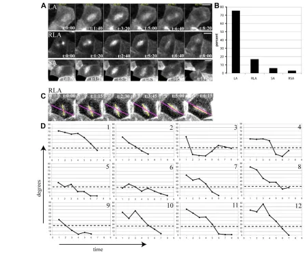
We found that the majority of spindles were already within 25° of the long axis at prometaphase/metaphase (Fig. 4A,B: LA, 75%; see Movie 4 in the supplementary material). The second most common spindle behaviour was rotation into the long axis (Fig. 4A,B; RLA, 16%; see Movie 5 in the supplementary material). Spindles that were set up in the short axis (SA) or rotated into the short axis (RSA; see Movie 6 in the supplementary material) were found only occasionally in 6% and 3% of cases, respectively (Fig. 4A,B). Thus, in the great majority of cases, spindles are already close to the long axis at the onset of metaphase, which indicates that spindle orientation in the Xenopusblastula takes place early in mitosis.
In order to show the temporal dynamics of any rotation movements that may occur, angle values between the long axis and the spindle axis were plotted against time for 12 cells from the RLA category, from three different embryos (Fig. 4C,D). The first movie frame analysed was always the first time point when the two centrosomes became clearly visible on opposite sides of the nucleus,
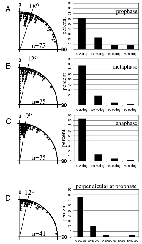
[image:6.612.55.501.58.428.2]and the last frame was one time-point before the onset of midbody constriction (Fig. 4C). These angle/time graphs show that overall spindle alignment is a gradual movement towards the long axis (Fig. 4D). Although oscillating changes of orientation can be observed between time-points in some cells (e.g. cells 3,4,10,12) oscillations generally do not involve large degree changes of spindle orientation (Fig. 4D). In most cases, spindles adjust in steps smaller than 20o between time points (time interval 92 seconds in cells 1-8 and 75 seconds in cells 9-12). Therefore, systematic oscillations with large angle changes in directionality do not occur in the Xenopusblastula. To exclude the possibility that tau-GFPexpression might have influenced microtubule dynamics and consequently spindle behaviour, these results were confirmed in immunostained fixed embryos. Fig. 5 shows angle measurements as absolute deviation of the spindle from the long axis at prophase (Fig. 5A), metaphase (Fig. 5B) and anaphase (Fig. 5C). The median deviation angle at prophase is 18°, at metaphase 12° and at anaphase 9°. To confirm specifically Fig. 4. Spindle alignment takes place early in the cell cycle; large angle oscillations do not occur.(A) Different spindle orientation
behaviour is shown in frames from 4D series of tau-GFPinjected embryos; LA, spindle is already set up within 25° of the long axis at
D
E
V
E
LO
P
M
E
N
T
that the spindle also aligns early with the long axis in cells that divide perpendicularly, measurements were taken in cells with a perpendicular centrosome orientation at prophase, in cross-section stacks. Fig. 5D shows that the median angle at prophase is 12° in these cells.
Astral microtubules are required for spindle orientation
To test the role of astral microtubules in spindle orientation, we have used the chemical microtubule polymerisation inhibitor nocodazole to reduce astral microtubules (Fig. 6A,D). Embryos at the 256-cell stage were incubated in nocodazole for 25 minutes (the average cell cycle time in the Xenopusblastula), and spindle orientation was
analysed at anaphase onset in z-series of fixed embryos that were whole-mount stained for ␣-tubulin. Angle measurements between the spindle axis and the long axis of a cell were carried out as above. Reduction of astral microtubules leads to an increase of the observed median angle to 23°, indicating a trend towards randomisation of spindle orientation when compared with the controls (Fig. 6B,C,E,F). Thus, in the Xenopusblastula, astral microtubules are necessary for spindle alignment with the long axis of a cell.
Astral microtubules make contact with the cortex between anaphase and the end of the next prophase
To determine the time window in the cell cycle when cell shape is most likely to be monitored by astral microtubules, we looked at the quantitative dynamics of astral microtubules in vivo using EB1-GFP injected embryos. EB1 belongs to a highly conserved family of proteins that binds to microtubule plus-ends and centrosomes (Mimori-Kiyosue and Tsukita, 2003; Liakopoulos et al., 2003; Piehl and Cassimeris, 2003; Morrison et al., 1998; Rogers et al., 2002). Movies were generated from projections of image stacks that were collected on a spinning disk confocal microscope (Fig. 7A, see Movie 7 in the supplementary material). The mitotic stage was estimated by the signal from the spindle (Fig. 7A) or by co-injecting Histone-GFP to visualise DNA (data not shown) and the EB1 signals were counted (Fig. 7B). A period of EB1 reduction was observed during late prophase/prometaphase followed by complete absence of EB1 signal in metaphase, which could explain the observed lack of extensive spindle movements during this time period. Conversely, the time period of maximum astral microtubule density is between anaphase and prophase of the next cell cycle (see also Morrison and Askham, 2001). In particular, the high density of astral microtubules in prophase is consistent with our previous results that the main orientation event has already taken place by prophase.
To exclude the possibility that this observed reduction of EB1 plus end signals is due to a loss of binding activity of EB1 to microtubules rather than to a reduction of microtubules, we have examined the distribution of astral microtubules in fixed cells. We could confirm a reduction of astral microtubules during prometaphase and metaphase in fixed cells (Fig. 7C).
Interestingly we observed that the centrosomes begin to separate at late anaphase, and are already several micrometers apart before cytokinesis is completed (Fig. 7C, arrow in anaphase 2). This is also the time period of maximum astral microtubule density and it is therefore possible that already centrosome separation takes place according to the long axis of a cell. This would explain the observation that the majority of spindles are already set up in the correct orientation at prometaphase.
DISCUSSION
The Xenopusblastula exhibits many of the characteristics expected from a developmental system that uses oriented divisions to generate cell fate diversity. First, cells of the Xenopusblastula are polarised; they exhibit the hallmarks of epithelial cell polarity with conserved molecular markers distributed on the apical or basolateral membrane (Chalmers et al., 2003; Chalmers et al., 2005; Muller and Hausen, 1995; Gawantka et al., 1992). Second, conserved genes play a role in controlling polarity as they do in other systems; misexpression and/or loss of aPKC and Lgl2 function alters the proportion of apical versus basolateral membrane (Chalmers et al., 2005). Third, divisions where the spindle is oriented along the apicobasal axis are fate asymmetric (Chalmers et al., 2003). Such divisions generate cells with distinct fates throughout the blastula and in particular in
0 10 20 30 40 50 60 70 80 90
0-20deg 20-40deg 40-60deg 60-80deg
prophase 0 10 20 30 40 50 60 70 80 90
0-20deg 20-40deg 40-60deg 60-80deg
metaphase 0 10 20 30 40 50 60 70 80 90
0-20deg 20-40deg 40-60deg 60-80deg
anaphase 0 10 20 30 40 50 60 70 80 90
0-20deg 20-40deg 40-60deg 60-80deg 80-90deg
perpendicular at prophase
[image:7.612.52.291.60.471.2]A
B
C
D
percent percent12
o
percent percent9
o
18
o
n=75
n=75
n=75
n=41
12
o
D
E
V
E
LO
P
M
E
N
T
the animal region, they have an intrinsic difference in competence for neuronal differentiation (Chalmers et al., 2002). Although the molecules underlying these fate differences are only just beginning to be elucidated (Chalmers et al., 2006), it is clear that divisions at the blastula stage bear all the hallmarks of typical fate-asymmetric divisions of polarised cells.
However, as we show in this paper, the Xenopusblastula differs from other systems of oriented divisions in that it appears to lack a specialised mechanism to orient the spindle according to polarity. Instead, cells divide according to their shape, by aligning the spindle with the long axis and bisecting this axis during division. Thus, the cells that are elongated in the apicobasal direction have a high propensity of dividing perpendicularly (bisecting the axis of polarity), and consequently generate daughter cells with different fates. Although the apical membrane domain with its resident proteins/protein complexes may prove to be important for cell fate determination (Gotz and Huttner, 2005; Chalmers et al., 2003; Chalmers et al., 2005), it does not seem to be important for spindle orientation.
Spindle orientation: default versus specialised mechanisms
Cell division where the spindle is set up along the long axis of the cell, is often thought to be a default mechanism, taking place when other cues of spindle orientation are absent (Honda, 1983). Division along the long axis was first formulated in the 19th century as a general rule of division by Hofmeister and Sachs, based on observations in plant cells, and studied experimentally by Hertwig and others in amphibian eggs (reviewed by Wilson, 1987). Only a few examples where cells divide according to their long axis during development were studied experimentally, such as some cells in the zebrafish embryo (Concha and Adams, 1998), the mouse zygote (Gray et al., 2004) and Xenopuseggs, which align their spindles into the long axis when mechanically deformed (Black and Vincent, 1988). Finally, normal rat kidney (NRK) in culture divide and bisect their long axis; as described for the blastula cells here, they also correct their spindle orientation to adjust to an experimentally imposed new long axis (O’Connell and Wang, 2000).
However, in many cases the influence of cell-shape is over-ridden. For example, during zebrafish early development, cells that are located in the dorsal epiblast divide such that the cleavage plane
bisects their short rather than their long axis, the opposite of what one would predict from geometric rules (Concha and Adams, 1998; Gong et al., 2004). In this case, ‘ignoring’ the long axis is thought to be mediated by the planar polarity pathway (PCP), (Gong et al., 2004), which has also been implicated in orienting cell divisions in DrosophilaSOPs (Gho and Schweisguth, 1998) and C. elegans embryos (Schlesinger et al., 1999). Activation of this pathway presumably overrides the default influence of the long axis. In most examples of asymmetric divisions of polarised cells, the influence of cell shape has not been studied directly but it is believed not to be important as many specialised cues for spindle orientation have been described. It is presumed that these specialised cues are dominant over any influence of cell shape in defining the spindle orientation (Tsou et al., 2002; Tsou et al., 2003a) (reviewed in Cowan and Hyman, 2004). In cultured cells, the influence of cell shape can also be over-ridden by the spatial distribution of extracellular matrix components (Thery et al., 2005).
Spindle rotations and timing of spindle alignment Many different cases have been described with regards to the timing of spindle alignment and the rotation behaviour of the spindle during mitosis. In Drosophilaneuroblasts, the spindles are set up parallel to the plane of the epithelium and rotate by 90° in metaphase to align with the apicobasal axis (Kaltschmidt et al., 2000), although more recent reports place the alignment earlier in the cell cycle (Siegrist and Doe, 2006). In zebrafish keel/rod stage embryos, the majority of the spindles are set up parallel to the plane of the epithelium but undergo a rapid 90° rotation in the neural keel region (Geldmacher-Voss et al., 2003). Extensive metaphase rotations have also been described in the rat (Adams, 1996) and mouse (Haydar et al., 2003) cortex, including (in the mouse) ‘flipping’ between the plane of the epithelium and the apicobasal axis. However, in the rat cortex, most of the rotations are confined within the plane of the epithelium, either parallel or anti parallel to the final division axis, suggesting that a preference for alignment is set up early in the cell cycle (Adams, 1996).
[image:8.612.52.359.59.269.2]We have reported here that in the Xenopus blastula, the final spindle orientation, which is aligned with the long axis, is already chosen at prophase, and that this is possibly achieved by the directed separation of duplicated centrosomes in the ‘correct’ direction. Spindles that are misaligned with respect to the long axis adjust with
D
E
V
E
LO
P
M
E
N
T
small angle steps towards it. In this sense, Xenopusblastomeres are more similar to the rat and chick retina, where spindles show only small rotations within a 30° band and the angle that the spindle assumes at metaphase entry tends to be the final angle at division (Tibber et al., 2004). Interestingly, similar to our case, but in contrast to the zebrafish retina (Das et al., 2003), in these systems the orientation of the spindle was found to be entirely random with respect to the anatomical landmarks of the retinal tissue (Tibber et al., 2004).
The reason for these differences in spindle behaviour across different systems is not known but it may ultimately be related to the mechanisms that align the spindle, such as the presence or absence of specialised orientation cues and the timing or mechanism by which astral microtubules interact with the cortex (e.g. Sanada and Tsai, 2005; Du and Macara, 2004).
Shape and mechanisms of spindle orientation Spindle orientation based on specialised cues depends on the interaction of microtubules with specialised cortical domains, which can be defined either by polarity or cell contact (Doe and Bowerman, 2001; Gonczy, 2002; Thery et al., 2005). It is thought that these specialised cortical sites or cortical domains act as attachment sites or modulate forces on microtubules to ‘pull’ the spindle in a preferred orientation. By contrast, the mechanisms
that orient the spindle according to cell shape are thought to rely on an overall balancing of forces acting on the spindle, which can only be achieved when the spindle is centered and aligned with the long axis (O’Connell and Wang, 2000). Different models, suggest either microtubule polymerisation – based pushing forces or motor-driven pulling forces (reviewed by Reinsch and Gonczy, 1998; Grill and Hyman, 2005; Vallee and Stehman, 2005). The common element is that forces should be proportional to microtubule length and that adjustment of spindle positioning takes place until a balance of forces on the spindle ends is achieved. Either of these models would be consistent with our observation that astral microtubules are important for spindle alignment in Xenopus.
Concluding remarks
[image:9.612.51.518.56.383.2]One of the attractive features of the developmental system we described here is its simplicity. Interpreting the overall cell shape simultaneously defines the plane and the orientation of division within the plane. Furthermore, there is no need for a specific mechanism to ‘select’ cells that will divide perpendicularly. As long as the dividing cells are polarised and polarity is important for fate, perpendicular divisions that are randomly distributed with respect to anatomical landmarks in the blastula will generate inner and outer cells that will follow different fates.
D
E
V
E
LO
P
M
E
N
T
In conclusion, a default mode of spindle orientation, featuring an element of spatial randomness, is sufficient for this system of polarised cells to generate different daughter cells. Thus, we propose that it is not obligatory to evoke a specialised mechanism for spindle orientation in all systems that generate cell fate asymmetry by division. Although well-studied examples from invertebrates have greatly influenced our thinking, perhaps we should expect more variety in the ways that different systems have adapted to control spindle orientation to generate cell fate diversity.
We thank Dr Jeremy Green for useful discussions and comments on the manuscript; Dr Andrea Brand and Dr Enrique Amaya for the tau-GFPplasmids; Dr Ewan Morrison for the EB1-GFPexpression plasmid; Alex Sossick for expert help with imaging; and Dr Chris Palmer for expert help with statistical analysis. N.P. is a Wellcome Trust Senior Research Fellow and B.S. was a Wellcome Trust Prize student. This work was funded by the Wellcome Trust.
Supplementary material
Supplementary material for this article is available at http://dev.biologists.org/cgi/content/full/133/19/3883/DC1
References
Adams, R. J.(1996). Metaphase spindles rotate in the neuroepithelium of rat cerebral cortex. J. Neurosci. 16, 7610-7618.
Betschinger, J. and Knoblich, J. A.(2004). Dare to be different: asymmetric cell division in Drosophila, C. elegans and vertebrates.Curr. Biol. 14, R674-R685.
Black, S. D. and Vincent, J. P.(1988). The first cleavage plane and the embryonic axis are determined by separate mechanisms in Xenopus laevis. II. Experimental dissociation by lateral compression of the egg. Dev. Biol. 128, 65-71.
Chalmers, A. D., Welchman, D. and Papalopulu, N.(2002). Intrinsic differences between the superficial and deep layers of the Xenopus ectoderm control primary neuronal differentiation. Dev. Cell2, 171-182.
Chalmers, A. D., Strauss, B. and Papalopulu, N.(2003). Oriented cell divisions asymmetrically segregate aPKC and generate cell fate diversity in the early Xenopus embryo. Development130, 2657-2668.
Chalmers, A. D., Pambos, M., Mason, J., Lang, S., Wylie, C. and Papalopulu, N.(2005). aPKC, Crumbs3 and Lgl2 control apicobasal polarity in early vertebrate development. Development132, 977-986.
Chalmers, A. D., Lachani, K., Shin, Y., Sherwood, V., Cho, K. W. Y. and Papalopulu, N.(2006). Grainyhead-like 3, a transcription factor identified in a microarray screen, promotes the specification of the superficial layer of the embryonic epidermis. Mech. Dev.(in press).
Colombo, K., Grill, S. W., Kimple, R. J., Willard, F. S., Siderovski, D. P. and Gonczy, P.(2003). Translation of polarity cues into asymmetric spindle positioning in Caenorhabditis elegans embryos. Science300, 1957-1961.
Concha, M. L. and Adams, R. J.(1998). Oriented cell divisions and cellular morphogenesis in the zebrafish gastrula and neurula: a time-lapse analysis.
Development125, 983-994.
Cowan, C. R. and Hyman, A. A.(2004). Asymmetric cell division in C. elegans: cortical polarity and spindle positioning. Annu. Rev. Cell Dev. Biol. 20, 427-453.
Das, T., Payer, B., Cayouette, M. and Harris, W. A.(2003). In vivo time-lapse imaging of cell divisions during neurogenesis in the developing zebrafish retina.
Neuron37, 597-609.
Doe, C. Q. and Bowerman, B.(2001). Asymmetric cell division: fly neuroblast meets worm zygote.Curr. Opin. Cell Biol. 13, 68-75.
Du, Q. and Macara, I. G.(2004). Mammalian Pins is a conformational switch that links NuMA to heterotrimeric G proteins. Cell119, 503-516.
Fesenko, I., Kurth, T., Sheth, B., Fleming, T. P., Citi, S. and Hausen, P.(2000). Tight junction biogenesis in the early Xenopus embryo. Mech. Dev.96, 51-65.
Gawantka, V., Ellinger-Ziegelbauer, H. and Hausen, P.(1992). Beta 1-integrin is a maternal protein that is inserted into all newly formed plasma membranes during early Xenopus embryogenesis. Development115, 595-605.
Geldmacher-Voss, B., Reugels, A. M., Pauls, S. and Campos-Ortega, J. A.
(2003). A 90-degree rotation of the mitotic spindle changes the orientation of mitoses of zebrafish neuroepithelial cells. Development130, 3767-3780.
Gho, M. and Schweisguth, F.(1998). Frizzled signalling controls orientation of asymmetric sense organ precursor cell divisions in Drosophila. Nature393, 178-181.
Gonczy, P.(2002). Mechanisms of spindle positioning: focus on flies and worms.
Trends Cell Biol. 12, 332-339.
Gonczy, P. and Hyman, A. A.(1996). Cortical domains and the mechanisms of asymmetric cell division. Trends Cell Biol. 6, 382-387.
Gong, Y., Mo, C. and Fraser, S. E.(2004). Planar cell polarity signalling controls cell division orientation during zebrafish gastrulation. Nature430, 689-693.
Gotz, M. and Huttner, W. B.(2005). The cell biology of neurogenesis.Nat. Rev. Mol. Cell Biol. 6, 777-788.
Gray, D., Plusa, B., Piotrowska, K., Na, J., Tom, B., Glover, D. M. and
Zernicka-Goetz, M.(2004). First cleavage of the mouse embryo responds to change in egg shape at fertilization.Curr. Biol. 14, 397-405.
Grill, S. W. and Hyman, A. A.(2005). Spindle positioning by cortical pulling forces. Dev. Cell8, 461-465.
Grill, S. W., Howard, J., Schaffer, E., Stelzer, E. H. and Hyman, A. A.(2003). The distribution of active force generators controls mitotic spindle position.
Science301, 518-521.
Hampoelz, B. and Knoblich, J. A.(2004). Heterotrimeric G proteins: new tricks for an old dog. Cell119, 453-456.
Hartenstein, V.(1989). Early neurogenesis in Xenopus: the spatio-temporal pattern of proliferation and cell lineages in the embryonic spinal cord. Neuron3, 399-411.
Haydar, T. F., Ang, E., Jr and Rakic, P.(2003). Mitotic spindle rotation and mode of cell division in the developing telencephalon. Proc. Natl. Acad. Sci. USA100, 2890-2895.
Honda, H.(1983). Geometrical models for cells in tissues. Int. Rev. Cytol.81, 191-248.
Huttner, W. B. and Kosodo, Y.(2005). Symmetric versus asymmetric cell division during neurogenesis in the developing vertebrate central nervous system.Curr. Opin. Cell Biol. 17, 648-657.
Izumi, Y., Ohta, N., Hisata, K., Raabe, T. and Matsuzaki, F.(2006). Drosophila Pins-binding protein Mud regulates spindle-polarity coupling and centrosome organization.Nat. Cell Biol. 8, 586-593.
Kaltschmidt, J. A., Davidson, C. M., Brown, N. H. and Brand, A. H.(2000). Rotation and asymmetry of the mitotic spindle direct asymmetric cell division in the developing central nervous system. Nat. Cell Biol. 2, 7-12.
Knoblich, J. A.(2001). Asymmetric cell division during animal development.Nat. Rev. Mol. Cell Biol. 2, 11-20.
Kusch, J., Liakopoulos, D. and Barral, Y.(2003). Spindle asymmetry: a compass for the cell. Trends Cell Biol. 13, 562-569.
Liakopoulos, D., Kusch, J., Grava, S., Vogel, J. and Barral, Y.(2003). Asymmetric loading of Kar9 onto spindle poles and microtubules ensures proper spindle alignment. Cell112, 561-574.
Lu, B., Jan, L. and Jan, Y. N.(2000). Control of cell divisions in the nervous system: symmetry and asymmetry. Annu. Rev. Neurosci.23, 531-556.
Mimori-Kiyosue, Y. and Tsukita, S.(2003). ‘Search-and-capture’ of
microtubules through plus-end-binding proteins (+TIPs). J. Biochem. 134, 321-326.
Morrison, E. E. and Askham, J. M.(2001). EB 1 immunofluorescence reveals an increase in growing astral microtubule length and number during anaphase in NRK-52E cells. Eur. J. Cell Biol. 80, 749-753.
Morrison, E. E., Wardleworth, B. N., Askham, J. M., Markham, A. F. and Meredith, D. M.(1998). EB1, a protein which interacts with the APC tumour suppressor, is associated with the microtubule cytoskeleton throughout the cell cycle. Oncogene17, 3471-3477.
Morrison, E. E., Moncur, P. M. and Askham, J. M.(2002). EB1 identifies sites of microtubule polymerisation during neurite development. Brain Res. Mol. Brain Res.98, 145-152.
Muller, H. A. and Hausen, P.(1995). Epithelial cell polarity in early Xenopus development. Dev. Dyn.202, 405-420.
O’Connell, C. B. and Wang, Y. L.(2000). Mammalian spindle orientation and position respond to changes in cell shape in a dynein-dependent fashion. Mol. Biol. Cell 11, 1765-1774.
Piehl, M. and Cassimeris, L.(2003). Organization and dynamics of growing microtubule plus ends during early mitosis. Mol. Biol. Cell 14, 916-925.
Reinsch, S. and Karsenti, E.(1994). Orientation of spindle axis and distribution of plasma membrane proteins during cell division in polarized MDCKII cells.J. Cell Biol. 126, 1509-1526.
Reinsch, S. and Gonczy, P.(1998). Mechanisms of nuclear positioning. J. Cell Sci.
111, 2283-2295.
Roegiers, F. and Jan, Y. N.(2004). Asymmetric cell division.Curr. Opin. Cell Biol.
16, 195-205.
Rogers, S. L., Rogers, G. C., Sharp, D. J. and Vale, R. D.(2002). Drosophila EB1 is important for proper assembly, dynamics, and positioning of the mitotic spindle.J. Cell Biol. 158, 873-884.
Sanada, K. and Tsai, L. H.(2005). G protein betagamma subunits and AGS3 control spindle orientation and asymmetric cell fate of cerebral cortical progenitors. Cell122, 119-131.
Schlesinger, A., Shelton, C. A., Maloof, J. N., Meneghini, M. and Bowerman, B.(1999). Wnt pathway components orient a mitotic spindle in the early Caenorhabditis elegans embryo without requiring gene transcription in the responding cell. Genes Dev.13, 2028-2038.
Siegrist, S. E. and Doe, C. Q.(2006). Extrinsic cues orient the cell division axis in Drosophila embryonic neuroblasts. Development133, 529-536.
Siller, K. H., Cabernard, C. and Doe, C. Q.(2006). The NuMA-related Mud protein binds Pins and regulates spindle orientation in Drosophila neuroblasts.
Nat. Cell Biol. 8, 594-600.
Suzuki, A. and Ohno, S.(2006). The PAR-aPKC system: lessons in polarity. J. Cell Sci. 119, 979-987.
D
E
V
E
LO
P
M
E
N
T
Bornens, M.(2005). The extracellular matrix guides the orientation of the cell division axis. Nat. Cell Biol. 7, 947-953.
Tibber, M. S., Kralj-Hans, I., Savage, J., Mobbs, P. G. and Jeffery, G.(2004). The orientation and dynamics of cell division within the plane of the developing vertebrate retina. Eur. J. Neurosci. 19, 497-504.
Tsou, M. F., Hayashi, A., DeBella, L. R., McGrath, G. and Rose, L. S.(2002). LET-99 determines spindle position and is asymmetrically enriched in response to PAR polarity cues in C. elegans embryos. Development129, 4469-4481.
Tsou, M. F., Hayashi, A. and Rose, L. S.(2003). LET-99 opposes Galpha/GPR signaling to generate asymmetry for spindle positioning in response to PAR and MES-1/SRC-1 signaling. Development130, 5717-5730.
Vallee, R. B. and Stehman, S. A.(2005). How dynein helps the cell find its center: a servomechanical model. Trends Cell Biol. 15, 288-294.
Wilson, E. B.(1987). Growth, cell division and development. In Genes, Cells and Organisms, Great Books in Experimental Biology. 3rd edn. (ed. J. A. Moore), pp. 980-1034. New York, London: Garland Publishing.
Wodarz, A.(2002). Establishing cell polarity in development. Nat. Cell Biol. 4, E39-E44.
Wodarz, A. and Huttner, W. B.(2003). Asymmetric cell division during neurogenesis in Drosophila and vertebrates. Mech. Dev.120, 1297-1309.
Zieba, P., Strojny, P. and Lamprecht, J.(1986). Positioning and stability of mitotic spindle orientation in the neuroepithelial cell. Cell Biol. Int. Rep.10, 91-100.