D
E
V
E
LO
P
M
E
N
T
INTRODUCTION
Morphogen signaling pathways are important components of developmental programs throughout the animal kingdom (Tabata and Takei, 2004). These pathways control many aspects of development, including determination of organ size and morphology. In contrast to the enormous morphological diversity found in animal body parts, only a handful of morphogen systems are known. This discrepancy predicts the existence of mechanisms that vary the activities of these signaling pathways in generating the range of animal structures seen in nature.
Selector genes, such as the Hox family of transcription factors, create differences between tissues by altering gene expression (Mann and Carroll, 2002). We hypothesized that these transcriptional regulators might control organ size and morphology by regulating morphogen production, distribution, and/or transduction. In support of this idea, we and others have previously reported that the distribution of the morphogen Decapentaplegic (Dpp) is different in the developing wing and haltere (Crickmore and Mann, 2006; de Navas et al., 2006; Makhijani et al., 2006) (see Fig. S1 in the supplementary material), which are two serially homologous flight appendages in Drosophilathat differ owing to the activity of a single selector gene, Ultrabithorax(Ubx) (Lewis, 1978). Ubxis expressed and required in all haltere cells but not in wing cells (Beachy et al., 1985; Lewis, 1978). In the primordia of both structures (the wing and haltere imaginal discs), Dpp is secreted from a group of specialized cells called the AP organizer, a stripe of anterior cells that is adjacent to the anterior (A)-posterior (P) compartment boundary. AP organizer cells are instructed to produce Dpp in response to the short-range morphogen Hedgehog (Hh), which is secreted from neighboring P-compartment cells (Basler and Struhl, 1994; Guillen et al., 1995; Tabata et al., 1995).
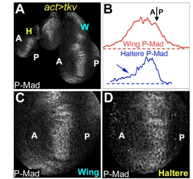
The Dpp pathway differs in the haltere and wing in several respects. For example, higher levels of Dpp are made in the wing organizer as compared with the haltere organizer (Crickmore and Mann, 2006; de Navas et al., 2006; Makhijani et al., 2006) (see Fig. S1 in the supplementary material). In addition, Dpp made in the wing is able to travel further from AP organizer cells than is Dpp made in the haltere. One reason for the decreased mobility of Dpp in the haltere is that the levels of the type-I Dpp-receptor Thickveins (Tkv) are higher in the medial haltere as compared with the medial wing. Higher levels of Tkv in the haltere limit the spread of Dpp, thus narrowing the Dpp activity gradient (Crickmore and Mann, 2006), which can be visualized with an antibody directed against the phosphorylated form of Dpp’s downstream transcription factor, Mothers against Dpp (P-Mad) (Fig. 1A and see Fig. S1 in the supplementary material) (Tanimoto et al., 2000). The Tkv-mediated narrowing of the Dpp activity profile in the haltere contributes to the smaller size of this appendage as compared with the wing (Crickmore and Mann, 2006).
In addition to receptors, a second class of molecules, heparin sulfate proteoglycans (HSPGs), have been reported to affect morphogen diffusion (Lin, 2004). Unlike receptors, which are selective for specific ligands, individual HSPGs influence the mobility of multiple morphogens (Lin, 2004). In this report we show that, in the haltere, Ubxcontrols the expression of one such HSPG gene, the glypican dally. The most striking aspect of this regulation is P compartment-specific repression of dally, which Ubxexecutes in conjunction with the posterior selector gene engrailed(en). By carrying out experiments in both the wing and haltere in which we vary Dally levels in a compartment-specific manner, we demonstrate that P-specific repression of dallydecreases the P:A compartment size ratio as well as the final size of the adult appendage. As Dally has been shown to regulate all three known morphogen systems in the wing (Wg, Hh and Dpp) (Han et al., 2005; Lin, 2004), alteration of dally expression by Ubx is expected to have important consequences for these signaling pathways. Here, we examine the consequences of asymmetric dallyexpression on Dpp signaling in the haltere as compared with the wing. We show that by downregulating dallyexpression in the P compartment, Ubxbiases
Hox control of morphogen mobility and organ development
through regulation of glypican expression
Michael A. Crickmore1and Richard S. Mann2,*
Animal bodies are composed of structures that vary in size and shape within and between species. Selector genes generate these differences by altering the expression of effector genes whose identities are largely unknown. Prime candidates for such effector genes are components of morphogen signaling pathways, which control growth and patterning during development. Here we show that in Drosophila the Hox selector gene Ultrabithorax(Ubx) modulates morphogen signaling in the haltere through transcriptional regulation of the glypican dally. Ubx, in combination with the posterior selector gene engrailed(en), represses dally expression in the posterior (P) compartment of the haltere. Compared with the serially homologous wing, where Ubxis not expressed, low levels of posterior dallyin the haltere contribute to a reduced P compartment size and an overall smaller appendage size. We also show that one molecular consequence of dallyrepression in the posterior haltere is to reduce Dpp diffusion into and through the P compartment. Our results suggest that Dpp mobility is biased towards cells with higher levels of Dally and that selector genes modulate organ development by regulating glypican levels.
KEY WORDS: Drosophila, P-Mad, BMP, dally, Organ size, Decapentaplegic, HSPG Development 134, 327-334 (2007) doi:10.1242/dev.02737
1Department of Biological Sciences, Columbia University, New York, NY 10027, USA. 2Department of Biochemistry and Molecular Biophysics, Columbia University, HHSC 1104, 701 W. 168th Street, New York, NY 10032, USA.
*Author for correspondence (e-mail: rsm10@columbia.edu)
D
E
V
E
LO
P
M
E
N
T
the diffusion of Dpp away from P cells in the haltere, which is likely to contribute to the decreased P:A size ratio and overall reduced size of the haltere compared with the wing. Interestingly, Dpp signaling itself also contributes to dally repression in the P compartment, demonstrating the presence of a self-reinforcing loop restricting Dpp diffusion into P cells. These results provide novel insights into the control of morphogen signaling by HSPGs and give further support to the hypothesis that selector genes control organ shape and size by regulation of morphogen signaling, a task which is accomplished by the spatial and quantitative regulation of morphogen signaling pathway components.
MATERIALS AND METHODS
Fly stocks
Both UAS-dallylines used in this paper were produced in the lab of H.
Nakato (University of Minnesota, Minneapolis, MN). The UAS-dallystrong
line is an insert on chromosome III, whereas the UAS-dallyweakline is an
insert on the X chromosome. Although we presume that UAS-dallystrongand
UAS-dallyweakproduce high and low quantities of Dally protein, respectively,
the names ‘strong’ and ‘weak’ refer to the effects we observe on P-Mad, Wg and Hh staining.
Dpp::GFP was driven in the AP organizers of the wing and haltere during
larval stages by crossing ptc-Gal4; Gal80tsflies to UAS-dpp::GFPflies. The
cross was raised at 18°C for two days and then transferred to 30°C until dissection.
UAS-dallyRNAiflies were obtained from Barry Dickson and the RNAi
group at IMBA (Vienna, Austria). Embryos from en-Gal4 UAS-dallyRNAi
and ci-Gal4 UAS-dallyRNAi crosses were collected at 25°C and shifted to
30°C after 24-48 hours.
Other stocks used: en-Gal4 UAS-GFP; en-Gal4 UAS-lacZ; hh-Gal4
UAS-GFP; ci-Gal4 UAS-GFP(II); FRT42D enE; hsflp; FRT42D Ub-GFP; dpp-lacZ10638; dally-lacZP2; FRT2A dally80; hsflp; FRT2A Ub-GFP; hsflp; Act>y>Gal4; UAS-GFP; tkv-lacZP906; FRT82B Ubx9-22; hsflp; FRT82B Ub-GFP; hsflp; act>y>Gal4 UAS-GFP; dally-lacZP2/TM6B; UAS-dad; UAS-tkvQD(III); UAS-tkv(III); UAS-tkv; dally-lacZP2; tub-Gal4/TM6B; UAS-GFP; act-Gal4; ptc-Gal4 UAS-GFP.
mRNA and protein detection
Standard procedures were followed for antibody staining except for the extracellular GFP staining for which the protocol of Belenkaya et al. (Belenkaya et al., 2004) was used.
Antibodies used: rabbit GFP 1:1000 (Molecular Probes), mouse anti-Wg 1:250 (Developmental Studies Hybridoma Bank, University of Iowa,
Iowa City, IA), rabbit anti--Gal 1:10,000 (Cappel), guinea pig anti-P-Mad
1:1250 (E. Laufer and T. Jessell, Columbia University, New York, NY), mouse Nub 1:10 (S. Cohen, EMBL, Heidelberg, Germany), mouse anti-Ubx 1:20, mouse anti-En 1:5 (Hybridoma Bank), rabbit anti-Hh 1:1000 (T. Tabata, University of Tokyo, Tokyo, Japan), mouse anti-Dlp 1:50 (Hybridoma Bank).
dallyin situ hybridization was performed with a DIG-labeled probe
generated using T7 RNA Polymerase to transcribe antisense dallycDNA
from PS Bluescript (a gift from H. Nakato).
Size and P:A ratio measurements
[image:2.612.231.562.61.308.2]All measurements were taken as pixel counts using Adobe Photoshop. For adult wings and halteres, only the blade or capitellum of females were measured. All measured animals were raised under non-crowding conditions. Eggs were collected for 2 hours and grown for 24-48 hours. Approximately 50 first-instar larvae were transferred to fresh tubes and
Fig. 1. dallyexpression and Dpp signaling are reduced in the posterior haltere.
(A-A⬙) Comparison of dally-lacZand P-Mad staining in the wing and haltere. In the wing, dally-lacZis expressed similarly on both sides of the AP organizer (A⬘). In the haltere, dally-lacZ expression is low in the P compartment, especially in P cells abutting the AP boundary (arrow). P-Mad staining in the wing is detected in two slopes that peak at either side of the AP organizer, whereas in the haltere, P-Mad is detected in a single stripe (A⬙). (B) dally-lacZ intensity traces for the wing and haltere discs shown in A. The approximate position of the AP boundary is indicated here (and in D,F) by an arrow. Note the symmetry in dallyexpression in the wing with respect to the AP organizer (just left of the arrow), as compared with the asymmetry in the haltere. Also note the generally lower dallylevels in AP organizer cells and the hyper-repressed levels in the P cells abutting the AP boundary in the haltere. (C-C⬙) The repression of dally-lacZin the haltere correlates with very low P-Mad staining in the P compartment. The yellow line marks the AP compartment boundary. Note that strong P-Mad
D
E
V
E
LO
P
M
E
N
T
grown until dissection at wandering or after eclosing. P:A size ratios were calculated by measuring the fraction of GFP-expressing cells in the Nubbin
domain of animals containing en-Gal4or ci-Gal4in combination with either
UAS-GFPalone, or withUAS-GFPplus UAS-dallyRNAi orUAS-dallyweak. The wing discs shown for the wing P:A ratio experiments were stained and imaged together and are therefore directly comparable. All sizes and ratios were measured without knowledge of the genotype. Error bars are s.e.m.
Intensity plots
Images were imported into ImageJ and pixel intensities measured by boxing a subset of the pouch regions of the discs.
RESULTS
Asymmetric Dpp signaling in the haltere
Upon comparing Dpp signaling readouts in the wing and haltere, we noticed that, in addition to a general narrowing of Dpp pathway activity (Crickmore and Mann, 2006), Dpp signaling was also asymmetric relative to its source (the AP organizer) in the haltere. Specifically, the P-Mad signal was stronger anterior to the AP organizer (roughly demarcated by the domain of peak P-Mad staining) than it was posterior to the organizer (Fig. 1A,B,D and see Fig. S1 in the supplementary material). To test if this asymmetry is due to asymmetric ligand distribution or differences in signal transduction, we used an extracellular staining protocol to examine the distribution of a Dpp::GFP fusion protein following its expression in AP organizer cells (Belenkaya et al., 2004; Entchev et al., 2000; Teleman and Cohen, 2000) (Fig. 1C,E). In wing cells, Dpp::GFP was detected in a broad gradient on both sides of the AP organizer (Fig. 1C and see Fig. S2 in the supplementary material). In the haltere, the distribution of Dpp::GFP is limited in both directions owing to high tkvexpression levels (Crickmore and Mann, 2006), but this restriction is stronger in the P direction (Fig. 1C,E). Dpp::GFP spread was abruptly halted a few cell diameters posterior to the haltere AP compartment boundary, contrasting with a tapering signal seen in the anterior direction. By contrast, the Gal4 driver used to express Dpp::GFP (ptc-Gal4) drove nearly symmetrical expression of a UAS-GFP transgene (see Fig. S1F,G in the supplementary material), demonstrating that the distribution of Dpp::GFP in the haltere is not due to asymmetric activity of the ptc-Gal4 driver. In both the wing and haltere, the pattern of extracellular Dpp::GFP was very similar to the P-Mad pattern, suggesting that Ubx does not affect Dpp signal transduction downstream of ligand binding, at least as detected with the anti-P-Mad antibody. Furthermore, in both the wing and haltere, a similar coincidence of extracellular Dpp::GFP and P-Mad patterns was observed when Dpp::GFP was expressed in clones (Crickmore and Mann, 2006). The correlation between the P-Mad and extracellular Dpp::GFP patterns in both the wing and haltere allows us to infer extracelluar Dpp ligand distribution by visualizing P-Mad in the proceeding experiments.
In the wing, Dpp::GFP distribution and P-Mad staining were also asymmetric, owing to slightly higher levels of Tkv in the P compartment, which impedes diffusion (see Figs S1, S2 in the supplementary material) (Tanimoto et al., 2000). By contrast, because Tkv levels are similar on both sides of the AP boundary of the haltere (see Fig. S1D in the supplementary material) (Crickmore and Mann, 2006; de Navas et al., 2006; Makhijani et al., 2006), Tkv levels are unlikely to account for the Dpp signaling asymmetry in this appendage. We tested this idea directly by providing uniform levels of UAS-tkv to both the haltere and wing. Under these conditions, P-Mad staining became symmetric in the wing, but remained asymmetric in the haltere (Fig. 2). These results suggest
that the more-restricted P-Mad staining in the P compartment of the wild-type haltere is due to a tkv-independent and haltere-specific anterior bias in the diffusion of Dpp.
[image:3.612.333.530.60.243.2]Adallyexpression asymmetry in the haltere In addition to receptors, the glypicans dally anddally-like(dlp) encode cell surface molecules known to affect morphogen signaling, and are thus candidates for generating the asymmetric Dpp distribution in the haltere (Lin, 2004). Although Dlp expression differs between the wing and haltere, its levels were symmetric along the AP axes of both tissues (see Fig. S1H in the supplementary material), making it unlikely that dlpis responsible for asymmetric Dpp signaling in the haltere. In the wing disc, dallyexpression, as monitored by a dally-lacZenhancer trap (Fig. 1A) (Fujise et al., 2001) and in situ hybridization (see Fig. S3 in the supplementary material) (Fujise et al., 2001), was high in the AP and dorsoventral (DV) organizers and was largely symmetric with respect to the source of Dpp production at the AP organizer (Fig. 1A and see Fig. S3 in the supplementary material) (Fujise et al., 2001). By contrast, in the haltere, dally expression was asymmetric with respect to the AP organizer: dallylevels were much lower on the P side of the AP organizer than they were on the A side (Fig. 1A,B,D and see Fig. S3 in the supplementary material). Most strikingly, dally-lacZexpression was almost undetectable in P cells that lie immediately adjacent to the AP boundary in the haltere (Fig. 1B,D). We refer to these cells as the domain of dally hyper-repression. In the A compartment of the haltere, dallyexpression was intermediate medially and high laterally. These findings suggest that differences in dallyexpression between the wing and haltere may contribute to the asymmetric Dpp signaling observed along the AP axis of the haltere.
D
E
V
E
LO
P
M
E
N
T
dallylevels and Dpp signaling
dallyis known to influence the diffusion of Wg, Hh and Dpp, all of which function as morphogens in the wing (Han et al., 2005; Lin, 2004) (see Fig. S4 in the supplementary material). Thus, the altered pattern of dallyexpression in the haltere might influence some or all of these pathways. Because of its role in appendage growth, we examined in detail how the pattern of dallyexpression in the haltere affects Dpp signaling.
Dpp signaling is promoted by Dally activity. For example, P-Mad levels are increased in Dally-overexpressing clones (Fujise et al., 2003) (see Fig. S4B in the supplementary material) and decreased in dally-mutant clones (Belenkaya et al., 2004) (see Fig. S4A in the supplementary material). We therefore hypothesized that the dally expression pattern in the haltere (in particular the domain of dally hyper-repression) might be responsible for the reduced Dpp diffusion and signaling seen in the P compartment of the haltere. Strong support for this idea comes from the observation that increasing Dally levels in the P compartment by driving UAS-dally with en-Gal4caused peak P-Mad staining to extend far into the P compartment of the haltere (Fig. 3B,C and see Fig. S5 in the supplementary material) (Makhijani et al., 2006). Similar results were obtained by expressing UAS-dallywith hh-Gal4(data not shown). Interestingly, the severity of this result depended on the UAS-dallyconstruct used, which is likely to reflect the different levels of Dally they produce (see Materials and methods). Another interesting observation derived from this experiment is that, in addition to increasing posterior Dpp signaling, driving either of the UAS-dally constructs with en-Gal4 or hh-Gal4 created a non-autonomous reduction in P-Mad labeling in the A cells immediately adjacent to the AP boundary (Fig. 3B,C and see Fig. S5 in the supplementary material; data not shown). Thus, increased Dally levels in the P compartment cause an increase in posterior Dpp signaling, and seem to do so at the expense of A compartment signaling. This observation suggests that the Dpp produced in the A compartment more readily travels into or is retained within the cells that express higher levels of dally. The non-autonomous reduction in P-Mad levels in the A cells of these discs argues against a role for Dally in promoting the stability of Dpp, as such effects would be predicted to be strictly cell-autonomous. We conclude that artificially high posterior Dally levels bias Dpp mobility in the posterior direction. By extension, these data suggest that the domain of dallyhyper-repression in the P compartment of the wild-type haltere functions to hinder the posterior diffusion of Dpp, thereby biasing its diffusion in the anterior direction. We further tested this idea by monitoring P-Mad levels in wing discs in which Dally levels were reduced in the P compartment. Consistently, driving UAS-dallyRNAiwith en-Gal4decreased the fraction of P-Mad staining detected in P cells (Fig. 4B). Thus, in this experiment, as in the wild-type haltere, Dpp appears to diffuse from its source in a direction that is determined by the levels of Dally in the surrounding tissue. We have observed similar non-autonomous alterations in P-Mad staining in cells surrounding UAS-dally-expressing cells when driven with ap-Gal4 or in flip-out clones (data not shown), demonstrating that the effects of altering Dally levels are not region-specific.
Consequences of posterior dallyrepression for appendage development
If dally repression in the posterior haltere alters morphogen signaling, it would be predicted to influence the development of the appendage. Below we show that asymmetric dallyexpression levels do indeed impact organ development. For example, Dpp target gene
expression in the haltere changes in accordance with the altered Dpp signaling levels seen when UAS-dallyis supplied to posterior haltere cells (see Fig. S6 in the supplementary material).
[image:4.612.341.528.59.419.2]Interestingly, the P:A compartment size ratio of a wild-type haltere (~0.48) is much less than that of a wild-type wing (~1), possibly owing to decreased morphogen signaling in the haltere P compartment. Consistent with this idea, drivingUAS-dallyweakwith en-Gal4increased the P:A size ratio of the appendage-forming region of the haltere disc from 0.48 to 0.59 (Fig. 3D). In addition to
D
E
V
E
LO
P
M
E
N
T
the increased P:A size ratio, we found that the overall size of the adult haltere was reproducibly 5% larger in en-Gal4 UAS-dallyweak
animals compared with control animals (Fig. 3E). These limited effects on relative compartment size and entire organ size, when compared with the dramatic effects on Dpp signaling in en-Gal4 UAS-dallyflies, suggest that Ubxworks through means in addition to dallyrepression to limit the P:A size ratio and overall size of the haltere. For example, Ubxalso represses wgexpression along the DV boundary of the haltere P compartment (Weatherbee et al., 1998) (see Fig. S1E in the supplementary material). Thus, wgexpression might become limiting for growth when dally expression is increased.
In order to understand the consequences of dally regulation independently from the influences of Ubx, we mimicked the dally expression asymmetry seen in the haltere by knocking down Dally levels in the P compartment of the wing, using en-Gal4to drive UAS-dallyRNAi. As mentioned above, P-Mad staining is decreased in the P compartments of these discs (Fig. 4B). In addition, the P:A size ratio of the appendage-forming region of these discs is decreased from 1.04 to 0.65 (Fig. 4C), consistent with the idea that reduced dallyexpression in the posterior compartment contributes to a smaller P:A size ratio. This P:A ratio decrease was also visualized in the adult wing as a posterior shift in vein L4, which lies just posterior to the AP compartment boundary (Fig. 4D). Further, adult en-Gal4 UAS-dallyRNAiwings were 19% smaller than control wings (Fig. 4D,E). These results suggest that, when analyzed independently of other Ubx-mediated changes in gene expression, reducing dally levels in the P compartment has a significant influence on P:A proportioning and final appendage size.
To test whether the P:A ratio and/or overall size altering capabilities of dallyrepression are specific to P cells, we knocked-down Dally levels in the A compartment of the wing by driving UAS-dallyRNAiwith ci-Gal4. P-Mad levels were increased in the P compartments of these discs as compared with control discs, which were co-stained and imaged identically and were therefore directly comparable (Fig. 4A,F). We interpret this observation to suggest that Dpp mobility is biased away from A cells as a consequence of low Dally levels in this compartment. P-Mad was also increased in the AP organizers (dpp-expressing cells) of these discs (Fig. 4, compare F with A). We suggest that, because Dally levels are lower throughout the A compartments of these discs, Dpp signaling is more tightly restricted to the cells that produce Dpp. The P:A ratio of ci-Gal4 UAS-dallyRNAidiscs increased from 0.93 to 1.39 and vein L4 was shifted anteriorly in adult wings, findings that complement the reduction of the P:A ratio of en-Gal4 UAS-dallyRNAi discs (Fig. 4G,H). However, in contrast to the en>dallyRNAi experiments (Fig. 4D,E), the final size of ci>dallyRNAiwings was not significantly different from that of control wings (Fig. 4H,I). This result suggests that knocking down Dally levels from either compartment of the wing is able to shift the P:A size ratio, but only a P-specific reduction in Dally levels reduces overall organ size. These data support the conclusion that the Ubx -mediated repression of dallyin the P compartment of the wild-type haltere contributes to the reduced P:A size ratio and overall reduced size of this appendage.
Regulation of dallyin the haltere
[image:5.612.52.299.59.484.2]We next turned our attention to how Ubx generates the haltere pattern of dallyexpression. dallyis activated by Hh and Wg (Fujise et al., 2001) and repressed by Dpp (Fig. 5B) (Fujise et al., 2003). In the wing, Dpp signaling, as monitored by P-Mad levels, peaked in the cells on either side of the AP organizer, whereas the cells that
D
E
V
E
LO
P
M
E
N
T
produce Dpp had lower levels of Dpp signaling (see Fig. S1A,B in the supplementary material). By contrast, Hh signal transduction was highest in the AP organizer, where Dpp signaling is low (see Fig. S1A,B in the supplementary material). Consequently, in the wing, dallyexpression is high in the AP organizer (due to positive Hh input) and low on either side of the organizer (due to negative Dpp input) (Fig. 5A). Dpp signaling also repressed dallyin the haltere (Fig. 5B,F).However, because of high Tkv levels in the medial haltere, Dpp diffusion is limited, resulting in peak Hh and Dpp signaling coinciding in the same cells (Crickmore and Mann, 2006). Therefore, opposing positive (Hh) and negative (Dpp) inputs into dallyoccur in AP organizer cells of the haltere, which is likely to contribute to the intermediate dallylevels observed in this region
[image:6.612.53.478.52.419.2]of the disc (Fig. 5A⬘). To test this idea, we expressed uniform tkv levels in the wing to mimic the haltere tkvpattern. This manipulation caused the peak Hh and Dpp signaling domains to align, as shown by the single stripe of P-Mad staining in the AP organizer (Fig. 2A, Fig. 5C) (Crickmore and Mann, 2006). The resulting dally-lacZ pattern in the A compartment is similar to that found in the A compartment of the wild type haltere (Fig. 5C). Thus, we conclude that the Ubx-dependent upregulation of tkvin the haltere is sufficient to account for the A-compartment differences in dally expression between the wing and haltere. Interestingly, a similar tkv-mediated effect also impacts upon the transcription of dpp, which, like dally, is activated by Hh signaling and repressed by Dpp signaling (Fig. 5G) (Crickmore and Mann, 2006).
D
E
V
E
LO
P
M
E
N
T
The low dally-lacZlevels in the P compartment of the wild-type haltere suggest that the combination of Ubxand the posterior selector gene en represses dally. Accordingly, dally-lacZ was derepressed in Ubx–or en–clones in the posterior haltere (Fig. 5D,E). We also note that, as previously reported, DV wg was repressed in the P compartment of the haltere (Weatherbee et al., 1998) (see Fig. S1E in the supplementary material), removing a positive input into dallyexpression. The strongest repression of dallyin the haltere (dallyhyper-repression) occurred in the P cells immediately adjacent to the A compartment. These were the only cells to show detectable P-Mad staining in the posterior haltere (Fig. 1B), suggesting a role for Dpp signaling in dallyhyper-repression. In support of this idea, dallywas derepressed in this region when Dpp signaling was clonally inhibited (Fig. 5F). Interestingly, the repression of dallyby Dpp in the posterior haltere demonstrates that the AP asymmetry in Dpp diffusion is self-reinforcing because any Dpp that is transduced in the P compartment further contributes to dally repression. In summary, as depicted in Fig. 5G, dally is repressed in the P compartment of the haltere by the convergence of enand Ubxand the absence of DV wg. The domain of dally hyper-repression results from an additional repressive input from Dpp signaling. As this stripe of P cells in which dallyis hyper-repressed abuts the Dpp producing cells of the A compartment, we suggest that it is responsible for biasing the movement of Dpp in the anterior direction and for the ensuing consequences to haltere development.
DISCUSSION
The fundamental importance of morphogen signaling pathways in controlling the growth and patterning of tissues has lead to the hypothesis that selector genes alter morphogen signaling landscapes to create structures of different shapes and sizes. In our previous work, we showed how the upregulation of the Dpp receptor, thickveins, in the haltere causes an overall decrease in Dpp mobility as compared with the wing, and consequently contributes to the small size of the haltere (Crickmore and Mann, 2006). Here we show that the HSPG dallyis repressed in the P compartment of the haltere and that this regulation decreases the P:A ratio and overall size of the haltere. We show that posterior dallyrepression causes Dpp diffusion to be biased away from P cells, generating an AP asymmetry in Dpp signaling. The findings reported here therefore provide another instance wherein Ubxcontrols the extracellular signaling environment of the developing haltere and thereby distinguishes it from the wing.
The movement of most or all signaling molecules through tissues is regulated by HSPGs, including glypicans such as dally. In contrast to receptors, HSPGs control the distribution of multiple signaling molecules. Regulation of HSPG expression and activity by selector genes is therefore a potentially very powerful mechanism for shaping signaling pathway activation profiles and molding organ shapes and sizes. However, the promiscuity of HSPGs also makes it difficult to assign the morphological consequences of their expression patterns to the alteration of individual signaling pathways. Indeed, we think it likely that the altered dallyexpression pattern in the haltere has implications for Hh, Wg and Dpp signaling (see Fig. S4 in the supplementary material) (Han et al., 2005; Lin, 2004), all of which control growth and patterning. Here, we have focused on the relationship between dallyexpression and Dpp signaling.
It has been shown that Dpp signaling is increased in dally+clones (Fujise et al., 2003) and decreased in dally–clones (Belenkaya et al., 2004) (see Fig. S4 in the supplementary material). These and other
findings have suggested that Dally participates in the control of Dpp mobility. Our results add to these earlier observations by suggesting that variations in the levels of Dally between the cells of a tissue influence the direction and extent of Dpp diffusion. Specifically, we propose that in addition to simply being promoted by Dally, Dpp mobility is biased towards cells with higher Dally levels. This idea derives mainly from the observation that Dally can influence Dpp movement in a cell-non-autonomous manner. For example, we found that when Dally levels are increased in the haltere P compartment, there is a shift in Dpp signaling from the A to the P compartments, as visualized by the levels of P-Mad. Similarly, knocking down Dally levels in the P compartment of the wing influences the extent and levels of P-Mad in the A compartment. If discontinuities in Dally levels can non-autonomously influence Dpp signaling across compartment borders, it follows that differences in Dally levels between cells within a compartment can also shape the Dpp signaling landscape. This might be important for wild-type wing development, where graded Dpp signaling represses dally, resulting in an inverse dallygradient that increases towards the lateral edge of the disc (Fig. 1A). We suggest that this inverse dally gradient helps to attract Dpp to more lateral regions of the disc. Accordingly, in a dally-mutant wing disc, the Dpp gradient is less broad than in a wild-type wing disc (data not shown) (Fujise et al., 2003). It is possible that other HSPGs control the mobility of signaling molecules in a similar manner.
We have shown that altering dallylevels in either the A or P compartment changes relative compartment size, but that only P compartment dally levels are relevant for total organ size. We suggest two possible explanations that link the P-specific dally repression seen in the haltere to a reduction in final organ size. Both of these scenarios (which are not mutually exclusive) focus on the role of P cells in producing Hh, which diffuses into A cells to instruct Dpp production and, consequently, controls final organ size. Importantly for both models, we find that there is in fact less Hh detected in the P compartment of the wild-type haltere as compared with the wing (see Fig. S1 in the supplementary material). In the first model, the repression of dallyreduces overall Hh production simply by reducing the size of the P compartment, which is a consequence of reduced Dpp signaling. In this scenario, fewer Hh-producing P cells result in less total Hh production from the P compartment, and therefore less Dpp produced in the A compartment. The logic of this potential mode of size regulation is interesting: a selector gene (Ubx) restricts growth factors (Wg and Dpp) from the pool of cells (the P compartment) that produces another growth factor (Hh). In the second scenario, dallyrepression may directly reduce the amount of Hh in the P compartment that can be transported into the A compartment. In support of this idea, we found Hh staining to be reduced in clones of cells where Dally levels are reduced through UAS-dallyRNAi(see Fig. S4 in the supplementary material).
D
E
V
E
LO
P
M
E
N
T
are combined with those of our earlier work showing that the levels of both Dpp and its receptor are regulated differently in the haltere and wing (Crickmore and Mann, 2006; de Navas et al., 2006; Makhijani et al., 2006), and the observation that wgis repressed in the posterior haltere (Weatherbee et al., 1998) (see Fig. S1 in the supplementary material), a picture emerges in which selector genes alter the expression of multiple components of multiple signaling pathways to change morphogen signaling landscapes between tissues and thereby modify organ shapes and sizes. We hypothesize that the summation of all signaling pathway changes may be sufficient to understand the size and shape differences between fundamentally similar epithelia such as the wing and haltere imaginal discs.
We thank K. Irvine, L. Johnston, X. Lin, H. Nakato, G. Struhl, the Bloomington Stock Center and the Developmental Studies Hybridoma Bank for fly stocks and reagents; H. Nakato and Y. Hayashi for advice on dallyin situ
hybridisation; Barry Dickson and the RNAi group at IMBA for providing UAS-dallyRNAiflies; and D. Rogulja for comments on the manuscript. This work was supported by a grant from the NIH to R.S.M. M.A.C. was supported by NIH training grants DK07328 and GM008798.
Supplementary material
Supplementary material for this article is available at http://dev.biologists.org/cgi/content/full/134/2/327/DC1
References
Basler, K. and Struhl, G.(1994). Compartment boundaries and the control of Drosophila limb pattern by hedgehog protein. Nature368, 208-214.
Beachy, P. A., Helfand, S. L. and Hogness, D. S.(1985). Segmental distribution of bithorax complex proteins during Drosophila development. Nature313, 545-551.
Belenkaya, T. Y., Han, C., Yan, D., Opoka, R. J., Khodoun, M., Liu, H. and Lin, X.(2004). Drosophila Dpp morphogen movement is independent of dynamin-mediated endocytosis but regulated by the glypican members of heparan sulfate proteoglycans. Cell119, 231-244.
Crickmore, M. A. and Mann, R. S.(2006). Hox control of organ size by regulation of morphogen production and mobility. Science313, 63-68.
de Navas, L. F., Garaulet, D. L. and Sanchez-Herrero, E.(2006). The
Ultrabithorax Hox gene of Drosophila controls haltere size by regulating the Dpp pathway. Development133, 4495-4506.
Entchev, E. V., Schwabedissen, A. and Gonzalez-Gaitan, M.(2000). Gradient formation of the TGF-beta homolog Dpp. Cell103, 981-991.
Fujise, M., Izumi, S., Selleck, S. B. and Nakato, H.(2001). Regulation of dally, an integral membrane proteoglycan, and its function during adult sensory organ formation of Drosophila. Dev. Biol. 235, 433-448.
Fujise, M., Takeo, S., Kamimura, K., Matsuo, T., Aigaki, T., Izumi, S. and Nakato, H.(2003). Dally regulates Dpp morphogen gradient formation in the Drosophila wing. Development130, 1515-1522.
Guillen, I., Mullor, J. L., Capdevila, J., Sanchez-Herrero, E., Morata, G. and Guerrero, I.(1995). The function of engrailed and the specification of Drosophila wing pattern. Development121, 3447-3456.
Han, C., Yan, D., Belenkaya, T. Y. and Lin, X.(2005). Drosophila glypicans Dally and Dally-like shape the extracellular Wingless morphogen gradient in the wing disc. Development132, 667-679.
Kirkpatrick, C. A., Dimitroff, B. D., Rawson, J. M. and Selleck, S. B.(2004). Spatial regulation of Wingless morphogen distribution and signaling by dally-like protein. Dev. Cell7, 513-523.
Kreuger, J., Perez, L., Giraldez, A. J. and Cohen, S. M.(2004). Opposing activities of Dally-like glypican at high and low levels of Wingless morphogen activity. Dev. Cell7, 503-512.
Lewis, E. B.(1978). A gene complex controlling segmentation in Drosophila. Nature276, 565-570.
Lin, X.(2004). Functions of heparan sulfate proteoglycans in cell signaling during development. Development131, 6009-6021.
Makhijani, K., Kalyani, C., Srividya, T. and Shashidhara, L. S.(2006). Modulation of Decapentaplegic gradient during haltere specification in Drosophila. Dev. Biol. doi:10.1016/j.ydbio.2006.09.029.
Mann, R. S. and Carroll, S. B.(2002). Molecular mechanisms of selector gene function and evolution. Curr. Opin. Genet. Dev. 12, 592-600.
Tabata, T. and Takei, Y.(2004). Morphogens, their identification and regulation. Development131, 703-712.
Tabata, T., Schwartz, C., Gustavson, E., Ali, Z. and Kornberg, T. B.(1995). Creating a Drosophila wing de novo, the role of engrailed, and the compartment border hypothesis. Development121, 3359-3369.
Tanimoto, H., Itoh, S., ten Dijke, P. and Tabata, T.(2000). Hedgehog creates a gradient of DPP activity in Drosophila wing imaginal discs. Mol. Cell5, 59-71.
Teleman, A. A. and Cohen, S. M.(2000). Dpp gradient formation in the Drosophila wing imaginal disc. Cell103, 971-980.