INTRODUCTION
The germline stem cells (GSCs) of the Drosophilaovary provide an excellent model system for studying the molecular mechanisms of stem cell regulation in vivo. In adult Drosophila females, two to three GSCs are easily recognized by their molecular markers (either a spherical spectrosome, or an extending fusome when GSCs are dividing) and their location at the apical region of the germarium in close contact with surrounding somatic cells, the terminal filament and cap cells, which together generate a specific micro-environment, or niche, for GSC regulation (Fig. 1A). The GSC divisions take place along the anteroposterior axis of the ovary to produce an anterior GSC, which remains attached to the niche cells, and a posterior cystoblast (Cb). The Cb divides precisely four times by incomplete cytokinesis to generate 16 interconnected cells that form the germline cyst of the follicle and sustain oogenesis.
Genetic analyses have revealed that the stem cell state of GSCs is maintained by both extrinsic and intrinsic mechanisms that repress their differentiation (Spradling et al., 2001). BMP ligands (Dpp, Gbb) from the niche cells (Song et al., 2004) maintain GSCs by suppressing Cb differentiation in the anteriormost cells. This is achieved by silencing the transcription of the bamgene, which encodes a GSC/Cb differentiation-promoting factor (Chen and McKearin, 2003a; Song et al., 2004). In the GSCs, BMP signaling activates cytoplasmic Mad and Medea, the DrosophilaSmads, and promotes their nuclear translocation. In the nucleus, the Smads complex physically interacts with both the bamsilencer element and nuclear lamin-associated protein (Ote), resulting in bam
transcriptional silencing (Chen and McKearin, 2003a; Jiang et al., 2008; Song et al., 2004). Thus, BMP/Dpp-dependent bam transcriptional control serves as the primary pathway for the regulation of GSC fate. Independent of the niche-based regulation of the bam silencing mechanism, the fate of GSCs is also intrinsically controlled by other GSC maintenance factors that repress their differentiation. It has been demonstrated that Pum/Nos-mediated and microRNA-Pum/Nos-mediated translational repression pathways are not required for bamsilencing, suggesting that these pathways act either downstream of or parallel to bamaction (Chen and McKearin, 2005; Jin and Xie, 2007; Park et al., 2007; Szakmary et al., 2005; Yang et al., 2007). Although it is proposed that the Pum/Nos-mediated and microRNA-mediated pathways repress the translation of key differentiation factors to prevent GSC differentiation, the targets of these translational pathways in GSCs are not identified. Thus, the intrinsic mechanisms that repress GSC differentiation are still poorly understood. In addition, as loss of function of the components in these translational repression pathways is not sufficient to completely cause bammutant germ-cell differentiation (Chen and McKearin, 2005), we speculate that the repression of GSC differentiation may also be controlled by other unknown intrinsic mechanisms.
Ubiquitin-mediated protein degradation plays a variety of roles in the regulation of many developmental processes (Glickman and Ciechanover, 2002). The enzymatic reaction of protein ubiquitylation is a coordinated three-step process involving three classes of enzymes known as E1 (Uba1 – FlyBase), E2 (UbcD4 – FlyBase) and E3. Firstly, E1 (Ubiquitin activating enzyme 1) catalyzes the formation of a thiolester bond linkage between the active-site cysteine residue on E1 and the C terminus of ubiquitin. Secondly, the activated ubiquitin (E1-Ub) is then transferred to E2 (Ubiquitin conjugating enzyme 4) via formation of an E2-Ub thiolester. Thirdly, E3 (ubiquitin ligase) promotes the transfer of the ubiquitin from E2-Ub to a lysine residue of the target protein through an isopeptide bond. Repeated cycles of this reaction can result in Development 136, 4133-4142 (2009) doi:10.1242/dev.039032
State Key Laboratory of Reproductive Biology and State Key Laboratory of Biomembrane and Membrane Biotechnology, Institute of Zoology, Chinese Academy of Sciences, Datun Road, Chaoyang, Beijing 100101, P.R. China.
*These authors contributed equally to this work †Author for correspondence (chendh@ioz.ac.cn)
Accepted 30 September 2009
Increasing evidence supports the idea that the regulation of stem cells requires both extrinsic and intrinsic mechanisms. However, much less is known about how intrinsic signals regulate the fate of stem cells. Studies on germline stem cells (GSCs) in the
Drosophilaovary have provided novel insights into the regulatory mechanisms of stem cell maintenance. In this study, we demonstrate that a ubiquitin-dependent pathway mediated by the Drosophila eff gene, which encodes the E2 ubiquitin-conjugating enzyme Effete (Eff), plays an essential role in GSC maintenance. We show that Eff both physically and genetically interacts with dAPC2, a key component of the anaphase-promoting complex (APC), which acts as a multisubunit E3 ligase and plays an essential role in targeting mitotic regulators for degradation during exit from mitosis. This interaction indicates that Eff
regulates the APC/C-mediated proteolysis pathway in GSCs. Moreover, we show that expression of a stable form of Cyclin A, but not full-length Cyclin A, results in GSC loss. Finally we show that, in common with APC2, Eff is required for the ubiquitylation of Cyclin A, and overexpression of full-length Cyclin A accelerates the loss of GSCs in the effmutant background. Collectively, our data support the idea that Effete/APC-mediated degradation of Cyclin A is essential for the maintenance of germline stem cells in
Drosophila. Given that the regulation of mitotic Cyclins is evolutionarily conserved between flies and mammals, our study also implies that a similar mechanism may be conserved in mammals.
KEY WORDS: APC/C complexes, Cyclin A, Stem cell fate determination, Ubiquitin-conjugating enzymes
Effete-mediated degradation of Cyclin A is essential for the
maintenance of germline stem cells in
Drosophila
Dongsheng Chen*, Qi Wang*, Haidong Huang*, Laixin Xia, Xiaoyong Jiang, Lijuan Kan, Qinmiao Sun and Dahua Chen†
D
E
V
E
LO
P
M
E
N
polyubiquitylation of the target protein, which is finally targeted for degradation by the 26S proteasome (Glickman and Ciechanover, 2002). The Drosophila effete (eff) gene encodes a class I ubiquitin-conjugating enzyme that was first shown to be required for proper telomere behavior (Cenci et al., 1997). Early studies also showed that effis required for proper cyst formation in ovary (Lilly et al., 2000). However, whether eff is involved in the regulation of GSC fate remains unknown.
Mitotic cyclins, in concert with their Cyclin-dependent kinase (CDK) partners, play important roles in the promotion of mitosis, such as entry into mitosis and the alignment of chromosomes at metaphase (Murray, 2004). The degradation of mitotic cyclins by the anaphase-promoting complex/cyclosome (APC/C), a large multisubunit E3 ligase, leads to inactivation of CDKs during the metaphase-anaphase transition and is essential for the completion of mitosis in all eukaryotes (Peters, 2002). In Drosophila, it has been shown that induction of stable versions of the three mitotic cyclins, Cyclin A, B and B3, arrests mitosis with different phenotypes, suggesting that degradation of distinct mitotic cyclins is required for specific mitotic events (Parry and O’Farrell, 2001). It has been shown that the asymmetric division of GSCs requires the activity of Cyclin B (Wang and Lin, 2005); however, it remains unknown whether the regulation of mitotic cyclins also contributes to fate determination in GSCs. In this study, we demonstrate that the effgene, which encodes an E2 ubiquitin-conjugating enzyme (Eff), is intrinsic to and essential for DrosophilaGSC maintenance. Moreover, we demonstrate that Eff both genetically and physically interacts with dAPC2, a key component of APC/C complexes. We also show that dCDC20/Fzy, a key regulator of APC/C complexes, is involved in GSC regulation. Finally, we show that constant expression of a stable form of Cyclin A results in the loss of GSCs. Notably, we show that in common with APC2, Eff is required for the ubiquitylation of Cyclin A, and overexpression of full-length Cyclin A accelerates the loss of GSCs in the eff mutant background. Thus, our data support the idea that Effete/APC-mediated degradation of Cyclin A is essential for the maintenance of GSCs in Drosophila.
MATERIALS AND METHODS
Drosophilastrains
Fly stocks used in this study were maintained under standard culture conditions. The w1118strain was used as the host for all P-element-mediated
transformations (Rubin and Spradling, 1982). The following strains were also used for experimentation: (1) bam86, a null bamallele (McKearin and
Ohlstein, 1995); (2) P{bamP-GFP}, which was described previously (Chen and McKearin, 2003b); (3) eff8, effs1782,effmer1andeff mer4, which are strong
alleles for the eff gene; (4) mr1and mr2, which are weak alleles for the dAPC2/mr gene; (5) fzy1, which is a null allele for the fzygene (from
Bloomington Stock Center); (6) P{nosP-gal4:vp16}, which was described previously (Van Doren et al., 1998).
Anti-Eff and anti-BamC antibodies
Anti-Eff and anti-BamC antibodies were generated by immunizing rabbits and mice with His6-Eff (full-length) and His6-Bam (full-length) recombinant proteins, respectively, produced in Escherichia coli.
Immunohistochemistry
Ovaries were prepared for immunohistochemistry as described previously (Chen and McKearin, 2005). The following primary antibody dilutions were used: rabbit anti-GFP (1:5000, Invitrogen); mouse anti-Hts (1:500, DSHB); rabbit Vasa (1:1000, Santa Cruz); mouse Eff (1:1000); mouse anti-BamC (1:2000); rabbit anti-anti-BamC (1:2000). The following secondary antibodies were used at a 1:200 dilution: goat anti-mouse Alexa fluor 568, goat anti-rabbit Alexa fluor 488 and goat anti-rat CY3 (Molecular Probes).
Germline clonal analysis
The FLP/FRT-mediated recombination method was used to generate effand other mutant GSC clones (Xie and Spradling, 1998; Yang et al., 2007). For example, to generate eff GSC clones, 3-day-old female flies with the genotype hs-flp/+; FRT82B, ubi-gfp/FRT82B, eff(hs-flp/+; FRT82B, ubi-gfp/FRT82B as the FRTcontrol) underwent heat-shock treatment at 37°C for 60 minutes twice per day. After 5 days of heat-shock treatment, ovaries were dissected for quantification of GSC clones at days 2, 6, 10, 14 and 18 following post-clonal induction. GSC clones were identified by the lack of GFP expression, as well as their attachment position to the base cells of the terminal filament or to cap cells.
Cell culture, immunoprecipitation and western blot analysis
S2 cells were cultured in Schneider’s Drosophila medium (Sigma). DNA construct transfection was performed using the Calcium Phosphate Transfection Kit (Specialty Media) according to the manufacturer’s instructions. Immunoprecipitation and western blot were performed using previously described protocols (Jiang et al., 2008; Zhang et al., 2006). The following antibodies were used: mouse Myc (Santa Cruz); rabbit anti-Myc (Santa Cruz); mouse anti-Flag (Sigma). Anti-Flag M2 affinity gel (Sigma) was used to purify anti-Flag antibody-antigen complexes.
dsRNAs synthesis
DNA fragments corresponding to effand morula mRNAs were synthesized in PCR reactions. The fragments were then fused to the T7 RNA polymerase-binding site at both 5⬘and 3⬘ends, which were used to generate the double-stranded RNAs (dsRNAs) in vitro by using the RiboMAX Large Scale RNA Production Systems (Promega). The primers used for the generation of dsRNAs are: eff, GCCGGACAGCCCTTATC (forward) CTGCCGTTGCTCTTGGT (reverse); mr, TGTTGGGCAGCATAGAC (forward) CATATCCGTCAGCCTTT (reverse).
In vivo ubiquitylation assay
For the in vivo ubiquitylation assay, S2 cells were treated with dsRNA and transfected according to standard protocols, 40 hours after transfection, 50
M MG132 (final concentration) was added into the media. Cells were harvested after 4-hour treatment of MG132 and lysed with lysis buffer (50 mM Tris, pH 7.5, 120 mM NaCl, 0.5% NP40). After immunoprecipitation with anti-Flag M2 affinity gel (Sigma), the immunoprecipitate was washed with lysis buffer containing 0.1% SDS and then subjected to immunoblot analysis. The following antibodies were used: mouse anti-Myc (Santa Cruz); rabbit anti-HA (Santa Cruz); anti-Ub (Santa Cruz); mouse anti-flag antibody and M2 affinity Gel (Sigma).
In vitro ubiquitylation assay for Cyclin A
His-Eff was purified from BL21 E.coli.Myc-APC2, Myc-APC11 and Flag-Cyclin A proteins were purified from S2 cell cultures by the immunoprecipitating method. To test whether the ubiquitylation of Cyclin A was supported by Eff in vitro, E1, E2 (His-Eff), E3 (APC2 and Myc-APC11), substrate (Flag-Cyclin A) and Ub were then incubated at 30°C for 2 hours in a 40 l ubiquitylation reaction (50 mM Tris-HCl pH 7.5, 2 mM dithiothreitol, 50 mM NaCl, 5 mM MgCl2, 2 mM ATP), with 0.2 g of E1,
10 g of ubiquitin (both from Upstate, USA), 0 or 0.4 g of His-Eff (E2), and 0 or 1.5 g of Flag–Cyclin A. Reactions were terminated with loading buffer and analyzed by western blotting with anti-Ub and anti-Flag antibodies.
RESULTS
Identification of Eff, a DrosophilaE2
ubiquitin-conjugating enzyme, as a GSC maintenance factor To identify new factors that regulate the self-renewal or differentiation of GSCs in the Drosophila ovary, we screened female sterile lines, or weak fertile lines with P-element insertion, available from Bloomington Stock Center. As described in previous studies, the typical characteristic of GSC maintenance defects is a reduction in germ-cell number. This eventually results in an empty germarium lacking germ cells, and a decline in the production of egg chambers (Forbes and Lehmann, 1998; Lin and
D
E
V
E
LO
P
M
E
N
Spradling, 1997; Maines et al., 2007). Based on these criteria, we identified a line with a P-element insertion in the third chromosome, P{PZ}eff8(Castrillon et al., 1993), which exhibits
severe defects in germline development, including the loss of germ cells. To systematically study the behavior of GSCs and early germ cells in the eff8mutant, we used anti-Vasa and anti-Hts antibodies
to visualize germ cells (Lasko and Ashburner, 1990) and fusomes (Zaccai and Lipshitz, 1996), respectively. In the tip of wild-type germarium, two or three GSCs were readily recognized by anti-Vasa antibody, and fusomes were morphologically spherical and anchored between the GSCs and cap cells (Fig. 1A,B). In addition, a normal germline lineage with sequentially differentiated cells marked by branched fusomes was also observed. However, in the 7-day-old ovaries from eff8homozygous females, about 30% of
mutant ovarioles (n>100) contained either empty or abnormal germaria (Fig. 1D). Furthermore, the germ-cell defect phenotype became much more severe with age (Fig. 1E-G). These findings suggest that the loss of effmay affect the maintenance of GSCs. To
determine whether the GSC maintenance defect associated with eff8was indeed due to the loss of efffunction rather than other
genetic backgrounds, we next analyzed the phenotypes resulting from removal of effin several allelic combinations, eff8/effs1782,
eff8/effmer1andeff8/effmer4. According to methods described in the
[image:3.612.52.355.57.518.2]previous study (Cox et al., 2000) (and see Fig. S1 in the supplementary material), we quantified the number of GSCs in the available effallelic combinations at days 1, 7 and 14 after eclosion. As shown in Fig. 1G, compared to wild type, the average number of GSCs in all effmutants either rapidly or progressively declined during the testing period, indicating that the loss of effresulted in the loss of GSCs. To further confirm this observation, we generated a transgene, P{effP-eff}, in which an effcDNA was placed under the control of a 5.8 kb effpromoter. The GSC loss phenotype in differenteffallelic backgrounds was fully rescued by the transgene line, P{effP-eff} (see Fig. S2 in the supplementary material). Taken together, our findings indicate that the effgene plays an essential role in the maintenance of GSCs.
Fig. 1. Identification of an effmutant with defects in germ-cell development. (A)Schematic diagram of the germarium, with different cell types and organelles indicated as follows: cap cells (CpC), cystoblast cells (Cb), follicle cells (FC), fusome, germline stem cells (GSC), inner germarium sheath cells (IGC), spectrosome (SS) and terminal filament (TF). (B-F)Ovaries collected from wild-type w1118(B), eff
(C-F) flies at the different ages indicated were stained with anti-Vasa (green) and anti-Hts (red) antibodies. Anti-Hts was used to outline the germarium and the morphology of the fusome, whereas the staining of anti-Vasa was used to visualize all germ cells in the germarium and egg chambers. GSCs were identified by germ cells with anterior-positioned spectrosomes (indicated by arrows). (G)Quantitative analysis of the number of GSCs in effmutant; the x-axis shows the day of examination post-eclosion, and the y-axis shows the average number of GSCs per germarium in different allelic combination of eff mutant and wild-type control. (H-H⬙) Ovaries from wild type were stained with anti-Vasa (green) and anti-Eff (red) antibodies as indicated. Scale bars: 10m.
D
E
V
E
LO
P
M
E
N
The effgene is intrinsically required for GSC maintenance
The maintenance of GSCs requires both intrinsic and extrinsic signaling pathways. To characterize the role of eff in GSC maintenance, we first examined the pattern of Eff protein in germ cells. As shown in Fig. 1H, Eff was highly present in early germ cells including GSCs, which is consistent with the data in the previous study (Buszczak et al., 2007). We then used the FLP/FRT-mediated mitotic recombination technique (Xie and Spradling, 1998; Xu and
Rubin, 1993) to generate marked eff mutant GSC clones, and examined the role of effin GSC maintenance by calculating the loss rates of the marked GSC clones as described previously (Yang et al., 2007). The loss rates of the marked GSCs were measured from the genotypes of female flies, hs-flp;FRT82B, eff/FRT82B, ubi-gfp and hs-flp;FRT82B/FRT82B, ubi-gfp, at days 2, 6, 10, 14 and 18 after heat-shock treatments (AHST). The marked FRT control and effmutant GSCs were recognized by a lack of gfpexpression in nuclei and also spectrosomes, which are anchored between GSCs and cap cells. As shown in Fig. 2, in the FRT control, the initial rate of the marked GSC clones was measured as 49.7% (n167) from tested samples at day 2 AHST, and 39.1% (n120) of the marked control GSCs were present at day 18 AHST. This suggested that only 21.3% of the marked GSCs were lost during the 18 day ASHT period. In contrast, under the same experimental conditions, 41.4% (n152) of the marked eff8mutant
GSCs were measured at day 2 AHST, but only 2.5% (n97) of the marked eff8mutant GSCs were detectable at day 18 AHST. This
suggested that 93.6% of marked eff8mutant GSC clones were lost
during the 18-day AHST period. Similar results were obtained from the clonal analysis of the other two alleles,effs1782and effmer1(Fig.
2K). Taken together, these findings demonstrated that effplays an essential and intrinsic role in GSC maintenance.
The loss of GSCs in eff mutants could be a result of either their death or their premature differentiation. To distinguish these two possibilities, we examined the rate of apoptosis in the effmutant GSC clones by TUNEL assay as described in our previous study (Yang et al., 2007). We found that there was no evidence for increased apoptosis with eff mutant GSCs (see Fig. S3 in the supplementary material). We conclude that intrinsic activity of the effgene is essential for GSC self-renewal.
To further determine whether extrinsic activity of eff is also required for GSC self-renewal, we generated a new transgene, P{UASp-eff}, in which expression of an eff cDNA was driven by theUASppromoter (Rorth, 1998). Using the Gal4-UAS system, we expressed Eff protein specifically in germ cells by combining P{UASp-eff} with somatic promoters, C587 or P{ptc-gal4}. As shown in Fig. 2I, we observed that the effphenotype could not be rescued in effmutant flies carrying the C587 and P{UASp-eff} transgenes. Similar results were observed in P{ptc-gal4}; effovaries, indicating that somatic expression of Eff could not rescue the loss of GSCs in effmutants. However, when Eff protein was expressed specifically in germ cells by combining P{UASp-eff} with P{nosP-gal4:vp16}, we found that this combination of P{nosP-gal4:vp16} and P{UASp-eff} was sufficient to fully rescue the GSC loss in theeffmutant ovaries (Fig. 2J). Taken together, these data indicate that eff is an intrinsic, rather than extrinsic, factor for the control of GSC self-renewal.
eff is not required for bamtranscriptional silencing
To determine whether effis involved in BMP/Dpp-dependent bam silencing, we examined bamexpression pattern in effovaries by anti-bamC antibody staining, and by scoring the expression of bam transcriptional reporter-P{bamP-GFP}. We noted that most germaria from the newly eclosed eff mutants contained a normal number (two or three) of GSCs carrying spectrosomes, which anchored between the GSCs and cap cells. The putative GSCs in the eff mutants were all, in common with the wild-type control, BamC and GFP negative (see Fig. S4A-D in the supplementary material), suggesting that effis dispensable for bamsilencing.
[image:4.612.54.296.58.461.2]To test if the effgene functions in a bam-independent manner, we generated eff andbamdouble-mutant flies. In bam single-mutant ovaries from 10-day-old flies we observed that all germ Fig. 2. Eff is required intrinsically for GSC maintenance.GSC
clones were induced by FLP/FRT-mediated mitotic recombination in adult female flies. (A-G)Ovaries from FRT control flies (A-C) and FRT, eff flies (D-G) were collected at the indicated times following heat-shock treatment and stained with anti-GFP (green) and anti-Hts (red) antibodies; GSC clones (indicated by arrows) and cyst clones (indicated by arrowheads) were identified by the lack of GFP expression. (H-J)Ovaries from eff mutant (H), C587/UASp-eff; eff8/effmer1(I) and
UASp-eff; nosP-gal4:vp16, eff8/effmer1(J) female flies were stained with
anti-Vasa (green) and anti-Hts (red) antibodies. Scale bars: 10m. (K)Percentage of negatively GFP-marked GSC clones in FRT control and three FRT, effnull alleles at days 2, 6, 10, 14 and 18 following post-clonal induction. In contrast to wild-type-marked control GSCs (GFP–), the percentage of marked GSCs (GFP–) that lacked effwas reduced dramatically.
D
E
V
E
LO
P
M
E
N
cells carried either spherical spectrosomes or associated spherical fusomes between two germ cells (see Fig. S4E in the supplementary material). By contrast, bam, effdouble-mutant ovaries of the same age produced a distinct phenotype. Even though most of the germaria were morphologically tumorous, more than 90% (n>100) of the germaria contained many germ cell clusters with highly branched fusomes (see Fig. S4F in the supplementary material), suggesting that these cells were undergoing differentiation. Thus, eff function is positioned downstream of or parallel to bamaction.
Eff interacts both physically and genetically with dAPC2, a core component of APC/C complexes Next, we attempted to understand the molecular mechanism underlying the action of Eff in GSCs by searching for Eff-interacting partners. Given that Eff functions as an E2 ubiquitin-conjugating enzyme, we carried out an E2/E3-based small-scale candidate screen by performing yeast two-hybrid experiments in which Eff was used as the bait to screen Eff-interacting E3 (data not shown). Notably, we found that, among the candidates, a protein named dAPC2, which is encoded by the Drosophila morula (mr) gene, could strongly interact with Eff protein (see Fig. S5A in the supplementary material). To confirm this yeast two-hybrid interaction, we
investigated whether Eff interacts with dAPC2 in DrosophilaS2 cells by performing immunoprecipitation experiments. As shown in Fig. 3A,B, Eff and dAPC2 could co-immunoprecipitate each other in transfected S2 cells, suggesting that Eff and dAPC2 are physically associated. To test whether dAPC2 physically associates with endogenous Eff in germ cells, we generated a transgene, P{ nosP-myc:dAPC2}. Results from co-immunoprecipitation showed that endogenous Eff physically associated with Myc:dAPC2 (Fig. 3C), supporting further the argument that Eff interacts with dAPC2 in germ cells. In mitosis, the anaphase-promoting complex/cyclosome (APC/C), a multisubunit complex that functions as an E3 ligase, plays important roles in ubiquitylating mitotic regulators such as mitotic cyclins and thus targets them for degradation by 26S proteasome. During this process, APC2, a cullin domain-containing protein, has been shown to function as a key mediator of APC/C complex activity (Tang et al., 2001). To test whether effgenetically interacts with mr (dAPC2) in the regulation of GSCs, we quantified the number of GSCs in both effsingle-mutant and mr; eff double-mutant backgrounds at different time points (Fig. 3E,F). A weak allelic combination of mr (mr1/mr2) (Kashevsky et al., 2002)
[image:5.612.51.359.56.473.2]exhibited no apparent defect in GSC maintenance (data not shown). However, as shown in Fig. 3D-F, mr; eff double-mutant ovaries showed more rapid GSC loss than theeff mutant alone, suggesting
Fig. 3. Eff interacts with dAPC2 both in vitro and in vivo.(A)S2 cells were transfected with a combination of Myc epitope-tagged dAPC2 and Flag epitope-tagged Eff, or with Myc epitope-tagged dAPC2 alone as indicated. Forty-eight hours after transfection, lysates from transfected S2 cells were
immunoprecipitated with anti-Flag M2 affinity gel, and western blots were performed to analyze the presence of Myc epitope-tagged dAPC2 as indicated. The results showed that dAPC2 specifically associated with Eff. (B)S2 cells were transfected with a combination of Flag epitope-tagged Eff and Myc epitope-tagged dAPC2, or with Flag epitope-tagged Eff alone. Forty-eight hours after transfection, lysates from transfected S2 cells were immunoprecipitated with anti-Myc antibody. Western blots were performed to analyze the presence of Flag epitope-tagged Eff as indicated. The results showed that Eff specifically associated with dAPC2. (C)Ovarian extracts from transgene P{nosP-myc:dAPC2} and w1118
flies were immunoprecipitated with anti-Myc antibody. Western blots were performed by anti-Eff and anti-Myc antibodies to analyze the presence of Eff and Myc: dAPC2 proteins, respectively, as indicated. (D)Ovaries dissected from 7-day-old effsingle-mutant and mr;eff double-mutant females were stained with anti-Vasa and anti-Hts antibodies. Scale bars: 20m. (E)A quantitative analysis of the number of GSCs in eff single-mutant or mr; eff double-mutant ovaries. The x -axis shows the day of examination post-eclosion, and the y-axis shows the average number of GSCs per germarium in different genotypes. (F)The percentage of germaria types that carried two or three GSCs, 1 GSC or 0 GSC (see Fig. S1 in the supplementary material) were measured in eff single-mutant and mr; eff double-mutant ovaries.
D
E
V
E
LO
P
M
E
N
that dAPC2/mrenhances the phenotype of GSC loss in eff. Together, our results demonstrate that Eff interacts both physically and genetically with dAPC2.
Ectopic expression of a stable form of Cyclin A leads to loss of GSCs
It has been shown previously that eff and mitotic cyclins(e.g. cyclin A andcyclin B) play opposite roles in the formation of the germline cyst (Lilly et al., 2000). Given that the APC/C complex is required for exit from mitosis by degradation of mitotic cyclins, and that Eff interacts with dAPC2 both in vitro and in vivo in the maintenance of GSCs, we hypothesized that Eff may act in concert with the APC/C complex to regulate GSC fate through controlling the proper turnover of cyclins. To test this hypothesis, we generated a series of transgenes in which different forms of cyclin A, cyclin Band cyclin B3were under the control of the UASp promoter. P{UASp-cycA}, P{UASp-cycB} and P{UASp-cycB3} encoded full-length Cyclin A, Cyclin B and Cyclin B3, respectively, whereas P{UASp-cycA}, P{UASp-cycB} and P{UASp-cycB3} encoded truncated forms of Cyclin A, Cyclin B and Cyclin B3, respectively, which lacked the destruction box (D-box) at their N-terminus (Glotzer et al., 1991). The deletion of the D-box significantly increases the stability of the Cyclin proteins relative to the full-length cyclins (Sigrist et al., 1995). Using the UAS-GAL4 system, we used the germ-cell-specific promoter, P{
[image:6.612.51.325.58.462.2]nosP-gal4-Vp16}, to overexpress each full-length and truncated cyclin in germ cells under standard culture conditions. As shown in Fig. 4A-H, overexpression of cycA, cycB, cycB3, cycBorcycB3exhibited no apparent defect in GSCs. However, overexpression of cycAled to severe defects in germ-cell maintenance, in which most germaria were either empty or filled with very few differentiated germ cells (Fig. 4D). These observations indicated that proper turnover of Cyclin A may be important for GSC establishment and maintenance. To further confirm that overexpression of cycAdoes indeed cause GSC loss, we took advantage of the temperature-dependent activity of Gal4:Vp16 to control the expression level of cycA. Crosses were performed and the resulting progeny were initially raised at 18°C until adult eclosion, when they were shifted to the standard culture temperature (25°C). The number of GSCs per germarium from the control and overexpressing cycAfemales was quantified at days 1 and 5 after eclosion. As shown in Fig. 4I-K, in contrast to controls, the number of GSCs per germarium decreased in the ovaries from the overexpressing cycAfemales during the testing period. In addition, when we overexpressed cycAin bammutant background, we found that overexpression of cycA did indeed lead to bam cell differentiation, which was similar to the phenotype of overexpression of cycA in wild type (data not shown). Collectively, these data strongly support the argument that proper turnover of Cyclin A is important for GSC maintenance.
Fig. 4. Overexpression of a stable form of Cyclin A in germ cells leads to GSC loss.(A-H)Ovaries from wild type (A), nosP-Gal4:Vp16 (abbreviated as nosgvp here, B), nosgvp, UASp-cycA (C), nosgvp, UASp-cycA (D), nosgvp, UASp-cycB (E), nosgvp, UASp-cycB (F), nosgvp, UASp-cycB3 (G) and nosgvp, UASp-cycB3 (H) were dissected and stained with anti-Vasa and anti-Hts antibodies. (I,J)Ovaries from flies overexpressing a stable form Cyclin A (nosgvp, UASp-cycA) at day 1 (I) and day 5 (J) post-eclosion were stained with anti-Vasa and anti-Hts antibodies. Scale bars: 20m.
(K)Quantitative analysis of the number of GSCs in ovaries overexpressing a stable form Cyclin A (cycA). The x-axis shows the day of examination post-eclosion, and the y-axis shows the average number of GSCs per germarium at different days post-eclosion. Flies analyzed in I-K were shifted from 18°C to 25°C following eclosion.
D
E
V
E
LO
P
M
E
N
The key regulator of APC/C complexes, dCDC20, is essential for GSC maintenance
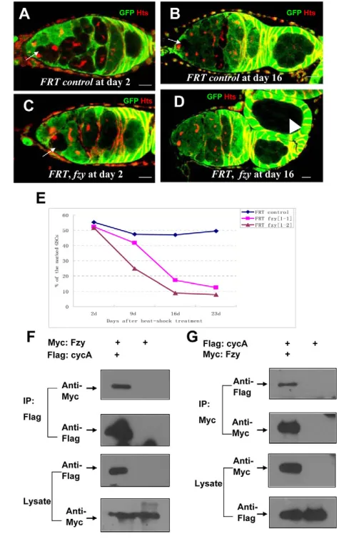
It has been shown that, in mitosis, mitotic cyclins are recognized by APC/C E3 ligase, which is activated by CDC20 protein (Zachariae and Nasmyth, 1999). In Drosophila, mutations in thefizzygene, which
encodes theDrosophilaCDC20 protein (dCDC20), lead to high-level expression of Cyclin A (Wolthuis et al., 2008). To assay whether dCDC20 forms a complex with Cyclin A, we performed co-immunoprecipitation experiments in S2 cells. As shown in Fig. 5F,G, in agreement with previous findings (Wolthuis et al., 2008), dCDC20 forms a complex with Cyclin A. However, no apparent association was observed between Cyclin A and dCDC20 proteins (data not shown), suggesting that dCDC20 associates with full-length Cyclin A through the D-box of Cyclin A. To further explore the biological function of dCDC20/fzyin GSCs, we used the FLP/FRT method to generate dCDC20/fzymutant GSC clones, and traced the rates of GFP-negative dCDC20/fzy–/–GSC clones for 3 weeks AHST. As
shown in Fig. 5A-E, for FRT controls, the rates of wild-type GSC clones declined slightly from 55.4 to 47.1% during the testing period. By contrast, the rate of dCDC20/fzy–/–GSC clones dramatically
reduced from 51.8 to 9.0% under the same conditions, suggesting that, in addition to Eff being an important regulator of the APC/C complex, dCDC20 is also essential for the maintenance of GSCs.
Eff is required for the ubiquitylation and degradation of Cyclin A
Based on the observations described above, we hypothesized that Eff acts in conjunction with the APC/C complex to regulate the proper turnover of Cyclin A in GSCs. To test this hypothesis, we first tested whether Eff is sufficient for the ubiquitylation of Cyclin A in vitro. As shown in Fig. 6A,B, a ubiquitylation reaction in which Eff functioned as E2 in concert with the APC2 and APC11 complex, which has been shown to represent the minimal E3 ligase activity of APC (Tang et al., 2001), was sufficient to catalyze the ubiquitylation of Cyclin A. In the absence of Eff, the APC2 and APC11 complex alone showed the background activity. We then investigated whether Eff is involved in the ubiquitylation of Cyclin A in vivo. As shown in Fig. 6C,D, strong ubiquitylation of Cyclin A was observed when S2 cells were transfected with epitope-tagged Cyclin A and ubiquitin. However, the conjugation of HA-ubiquitin to Cyclin A was markedly reduced when the cells were also treated with the dsRNA of either eff(Fig. 6D) or dAPC2(see Fig. S5B in the supplementary material), suggesting that, as with dAPC2, eff is also important for the ubiquitylation of Cyclin A. To test whether effis involved in the stability of Cyclin A, we compared the expression of Cyclin A in the bamsingle-mutant ovaries with that in eff, bamdouble-mutant ovaries by a western blot assay. As shown in Fig. 6E, the expression of Cyclin A in eff, bammutant ovaries was higher than that in bamsingle-mutant ovaries. We then performed immunostaining experiments to examine the expression of Cyclin A in dividing eff GSC clones (4 days after clonal induction), which carried short-bar fusomes. As shown in Fig. 6F, 64.3% (n28) of dividing effGSC clones have strong Cyclin A expression, whereas only 39.5% (n38) of dividing control GSC clones expressing Cyclin A were observed. Taken together, these results support the view that effplays a role in the regulation of Cyclin A stability.
Overexpression of full-length Cyclin A accelerates
GSC loss ineffmutant ovaries
Given that Cyclin A is a substrate of the Eff protein, and that the fate of GSCs is sensitive to expression of the stable form of Cyclin A (CycA) rather than the corresponding full-length Cyclin A, we proposed that GSCs should be sensitive to full-length Cyclin A when effactivity is compromised or reduced. To substantiate this hypothesis, we quantified the number of GSCs in eff ovaries overexpressing full-length Cyclin A. As shown in Fig. 7, overexpression of Cyclin A in wild-type ovaries resulted in no apparent defect in GSC maintenance; the average number of GSCs per germarium was 2.2 (n>100), which Fig. 5. The cdc20/fzygene is essential for GSC maintenance.
(A-D)GSC clones were induced by heat-shock treatment in adult female flies. Ovaries from FRT control flies (A,B) and FRT, fzy flies (C,D) were dissected at day 2 and day 16 following heat-shock treatment and stained with anti-GFP (green) and anti-Hts (red) antibodies; GSC clones (indicated by arrows) and cyst clones (indicated by arrowheads) were identified by the absence of GFP expression. Scale bar: 10m. (E)Percentage of negatively GFP-marked GSC clones in FRT control and FRT, fzynull alleles at days 2, 9 16 and 23 after heat-shock treatment. (F)S2 cells were transfected with a combination of Myc epitope-tagged Fzy and Flag epitope-tagged CycA, or with Myc epitope-tagged Fzy alone as indicated. Forty-eight hours after transfection, lysates from transfected S2 cells were immunoprecipitated with anti-Flag M2 affinity gel, and western blots were performed to analyze the presence of Myc epitope-tagged Fzy as indicated. The results showed that Fzy specifically associated with CycA. (G)S2 cells were transfected with a combination of Flag epitope-tagged CycA and Myc epitope-tagged Fzy, or with Flag epitope-tagged CycA alone. Forty-eight hours after transfection, lysates from transfected S2 cells were immunoprecipitated with anti-Myc antibody. Western blots were performed to analyze the presence of Flag epitope-tagged CycA as indicated. The results showed that CycA specifically associated with Fzy.
D
E
V
E
LO
P
M
E
N
[image:7.612.52.293.61.438.2]was similar to that in wild-type ovaries. However, we found that the overexpression of full-length Cyclin A strongly enhanced the eff phenotype in GSCs. For example, in 6-day-old effsingle-mutant ovaries, about 27.9 and 31.4% of germaria contained 1 GSC or no GSCs, respectively, and the average number of GSCs per germarium was 1.2 (n204). However, for age-matched eff mutant overexpressing full-length Cyclin A, about 93.3% of germaria did not possess GSCs, and the average number of GSCs was only 0.08 (n104). These data suggest that GSCs are much more sensitive to overexpression of full-length Cyclin A in the effbackground than in the wild-type background, indicating that eff interacts with cyclin Ain vivo. These findings are consistent with our hypothesis that Eff functions in concert with the APC/CDC20 complexes to regulate GSC fate, probably through controlling the proper degradation of Cyclin A.
DISCUSSION
In this study, by using a combination of genetic and biochemical strategies, we demonstrate that the Eff/APC-mediated ubiquitylation pathway plays an important role in the regulation of GSCs in Drosophilaovary, probably through controlling the degradation of mitotic Cyclin A. Our results reveal a new role for the tight regulation of mitotic cyclins in the fate determination of stem cells, implicating that a similar mechanism may be conserved in mammals.
The Eff-mediated ubiquitylation pathway represses GSC differentiation in a Bam-independent manner
[image:8.612.52.333.58.470.2]The ubiquitin-mediated proteolysis mechanism, which is evolutionarily conserved for the regulation of protein turnover, has been shown to play important roles in numerous biological processes, such as the cell cycle, pattern formation and tissue homeostasis (Harper et al., 2002; Tang et al., 1997; Zachariae and Nasmyth, 1999). DrosophilaEff, which was initially identified as a class I E2 ubiquitin-conjugating enzyme encoded by the eff gene, has been shown to be involved in several cellular and developmental processes, including chromosome segregation, chromatin remodeling and protection against cell death (Cenci et al., 1997; Ryoo et al., 2002). In this study, we found that loss of eff function results in the depletion of GSCs, revealing a new role for eff in the regulation of GSC fate. Using germline clonal analysis and rescue experiments, we further defined that the eff gene is an intrinsic, rather than extrinsic, factor for the maintenance of GSCs. Previous studies have shown that Eff is involved in the rpr-induced apoptosis pathway through a physical interaction with DIAP1 that stimulates DIAP1 auto-ubiquitylation (Ryoo et al., 2002). Therefore, it is possible that loss of GSCs in effmutants may be due to reduced viability of GSCs. Our results
Fig. 6. Eff is required for Cyclin A ubiquitylation and stability. (A,B)An in vitro ubiquitin reaction was
reconstituted with a complex that contained E1, Eff, APC11, APC2 and the putative substrate, Cyclin A, as indicated (lane 1). In lane 2 and lane 3 the ubiquitin reaction lacked Cyclin A or Eff, respectively. Western blots were performed to analyze ubiquitylation products using the antibodies indicated. (C,D)For in vivo ubiquitylation assay, S2 cells were transfected with DNA combinations including Myc epitope-tagged Cyclin A and/or HA epitope-epitope-tagged Ub (Ubiquitin) with or without effdsRNA treatment as indicated. Western blots were performed to analyze the ubiquitination product of Cyclin A. (E)Ovarian extracts were probed with anti-Cyclin A antibodies. The left lane contains an extract from bam mutant ovaries and Cyclin A protein is indicated by an arrow. The right lane contains an extract from bam and eff double-mutant ovaries and shows a significant increase in the Cyclin A protein levels. Equal loading of the extracts was confirmed by western blotting with anti-beta actin antibody as a control. (F)GSC clones were induced by heat-shock treatment, and ovaries from FRT control flies and FRT, eff flies (as indicated) were dissected at day 4 following heat-shock treatment and stained with anti-Cyclin A (pink) and anti-Hts (red) antibodies. Dividing GSC clones (indicated by arrows) were identified by the absence of GFP expression and carrying short-bar fusomes. Scale bars: 10m.
D
E
V
E
LO
P
M
E
N
clearly show that eff–/–GSCs undergo differentiation rather than
apoptosis, thus supporting the idea that the role of eff is to repress the premature differentiation of GSCs.
The biochemical role of Eff in the regulation of GSCs In the ubiquitin pathway, E2 conjugating enzymes have much lower specificity compared with E3 ligases. Certain E2s are known to function together with distinct type E3 ligases for substrate ubiquitylation and degradation. Eff is involved in protein degradation mediated by various RING finger-containing E3 ligases [e.g. Sina, Neur and DIAP (Iap2 – FlyBase)], and regulates several signaling transduction pathways (Kuo et al., 2006; Ryoo et al., 2002; Tang et al., 1997). As Eff plays a role downstream of, or parallel to, bamfunction, it is important to know what biochemical functions Eff performs in the regulation of GSCs. In this work, we provided biochemical evidence showing that Eff not only physically interacts with the dAPC2 protein, but is also crucial for the ubiquitylation and degradation of Cyclin A. Moreover, our genetic analyses revealed that eff interacts with both dAPC2 and cyclin A with respect to the
regulation of GSCs. In addition, we also show that dCDC20/Fzy, a key regulator of APC/C complexes, is involved in GSC regulation. Together, these data strongly support our model in which Eff facilitates the E3 ligase function of APC/CDC20 to ensure the self-renewal of GSCs.
Early studies in Xenopusand clam extracts demonstrated that both UBC4, a homolog of Eff, and UBCx/E2-C equally supported APC-mediated ubiquitylation reactions in vitro (Aristarkhov et al., 1996; King et al., 1995; Yu et al., 1996). However, an in vivo study showed that these two classes of E2 are not functionally equivalent but exhibit distinct functions in mitotic cyclin degradation, suggesting that different E2 family members probably execute distinct functions (Seino et al., 2003). The Drosophilaortholog of UBCx/E2-C, Vihar E2, has been reported to be involved in Cyclin B degradation during the metaphase-anaphase transition (Mathe et al., 2004). In this work, we present both genetic and biochemical evidence that Eff, the Drosophilahomolog of UBC4, is essential for Cyclin A degradation in GSCs. Because the mitosis-related ubiquitin-conjugating enzyme, Vihar E2, is involved in APC/C-mediated ubiquitylation that potentially regulates Cyclin A degradation, it would be interesting to determine whether and/or how different E2 family members (e.g. Eff and Vihar E2) coordinately support specific APC-mediated mitotic cyclin destruction with respect to GSC regulation.
The relationship between the proper cell mitosis mediated by mitotic cyclins and the maintenance of stem cells
It has been shown that APC/C activity is required for the asymmetric localization of Miranda and its cargo proteins during neuroblast division (Slack et al., 2007). In Drosophilaovary, previous studies have demonstrated that Cyclin B plays important roles in GSC division and is essential role for GSC maintenance (Hsu et al., 2008; Wang and Lin, 2005). However, it still remains unexplored whether the tight regulation of cyclins is also required for the fate determination of GSCs. The regulatory roles of mitotic cyclins at the cellular level during mitosis have been explored in detail (Parry and O’Farrell, 2001). It has been reported that the sequential degradation of Cyclin A, Cyclin B and Cyclin B3 completes mitotic exit, which is mediated by APC/CDC20 in early M phase and by APC/Cdh1 during late M phase (Zachariae and Nasmyth, 1999). Interestingly, the expression of stable forms of each cyclin leads to distinct mitosis defects, suggesting that the degradation of distinct mitotic cyclins is responsible for specific steps of mitosis. However, the biological basis for the control of the cyclin destruction remains elusive. Given that the APC-mediated pathway plays important roles in the proper cell mitosis, as loss of function of components in the pathway results in upregulation of mitotic cyclins that cause mitosis delay/or arrest (Dawson et al., 1995; Harper et al., 2002; Sigrist et al., 1995), the question becomes whether the maintenance of GSCs requires the proper cell mitosis mediated by the regulation of mitotic cyclins. In this study, we showed that the forced expression of a stable form of Cyclin A leads to defects in GSC maintenance, suggesting that blocking mitotic progression may force germline stem cells to precociously differentiate, essentially altering their fate. Although the forced expression of a stable form of Cyclin B or Cyclin B3 does not give rise to any apparent defect in GSCs, one explanation is that stabilized Cyclin A may block cell-cycle progression more severely than the other stabilized Cyclins and prolonged M phase might be unfavorable for stem cell maintenance.
[image:9.612.52.295.61.386.2]Taken together, our findings support a mechanism underlying the fate determination of stem cells that is linked to the control of the proper cell mitosis. As the control of degradation of mitotic cyclins Fig. 7. Overexpression of the full-length version of Cyclin A
accelerates GSC loss ineffmutant ovaries. (A-E)Ovaries from 6-day-old eff8/effmer1mutant (A-C), UASp-cycA; nosgvp, eff8/effmer1(D)
and UASp-cycA; nosgvp (E) female flies were stained with anti-Vasa and anti-Hts antibodies. Scale bars: 20m. (F)Quantitative analysis of the percentage of germaria types in effmutants, and overexpression of full-length Cyclin A in wild-type or the eff mutant background are
indicated. The x-axis shows the day of examination post-eclosion, and the y-axis shows the percentage of types of germaria in different genotypes.
D
E
V
E
LO
P
M
E
N
is evolutionarily conserved between flies and mammals, it would be interesting to also determine whether the control of proper cell mitosis is important for the maintenance of stem cells from other organisms, including mammals.
Acknowledgements
We thank Drs Duojia Pan, Jin Jiang, Peng Jin, Xinhua Lin and Dennis McKearin for important discussions and critical readings of the manuscript. This work was supported by grants from the National Basic Research Program of China (2007CB947502, 2006CB944000 and 2007CB507400 to D.C.) and the Chinese NSFC (30630042 and 30825026 to D.C.; and 30871441 to D.S.C.).
Supplementary material
Supplementary material for this article is available at http://dev.biologists.org/cgi/content/full/136/24/4133/DC1
References
Aristarkhov, A., Eytan, E., Moghe, A., Admon, A., Hershko, A. and Ruderman, J. V.(1996). E2-C, a cyclin-selective ubiquitin carrier protein required for the destruction of mitotic cyclins. Proc. Natl. Acad. Sci. USA93, 4294-4299.
Buszczak, M., Paterno, S., Lighthouse, D., Bachman, J., Planck, J., Owen, S., Skora, A. D., Nystul, T. G., Ohlstein, B., Allen, A. et al.(2007). The Carnegie protein trap library: a versatile tool for Drosophila developmental studies. Genetics
175, 1505-1531.
Castrillon, D. H., Gonczy, P., Alexander, S., Rawson, R., Eberhart, C. G., Viswanathan, S., DiNardo, S. and Wasserman, S. A.(1993). Toward a molecular genetic analysis of spermatogenesis in Drosophila melanogaster: characterization of male-sterile mutants generated by single P element mutagenesis. Genetics135, 489-505.
Cenci, G., Rawson, R. B., Belloni, G., Castrillon, D. H., Tudor, M., Petrucci, R., Goldberg, M. L., Wasserman, S. A. and Gatti, M.(1997). UbcD1, a Drosophila ubiquitin-conjugating enzyme required for proper telomere behavior. Genes Dev.
11, 863-875.
Chen, D. and McKearin, D.(2003a). Dpp signaling silences bam transcription directly to establish asymmetric divisions of germline stem cells. Curr. Biol. 13, 1786-1791.
Chen, D. and McKearin, D. M.(2003b). A discrete transcriptional silencer in the bam gene determines asymmetric division of the Drosophila germline stem cell. Development130, 1159-1170.
Chen, D. and McKearin, D.(2005). Gene circuitry controlling a stem cell niche. Curr. Biol. 15, 179-184.
Cox, D. N., Chao, A. and Lin, H.(2000). piwi encodes a nucleoplasmic factor whose activity modulates the number and division rate of germline stem cells. Development127, 503-514.
Dawson, I. A., Roth, S. and Artavanis-Tsakonas, S.(1995). The Drosophila cell cycle gene fizzy is required for normal degradation of cyclins A and B during mitosis and has homology to the CDC20 gene of Saccharomyces cerevisiae. J. Cell Biol. 129, 725-737.
Forbes, A. and Lehmann, R.(1998). Nanos and Pumilio have critical roles in the development and function of Drosophila germline stem cells. Development125, 679-690.
Glickman, M. H. and Ciechanover, A.(2002). The ubiquitin-proteasome proteolytic pathway: destruction for the sake of construction. Physiol. Rev. 82, 373-428.
Glotzer, M., Murray, A. W. and Kirschner, M. W.(1991). Cyclin is degraded by the ubiquitin pathway. Nature349, 132-138.
Harper, J. W., Burton, J. L. and Solomon, M. J.(2002). The anaphase-promoting complex: it’s not just for mitosis any more. Genes Dev. 16, 2179-2206.
Hsu, H. J., LaFever, L. and Drummond-Barbosa, D.(2008). Diet controls normal and tumorous germline stem cells via insulin-dependent and-independent mechanisms in DrosophilaDev. Biol. 313,700-712.
Jiang, X., Xia, L., Chen, D., Yang, Y., Huang, H., Yang, L., Zhao, Q., Shen, L., Wang, J. and Chen, D.(2008). Otefin, a nuclear membrane protein, determines the fate of germline stem cells in Drosophila via interaction with Smad complexes. Dev. Cell14, 494-506.
Jin, Z. and Xie, T.(2007). Dcr-1 maintains Drosophila ovarian stem cells. Curr. Biol.
17, 539-544.
Kashevsky, H., Wallace, J. A., Reed, B. H., Lai, C., Hayashi-Hagihara, A. and Orr-Weaver, T. L.(2002). The anaphase promoting complex/cyclosome is required during development for modified cell cycles. Proc. Natl. Acad. Sci. USA99, 11217-11222.
King, R. W., Peters, J. M., Tugendreich, S., Rolfe, M., Hieter, P. and Kirschner, M. W.(1995). A 20S complex containing CDC27 and CDC16 catalyzes the mitosis-specific conjugation of ubiquitin to cyclin B. Cell81, 279-288.
Kuo, C. T., Zhu, S., Younger, S., Jan, L. Y. and Jan, Y. N.(2006). Identification of E2/E3 ubiquitinating enzymes and caspase activity regulating Drosophila sensory neuron dendrite pruning. Neuron51, 283-290.
Lasko, P. F. and Ashburner, M.(1990). Posterior localization of vasa protein correlates with, but is not sufficient for, pole cell development. Genes Dev. 4, 905-921.
Lilly, M. A., de Cuevas, M. and Spradling, A. C.(2000). Cyclin A associates with the fusome during germline cyst formation in the Drosophila ovary. Dev. Biol. 218, 53-63.
Lin, H. and Spradling, A. C.(1997). A novel group of pumilio mutations affects the asymmetric division of germline stem cells in the Drosophila ovary. Development
124, 2463-2476.
Maines, J. Z., Park, J. K., Williams, M. and McKearin, D. M.(2007). Stonewalling Drosophila stem cell differentiation by epigenetic controls. Development134, 1471-1479.
Mathe, E., Kraft, C., Giet, R., Deak, P., Peters, J. M. and Glover, D. M.(2004). The E2-C vihar is required for the correct spatiotemporal proteolysis of cyclin B and itself undergoes cyclical degradation. Curr. Biol. 14, 1723-1733.
Murray, A. W.(2004). Recycling the cell cycle: cyclins revisited. Cell116, 221-234.
Park, J. K., Liu, X., Strauss, T. J., McKearin, D. M. and Liu, Q.(2007). The miRNA pathway intrinsically controls self-renewal of drosophila germline stem cells. Curr. Biol. 17, 533-538.
Parry, D. H. and O’Farrell, P. H.(2001). The schedule of destruction of three mitotic cyclins can dictate the timing of events during exit from mitosis. Curr. Biol. 11, 671-683.
Peters, J. M.(2002). The anaphase-promoting complex: proteolysis in mitosis and beyond. Mol. Cell9, 931-943.
Rorth, P.(1998). Gal4 in the Drosophila female germline. Mech. Dev. 78, 113-118.
Ryoo, H. D., Bergmann, A., Gonen, H., Ciechanover, A. and Steller, H.(2002). Regulation of Drosophila IAP1 degradation and apoptosis by reaper and ubcD1. Nat. Cell Biol. 4, 432-438.
Seino, H., Kishi, T., Nishitani, H. and Yamao, F.(2003). Two ubiquitin-conjugating enzymes, UbcP1/Ubc4 and UbcP4/Ubc11, have distinct functions for ubiquitination of mitotic cyclin. Mol. Cell. Biol. 23, 3497-3505.
Sigrist, S., Jacobs, H., Stratmann, R. and Lehner, C. F.(1995). Exit from mitosis is regulated by Drosophila fizzy and the sequential destruction of cyclins A, B and B3. EMBO J. 14, 4827-4838.
Slack, C., Overton, P., Tuxworth, R. and Chia, W.(2007). Asymmetric localisation of Miranda and its cargo proteins during neuroblast division requires the anaphase-promoting complex/cyclosome. Development 134, 3781-3787.
Song, X., Wong, M. D., Kawase, E., Xi, R., Ding, B. C., McCarthy, J. J. and Xie, T.(2004). Bmp signals from niche cells directly repress transcription of a differentiation-promoting gene, bag of marbles, in germline stem cells in the Drosophila ovary. Development131, 1353-1364.
Spradling, A., Drummond-Barbosa, D. and Kai, T.(2001). Stem cells find their niche. Nature414, 98-104.
Szakmary, A., Cox, D. N., Wang, Z. and Lin, H.(2005). Regulatory relationship among piwi, pumilio, and bag-of-marbles in Drosophila germline stem cell self-renewal and differentiation. Curr. Biol. 15, 171-178.
Tang, A. H., Neufeld, T. P., Kwan, E. and Rubin, G. M.(1997). PHYL acts to down-regulate TTK88, a transcriptional repressor of neuronal cell fates, by a SINA-dependent mechanism. Cell90, 459-467.
Tang, Z., Li, B., Bharadwaj, R., Zhu, H., Ozkan, E., Hakala, K., Deisenhofer, J. and Yu, H.(2001). APC2 Cullin protein and APC11 RING protein comprise the minimal ubiquitin ligase module of the anaphase-promoting complex. Mol. Biol. Cell12, 3839-3851.
Van Doren, M., Williamson, A. L. and Lehmann, R.(1998). Regulation of zygotic gene expression in Drosophila primordial germ cells. Curr. Biol. 8, 243-246.
Wang, Z. and Lin, H.(2005). The division of Drosophila germline stem cells and their precursors requires a specific cyclin. Curr. Biol. 15, 328-333.
Wolthuis, R., Clay-Farrace, L., van Zon, W., Yekezare, M., Koop, L., Ogink, J., Medema, R. and Pines, J.(2008). Cdc20 and Cks direct the spindle checkpoint-independent destruction of cyclin A. Mol. Cell30, 290-302.
Xie, T. and Spradling, A. C.(1998). decapentaplegic is essential for the maintenance and division of germline stem cells in the Drosophila ovary. Cell94, 251-260.
Xu, T. and Rubin, G. M.(1993). Analysis of genetic mosaics in developing and adult Drosophila tissues. Development117, 1223-1237.
Yang, L., Chen, D., Duan, R., Xia, L., Wang, J., Qurashi, A., Jin, P. and Chen, D.
(2007). Argonaute 1 regulates the fate of germline stem cells in Drosophila. Development134, 4265-4272.
Yu, H., King, R. W., Peters, J. M. and Kirschner, M. W.(1996). Identification of a novel ubiquitin-conjugating enzyme involved in mitotic cyclin degradation. Curr. Biol. 6, 455-466.
Zaccai, M. and Lipshitz, H. D.(1996). Differential distributions of two adducin-like protein isoforms in the Drosophila ovary and early embryo. Zygote4, 159-166.
Zachariae, W. and Nasmyth, K.(1999). Whose end is destruction: cell division and the anaphase-promoting complex. Genes Dev. 13, 2039-2058.
Zhang, Q., Zhang, L., Wang, B., Ou, C. Y., Chien, C. T. and Jiang, J.(2006). A hedgehog-induced BTB protein modulates hedgehog signaling by degrading Ci/Gli transcription factor. Dev. Cell10, 719-729.