4287
Introduction
Recent reports demonstrating that stem cells of the central nervous system retain the potential to differentiate into non-neural progeny in vitro (Galli et al., 2000; Rietze et al., 2001) have drawn the attention of both the stem cell and neuroscience communities (Greco and Recht, 2003; Vescovi et al., 2002; Wagers et al., 2002; Weissman et al., 2001). Alternative explanations to enhanced neural stem cell (NSC) potentiality have been put forward (Alvarez-Dolado et al., 2003; Liu and Rao, 2003), but it is important to elucidate the broadest potential of tissue-derived stem cell populations because it challenges and deepens our basic concepts of stem cell potential and the mechanisms that govern lineage restriction, cellular differentiation and stem cell maturation. Moreover, adult tissue-derived stem cells provide an attractive source of stem cells for transplantation and tissue engineering efforts, especially if phenotypic potential is broader than believed.
NSCs in the central nervous system, and their in vitro model, neurospheres are restricted to a differentiation profile of neurons, astrocytes and oligodendrocytes (Davis and Temple, 1994; Reynolds et al., 1992; Reynolds and Weiss, 1992; Reynolds and Weiss, 1996; Tsai and McKay, 2000). Whether NSCs are tripotent in vivo is still unclear (Gabay et al., 2003; Pevny and Rao, 2003). However, NSCs expanded from either
cell-adherent or aggregate cultures differentiate into immunoreactive myocytes when co-cultured with either primary myoblasts, ES cells or when introduced into pre-implantation embryos (Clarke et al., 2000; Galli et al., 2000; Rietze et al., 2001; Tsai and McKay, 2000). The differentiation of NSC into myogenic derivatives has been reported to be density dependent in some microenvironments (Tsai and McKay, 2000) and dependent upon pre-existing myocytes in other environments (Galli et al., 2000; Rietze et al., 2001). It has also been reported that bone morphogenetic protein (BMP) induces smooth muscle differentiation of NSCs, and it has been suggested that BMP induces a transformation of central nervous system NSC into peripheral nervous system neural crest stem cells (Tsai and McKay, 2000).
Although studies using the neurosphere assay have begun to discern the molecular mechanism of specification of NSCs to specific neural sublineages (Hitoshi et al., 2002a; Hitoshi et al., 2002b; Tropepe et al., 2001), the molecular mechanisms of non-neural specification and differentiation of NSCs remain unknown. Thus far, studies demonstrating the myogenic differentiation potential of NSCs leave unanswered the question of whether myogenic and neurogenic differentiated progeny arise from a single multilineage neural stem cell (MLNSC), as well as the issue of which factors propagate these MLNSC populations and instruct myogenic differentiation. It Reports of non-neural differentiation of neural stem cells
(NSCs) have been challenged by alternative explanations for expanded differentiation potentials. In an attempt to demonstrate the plasticity of NSC, neurospheres were generated from single retrovirally labeled embryonic cortical precursors. In a defined serum-free insulin-containing media, 40% of the neurospheres contained both myogenic and neurogenic differentiated progeny. The number of NSCs displaying multilineage differentiation potential declines through gestation but does exist in the adult animal. In this system, insulin appears to function as a survival and dose-dependent myogenic differentiation signal for multilineage NSCs (MLNSC). MLNSC-derived
cardiomyocytes contract synchronously, respond to sympathetic and parasympathetic stimulation, and regenerate injured heart tissues. These studies provide support for the hypothesis that MLNSCs exist throughout the lifetime of the animal, and potentially provide a population of stem cells for cell-based regenerative medicine strategies inside and outside of the nervous system.
Movies available online
Key words: Plasticity, Neural stem cells, Myogenesis
Summary
Insulin acts as a myogenic differentiation signal for neural stem
cells with multilineage differentiation potential
Mahmud Bani-Yaghoub1,*, Stephen E. Kendall2, Daniel P. Moore2, Stephen Bellum2, Rebecca A. Cowling2, George N. Nikopoulos2, Chris J. Kubu1,†, Calvin Vary3 and Joseph M. Verdi1,2,‡
1The John P. Roberts Research Institute, 100 Perth Drive, London, ON, N6A 5K8, Canada
2Center for Regenerative Medicine, Maine Medical Center Research Institute, 81 Research Drive, Scarborough, ME 04074, USA 3Center for Molecular Medicine, Maine Medical Center Research Institute, 81 Research Drive, Scarborough, ME 04074, USA *Present address: Neurogenesis and Brain Repair, Institute for Biological Sciences, National Research Council of Canada, 1500 Montreal Road, Building M-54, Ottawa, ON, K1A 0R6, Canada
†Present address: USB Corporation, 2611 Miles Road, Cleveland, OH 44128, USA ‡Author for correspondence (e-mail: verdij@mmc.org)
Accepted 8 June 2004
Development 131, 4287-4298
Published by The Company of Biologists 2004 doi:10.1242/dev.01295
is also not known whether NSCs can generate biologically functional myogenic derivatives.
We have attempted to address these questions by demonstrating that differentiated neural and myogenic progeny can originate from a single isolated cell and can be propagated in serum-free insulin-containing media. MLNSCs arise as a consequence of insulin-mediated survival and not as a result of cell fusion events. Moreover, myogenic differentiation is insulin dose sensitive; low concentrations of insulin promote cardiomyocyte differentiation, whereas elevated concentrations induce skeletal muscle formation. At any dose, myogenic differentiation occurred at the expense of neural differentiation. MLNSC-derived cardiomyocytes were metabolically coupled, contracted spontaneously, responded to sympathetic and parasympathetic stimulation, and engrafted and differentiated appropriately when introduced into damaged cardiac muscle. These studies provide support for the hypothesis that NSCs have a broad differentiation potential, including phenotypes outside the neuroectodermal lineage. As such, MLNSCs may provide a population of cells suitable for regenerative medicine strategies inside and outside of the nervous system.
Materials and methods
Isolation of neural stem cells
CD1 mice were collected at E14 (observation of vaginal plug was E0.5) through adulthood (6-8 weeks old). Cerebral cortices were dissected free of meninges, dispersed (0.05% trypsin-EDTA) and passed through cell strainers to enrich for single cells. Cortical cells were grown for 6 days in suspension in a medium containing 65% neurobasal medium and 35% Dulbecco’s modified Eagle medium supplemented with either 20 µg/ml insulin (MB-media) or 10% fetal calf serum. In initial experiments, MB-medium was supplemented with 20 ng/ml bFGF (R&D Systems) during the first 48 hours and 2
µM 5-azacytidine (Sigma) during the second 48 hours and without supplementation the final 48 hours. After 6 days in culture, spheres were dissociated and grown on poly-D-Lysine-coated 24-well plates (one sphere per well) in MB-media without supplementation.
Flow cytometry
Embryonic cortical precursors were immunolabeled using monoclonal insulin receptor (IR) (Pharmingen) and FITC-GAM antibodies. In addition, cells were labeled with CD35 and anti-CD31 conjugated to rhodamine and PE, respectively. anti-CD31– CD35–
cells were sorted into IR high, low and non-expressing subpopulations using a BD FACS Vantage SE cell sorter.
Transfection and retroviral transduction
293GPG packaging cells were transfected with the AP2-IRES-EGFP retroviral construct (Galipeau et al., 1999). Enhanced green fluorescent protein (EGFP) retroviral particles were collected every 24 hours. Flow cytometric analysis of EGFP fluorescence was used to determine the viral titer (Galipeau et al., 1999). NSCs were infected with 1.231010cfu/ml EGFP-retroviral particles.
Southern blotting analysis
Clones were digested overnight in 400 µg/ml proteinase K at 50°C. The resulting DNA (25 µg) was digested with BglII and run on a 0.7% agarose TAE gel and transferred to a Nytran Plus membrane. Prehybridization, hybridization and washing of the resulting membrane were performed using standard methods.
RT-PCR analysis
RT-PCR analysis was performed according to published methods
(Verdi and Anderson, 1994) with the following parameters: 35 cycles of denaturing at 94°C (30 seconds), annealing at 53-58°C for 1 minute and elongation at 72°C for 1 minute. Primer sequences are available upon request.
Antibodies
The antibodies used included monoclonal antibodies to βIII-tubulin (Chemicon), microtubule associated protein 2a+2b, β-actin (Sigma), 47A skeletal myosin heavy chain (MHC), BA-G5 cardiac MHC (New England Biolabs); CD31, CD35, tropomyosin and troponin1 (Chemicon); and polyclonal antibodies to caspase 3 (Pharmingen), phospho-Akt (Ser473 and Thr308; New England Biolabs), glial fibrillary acidic protein, (Sigma), nestin. Secondary antibodies used were purchased from Jackson ImmunoResearch Laboratories.
Immunocytochemistry
Cells were fixed with 70% ethanol and 0.15 M NaCl. Non-specific binding sites were blocked with 10% goat serum and 0.1% Nonidet P-40 for 30 minutes. Cells were incubated 1 hour at room temperature with primary antibody prior to washing and application of the secondary antibody for 1 hour at room temperature. The immunoreactivity was examined with an Olympus IX70 fluorescent microscope.
For immunohistochemical analysis of tissues, specimens were fixed with 4% paraformaldehyde and embedded in paraffin wax. Serial 15
µm sections were dewaxed in xylene, hydrated in a graded ethanol series (100%, 90%, 70%, 50%), for 5 minutes per hydration and washed with PBS.
Western blot analysis
Cells were lysed in ice-cold buffer composed of 10 mM NaCl, 20 mM Tris (pH 8.0), 0.5 mM EDTA, 10% glycerol, 1% Nonidet P-40, 1 mM phenyl-methylsulfonyl fluoride, 1 mM sodium orthovanadate, 2
µg/ml leupeptin and 10 µg/ml aprotinin. Clarified protein lysate (30
µg) was separated on a reducing 15% SDS-PAGE and blotted onto Immobilon-P (Millipore). Membranes were blocked with 5% blotto/10 mM tris-saline pH 7.4 overnight. Membranes were incubated with either anti-phospho-Akt (1:4000), anti-caspase 3 (1:1000) or anti-β-actin (1:5000) antibody prior to incubation with horseradish peroxidase-conjugated goat anti-rabbit or anti-mouse antibodies. Immunoreactivity was detected using an enhanced chemiluminescent methodology (Amersham Biosciences).
DNA synthesis analysis
Cells were incubated with 10 µM BrdU in triplicates, at 37°C, 5% CO2for 6 or 12 hours after 24, 72 and 120 hours of expansion in MB
media. Cells were fixed in 4% paraformaldehyde (RT-20 minutes) and labeled with a monoclonal anti-BrdU, according to manufacturer’s specifications (Becton-Dickinson Biosciences). BrdU+ cells were
counted using an epifluorescent microscope.
Dye preloading
NSC-derived cardiomyocytes were incubated for 20 minutes in a solution containing: isotonic glucose, 0.1% calcein AM, 0.1% 1,1′ -dioctadecyl-3,3,3′,3′-tetramethylindocarbocyaine perchlorate (DiI) (Molecular Probes) at 37°C (Goldberg et al., 1995). Single preloaded (donor) cells were seeded over contracting NSC-derived cardiomyocyte clusters (one donor cell per cluster) and incubated for 2 hours at 37°C.
Dye microinjection
NSC-derived cardiomyocytes were incubated in cold medium 5 minutes before microinjection to slow contraction. A single cell within each cardiomyocyte cluster was pressure-microinjected with 10 mM 5(6)-carboxyfluorescein (Mr=376, Molecular Probes) using a
Live imaging of the NSC-derived cardiomyocytes
Time-lapse images of individual clusters were used to examine the contraction rate of cardiomyocyte clusters. Fluorescent images were taken using a 488 nm argon/krypton laser line on a Zeiss LSM 410 inverted confocal microscope. Optical sections were scanned at a speed of 32 seconds/image and collected continuously for up to 10 minutes. Focus, contrast and brightness settings remained constant during acquisition.
Organotypic cultures
Adult CD1 hearts were isolated and placed in Hanks balanced salt solution, freed of blood and placed in trans-well inserts for six-well Nunc plates. Organs were covered with MB-media such that a meniscus formed over the top of the explant. To induce injury, one ventricle was injured with an etched tungsten micro-needle. Immediately after injury, medium was refreshed and EGFP-tagged NSC (5000 cells grown in MB-media) were injected into the injured area. Organs were maintained in the culture for 10 days and then fixed for analysis. Manual counting of z-sections taken through the entire heart (n=5) was used to calculate the percentage of cardiac MHC+/EGFP+ cells
engrafted in the host.
Results
Clonal neurospheres differentiate into multiple germ line derivatives
In order to demonstrate that a single NSC is capable of generating progeny within both the myogenic and neurogenic lineages, embryonic cortical precursors were isolated and infected with an EGFP-retrovirus at a multiplicity of infection to facilitate single unique integrations. To ensure the founder cells were neural in origin and not mesodermal or hematopoetic derived, individual EGFP+ CD31–CD35–cells were sorted into 96-well suspension dishes and allowed to expand in a serum-free insulin-containing medium (‘MB-media’, see Materials and methods, Fig. 1A). Wells were examined under epifluorescence and phase contrast microscopy (to visualize EGFP- cells) 2 hours after plating, and any wells that contained two or more cells were discarded from analysis. After six days of expansion, during which clones grew from single founder cells to spheres often in excess of 150 cells (Fig. 1A-C), the neurospheres were dissociated and grown as adherent
cultures to complete the differentiation process. As demonstrated in Fig. 1D, a single EGFP-labeled cell was able to give rise to a differentiated colony that contained both muscle and neural progeny [see arrows demarking EGFP-expressing neurons in a cluster of EGFP-EGFP-expressing skeletal myosin heavy chain (MHC) immunoreactive cells]. Other combinations of muscle and neural progeny were also observed (Fig. 1E-H). Of the single EGFP+CD31–CD35–cells at E14
expanded in MB-media, 21 of 48 (44%) were composed of mixed lineage differentiated progeny (Table 1). By contrast, the differentiation outcomes of single EGFP+ cells expanded in MB-media containing serum were restricted to neuronal and glial differentiation events.
[image:3.612.250.567.73.453.2]The differentiation outcomes of the resulting myogenic clones can be further divided: all 21 contained neurons, 11 (54%) contained neurons and astrocytes, and 5 (24%)
Fig. 1. Single neural stem cells can differentiate into neurons, glia and myocytes. A
single retrovirally labeled E14 CD31–CD35–NSC was expanded in suspension in
MB-media for 6 days then transferred to an adherent surface and allowed to differentiate. Differentiated progeny were identified by immunostaining 14 days post-plating. (A-C) Epifluorescent images of an expanding EGFP+NSC. The resulting sphere was
dissociated and grown as an adherent culture for 14 days to complete the differentiation process. (D) Presented are epifluorescent images of EGFP+cells (green) and sk-MHC
immunoreactive cells (red). Arrowheads indicate a phenotypic EGFP+neuron in a
cluster of EGFP+sk-MHC+myocytes. Scale bar: 80 µm. (E-H) The observed
multilineage differentiation covered many combinations of differentiation possibilities. (E,F) Confocal z-sections of differentiated clones containing BAG5+cardiac myocytes
(red) and MAP2 immunoreactive neurons (green); (G,H) BAG5 cardiac myocytes (red) and GFAP+glial cells (green). To further verify that differentiated clones arose from a
single EGFP+cell, Southern analysis was performed on random differentiated progeny;
contained neurons, astrocytes and oligodendrocytes. This was surprising because a basic tenant of the neurosphere assay is that neurons, astrocytes and oligodendrocytes originate from a tripotent NSC. In these conditions, the transient exposure to basic fibroblast growth factor (bFGF) was insufficient to induce robust oligodendrocyte differentiation as previously reported (Gabay et al., 2003). The distribution of specific
myogenic progeny was also determined for these 21 myogenic clones grown in MB-media: 19 (90%) contained both skeletal and cardiac myocyte derivatives, 1 (5%) contained only cardiac MHC and tropomyosin immunoreactivity co-expressing cells and 1 (5%) contained only skeletal MHC immunoreactive cells. Smooth muscle immunoreactive cells were not observed in these studies. Our results demonstrate that a single neurosphere-forming cell harbors both myogenic and neurogenic differentiation potential.
To confirm that the sphere was generated from a single retrovirally infected cell, aliquots from five random wells prior to adherent culturing were subjected to Southern analysis. Every clone had a single unique integration event (Fig. 1I). Moreover, as shown in Fig. 1D, both neurogenic and myogenic EGFP expressing differentiated progeny were observed within a single clone, strongly suggesting that both lineages arose from one founding cell and not as an aggregate of EGFP+and EGFP–founders.
MLNSC progeny express markers of mature skeletal and cardiac muscle
[image:4.612.42.564.86.149.2]We further characterized the course of maturation of the myogenic progeny produced from the differentiation of MLNSCs. CD31– CD35– NSC did not express skeletal Table 1. Clonal analysis of NSC myogenic differentiation potential
Single cells plated Plating efficiency Clone survival day 6 Myogenic clones
Serum-containing medium 480 234±26 (53%) 185±35 (79%) 0.7±0.3 (0%)
Complete MB-media 576 188±29 (33%) 48±6 (26%)* 21±5 (44%)*
MB-media minus bFGF 554 170±10 (31%) 36±2 (29%)* 14±2 (39%)*
MB-media minus 5-aza 526 126±6 (24%) 45±6 (25%)* 12±5 (26%)*,‡
MB-media minus insulin 560 172±4 (31%) 9±4 (5%)*,† 1±0.6 (6%)†
Differentiation profile of NSCs originating from single EGFP+ CD31– CD34–NSCs. Surviving clones were counted on day 2 (plating efficiency) and day 6.
Fourteen days post-plating, clones were fixed and the resulting progeny were immunohistochemically identified and the number of percentage of clones containing myogenic derivatives determined (myogenic clones). Shown are the mean±s.d. of three experiments originating from distinct cell isolations.
*P<0.01 for comparisons to serum-containing media.
†P<0.01 for comparisons among different MB-media. ‡P<0.05 for comparisons among different MB-media.
Fig. 2. Neural stem cells can generate skeletal and cardiac myocytes.
In order to demonstrate the potential of NSCs, CD31– CD35–EGFP+
cortical progenitor cells were expanded for 6 days in MB-media prior to plating on an adherent surface to complete differentiation. By both reverse transcription polymerase chain reaction (RT-PCR) and
immunohistochemical analyses, differentiated progeny expressed markers of myogenic
[image:4.612.49.349.329.737.2](sk)-MHC or cardiac-MHC immunoreactivity prior to differentiation in MB-media (data not shown). However, during differentiation, neurospheres grown in MB media began to express markers identifying some progeny as potential skeletal (Fig. 2A-D) or cardiac myocytes (Fig. 2A,E). In a limited time course, it was demonstrated that MyoD expression preceded that of myogenin (data not shown) and subsequent tissue type-specific expression (Fig. 2A). Differentiating clones expressed sk-MHC immunoreactivity as early as day 9 and formed clusters of skeletal myoblasts and slender myofibers that joined those of other clusters (Fig. 2B-D). In addition to formation of skeletal muscle, NSC cultures also differentiated into cardiac myocytes with the appearance of terminal differentiation markers beginning (~day 13) after the onset of skeletal muscle terminal differentiation (~day 9). NSC-derived cardiac myocytes grew clusters of cells expressing cardiac α-actin (data not shown), cardiac-MHC and connexin43 (Cx43) (Fig. 2E), and these clusters over time (~3 weeks) began to contract synchronously (see Movies 1-3 at http://dev.biologists.org/supplemental). This contraction was used as an additional phenotypic marker to distinguish skeletal and cardiac myocytes, as well as Cx43, a standard marker to distinguish between the two cell types.
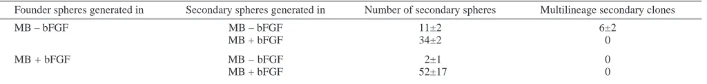
Multilineage differentiation is maintained in secondary stem cell colonies
Serial subcloning experiments of MLNSC spheres were conducted in order to assess the self-renewal potential of MLNSCs. In these experiments (and all experiments that follow, unless stated otherwise), bFGF and 5-azacytidine treatments were deleted from the expansion protocol. Expanding EGFP+ CD31– CD35– founder spheres were dissociated on day 6 and a some were grown as adherent cultures to complete the differentiation process. The remainder of each clone was seeded at 4 cells per well of a 96-well dish to generate secondary spheres. After 10 days of expansion, the process was repeated to generate tertiary spheres. Secondary spheres (21 of 54, 38%) originating from MLNSCs retained multilineage differentiation potential, as did tertiary clones (16 of 51, 31%) resulting from multilineage secondary clones. By contrast, NSCs grown in serum (n=43) or originating from neural restricted founders in MB-media (n=26) failed to produce secondary clones with myogenic potential. This suggests that the founder cell was capable of self-renewal in culture and that the differentiation potential of the founder sphere-forming cell is maintained in the stem cell population. Southern analysis of the resulting primary, secondary and tertiary clones confirmed that the subclones contained the same unique integration event (Fig. 1I). In one case, the integration site of the founder, and secondary differentiated progeny was
cloned and sequenced (data not shown) to further validate the Southern analysis.
A criterion that defines stem cell populations is the existence of the population throughout the lifetime of the animal (Weissman et al., 2001). We examined the prevalence of MLNSCs within cortical precursors during gestation and within the adult counterpart, the frontal ventricular subependymal layer (6 weeks post partum). The absolute number and percentage of MLNSCs declined through development (Table 2). However, MLNSCs could be isolated from the adult animal, suggesting that the cell population exists for the lifetime of the animal and thus meets this criterion defining stem cells.
MLNSCs become primitive neural-restricted stem cells
[image:5.612.316.567.84.184.2]Although multilineage secondary clones are generated from primary multilineage spheres, the percentage of secondary and tertiary spheres with myogenic differentiation capacity declines through successive subclonings, suggesting that MLNSCs are depleted during expansion. We inquired whether MLNSCs gave rise to lineage-restricted neurospheres. As bFGF supports the survival, expansion and differentiation of NSC population (Bartlett et al., 1998; Kilpatrick and Bartlett, 1993; Kilpatrick and Bartlett, 1995; Reynolds and Weiss, 1992), MB-medium was supplemented with 40 ng/ml bFGF to support the survival and expansion of NSC populations (Table 2). Although the number of primary spheres and the numbers of cells per sphere were significantly higher in the presence of bFGF, the absolute number of multilineage spheres remained constant. This suggests that MLNSCs are a subset of the bFGF-responsive NSC population. It is also possible that MLNSCs give rise to classical bFGF-dependent neurospheres during expansion. Indeed, serial subcloning of primary MLNSCs
Table 2. MLNSCs exist through adulthood
MB-media MB-media
minus bFGF MLNSC plus bFGF MLNSC
E14 128 49 (38%) 551 41 (7%)
E15 133 41 (31%) 453 53 (12%)
E16 131 54 (41%) 503 39 (8%)
E17 107 56 (52%) 381 51 (13%)
E18 39 8 (21%) 492 11 (2%)
P1 51 9 (17%) 313 10 (3%)
Adult 47 5 (11%) 278 6 (2%)
(6-8 weeks old)
The number of clonally derived MLNSC spheres declines through development. Clonal spheres were generated in MB-media in the presence and absence of 40 ng/ml bFGF. Presented are composites of three experiments originating from distinct cell isolations.
Table 3. MLNSC give rise to bFGF-responsive neural-restrictive stem cells
Founder spheres generated in Secondary spheres generated in Number of secondary spheres Multilineage secondary clones
MB – bFGF MB – bFGF 11±2 6±2
MB + bFGF 34±2 0
MB + bFGF MB – bFGF 2±1 0
MB + bFGF 52±17 0
[image:5.612.59.571.655.716.2]demonstrated that MLNSCs gave rise to both secondary MLNSC and bFGF-dependent spheres whose differentiation potential was restricted to neural differentiation outcomes (Table 3).
Insulin enhances the cell survival of MLNSC
We explored the potential mechanism by which insulin enriched MLNSCs in vitro. Insulin could be acting as an agent that selectively promotes the proliferation of MLNSCs or as a requirement for the survival of MLNSCs, or both. To begin to address this question, NSCs were cultured in MB-media with insulin, without insulin or with insulin plus an insulin receptor antagonist, quercetin. Abrogation of insulin signaling either by the removal of insulin or by the addition of quercetin resulted in the loss of neurospheres, relative to spheres in insulin-containing MB-media with fewer than 40% surviving beyond 120 hours (Fig. 3A). The percentage of cells within neurospheres incorporating BrdU under the same conditions and time course did not decrease as dramatically as the change in sphere number (Fig. 3B,C) suggesting that insulin was supporting the survival and not the enhanced
expansion of MLNSCs. Only with 8 hours of BrdU exposure at 120 hours of growth, a time point when less than one-half of the original neurospheres survived, was a larger difference in BrdU incorporation observed relative to insulin-containing media.
The role of insulin as a putative survival factor for MLNSC is further supported by evidence of the activation of Akt, which is necessary and sufficient to mediate insulin-dependent survival (Datta et al., 1996; Datta et al., 1999). Activated Akt [phospho-Akt (pS473)] was prevalent in NSC cultures grown in media but not in cultures containing serum or MB-media plus quercetin (Fig. 3D). In addition, NSCs grown in MB-media but not in MB-media containing quercetin, maintained caspase 3 in its inactivated form (Fig. 3E), a result that suggests NSCs undergo apoptosis in the absence of insulin signaling. Clearly, our data support the notion that insulin acts as a survival factor for MLNSCs.
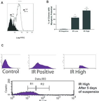
As insulin signaling appeared to be crucial for the formation of MLNSC neurospheres, we used insulin receptor (IR) to further demarcate the MLNSC population. We prospectively isolated CD31– CD35– IR+ embryonic cortical precursors at embryonic day 14 and found that 65% of the population was IR expressive (Fig. 4A). Neurospheres generated from the IRhigh (the brightest 20% of the IR+cells) population generated more multilineage differentiated progeny in comparison to neurospheres generated from the IRneg and IRlow (the lowest 20% of IR+cells) populations (Fig. 4B). Although a minimum number of insulin receptors were required for cells to differentiate into myocytes, cells with more receptors were more competent to generate myocytes, suggesting a role for insulin beyond that of a survival factor.
[image:6.612.50.293.317.742.2]It was curious that among the IRhigh population, only 40% of the clones went on to initiate myogenic differentiation (Fig. 4B). One explanation is that we were still assaying a non-homogenous population. However, the levels of IR receptor expression may also change as a consequence of expansion prior to the onset of differentiation. bFGF induces olig2 expression during neurosphere expansion (Gabay et al., 2003), and thus it is plausible that either transient bFGF exposure or cell-cell signaling within the clone may also alter the
Fig. 3. Survival of MLNSC in insulin correlates with insulin-induced
Akt activation. Cell recovery and BrdU assays were performed on cultures of CD31– CD35– EGFP+ neurospheres grown in MB-media,
MB-media minus insulin or MB-media plus 100 µM quercetin. (A) Sphere recovery was determined by counting the number of spheres daily and normalizing to the number of spheres grown in complete MB-media. Presented is the mean±s.d. for three experiments from distinct cellular isolations. (B) Proliferation is unaltered in BM-media. BrdU incorporation assays were conducted on neurospheres grown in MB-media, MB-media minus insulin and MB-media plus 100 µm quercetin. Little change in BrdU
expression of IR during expansion and, consequently, the differentiation outcomes. Indeed, a significant change in the IR expression profile was observed (Fig. 4C) when spheres originating from the IRhigh subpopulation were resorted after 6 days of expansion. The distribution of the population was Gaussian with cells expressing all levels of IR immunoreactivity, 70% of the cells expressing IR levels outside of the IRhigh founder population. Moreover, ~47% of the resorted population expressed undetectable levels of IR expression.
We examined the differentiation profiles of NSC subsets after 6 days of expansion based on IR immunoreactivity. IRneg cells gave rise exclusively to clones with neural differentiated progeny (61±8% neuron + astrocytes; 22±11% neurons only; astrocytes only 16±4%). IRlow cells gave rise to multilineage clones. Of the 227 clones examined, 83 contained myogenic progeny, of which 36 (47±14%) contained neurons, astrocytes, and both skeletal and cardiomyocytes. The IRhigh cells gave rise to the broadest differentiation profile, composed of all combinations of differentiation outcomes including oligodendrocytes that were not present in the IRneg or IRlow differentiated progeny. Eighty-one out of 102 multilineage clones (80±15%) were composed from neuronal, astrocytes and myogenic progeny; of the 81 clones, 17 also contained oligodendrocytes (21±7%). Importantly, all three IR subpopulations contain multipotential cells, but myogenic potential was contained only within IR expressive cells, and
higher percentage of myogenic differentiation correlated with higher levels of IR expression.
Insulin is a dose-dependent instructive myogenic differentiation signal for NSC
Because sorting NSCs by IR levels suggested the possibility that insulin was acting as more than a survival agent for MLNSCs, we examined the propensity of MLNSCs to produce clones with myogenic progeny as the insulin dose varied (Fig. 5A). The percentage of clones expressing myogenic derivatives increased in a dose-dependent manner. Surprisingly, the type of muscle cells generated was also dose sensitive. At lower insulin concentrations, both cardiomyocyte and skeletal muscle differentiation were equally favored within a particular clone. Increasing the concentration of insulin strongly favored skeletal muscle differentiation at the expense of cardiomyocyte differentiation (Fig. 5B). In all cases, myogenic differentiation occurred at the expense of neuronal and glial differentiation as the percentage of neurons and glial progeny within myogenic clones was reduced.
We next looked at the dose response of the subpopulations of IR-expressing cells (Fig. 5C,D). IRlow cells were more apt to form clones containing cardiomyocytes at lower concentrations of insulin. A higher concentration of insulin increased the likelihood of both skeletal and cardiomyocytes existing within a clone. IRhigh NSCs in low doses of insulin generated clones that contained both skeletal and
IR Negative IR Low IR High 0
10 20 30
40 **
*
%
o
fC
lo
n
e
s
wi
th
My
oge
n
icP
ot
e
n
tia
l
IR low (16.0%)
IR high (48.5 %)
Log FITC
100
101
102
103
B A
[image:7.612.51.393.388.738.2]C
Fig. 4. Myogenic differentiation potential
is correlated with insulin receptor expression. In an attempt to correlate the level of insulin signaling with myogenic differentiation potential, fluorescence activated cell sorting (FACS) was used to identify subpopulations of NSCs, based on IR expression. (A) FACS analysis showing two subpopulation of NSC based on IR expression. (B) To further correlate IR expression with myogenic potential, neurospheres were generated from EGFP-labeled IRhigh and IRlow cells and allowed
to expand and differentiate in MB-media. Presented is the mean±s.d. of three experiments demonstrating that myogenic potential correlates with insulin receptor expression. Note that few multilineage clones were established from IR negative founders. *P<0.05; **P<0.01. (C) IRlow
NSC arise from IRhigh cells during
expansion. E14 IRhighcortical progenitor
cells were isolated and a fraction frozen down (control) for comparison to the remainder that was expanded for 5 days, to assess the changes of IR expression during expansion. After 5 days of expansion in MB-media, the cells were re-sorted to examine the IR expression profile. The re-distribution of IR+cells
now includes IRlow-expressing and IRneg
cells. R1, IRlowpopulation; R2, IRhigh
cardiomyocytes, and the production of skeletal myocytes increased at the expense of cardiomyocyte differentiation at elevated insulin concentrations (Fig. 5D). This, combined with our observation that either population grown in the presence of quercetin was unable to generate myogenic progeny (data not shown), supports the contention that insulin is a dosage-sensitive instructive myogenic differentiation factor in vitro.
We challenged the myogenic signal by co-exposing NSCs to insulin plus either EPO, a neuronal enhancer (Shingo et al., 2001), or CNTF (Johe et al., 1996), a promoter of glial differentiation, after the fourth day of expansion. In all cases, the myogenic differentiation signals of insulin were subordinate to EPO, LIF and CNTF (Table 4). This may be one explanation of why myogenic differentiation does not occur in vivo despite the observations that subsets of NSCs maintain the potential.
We next examined whether insulin-sensitive MLNSC existed in other areas of the developing brain known to harbor NSCs. The subgranular zone (SGZ) of the hippocampus was
dissected from E14-P1 mice. SGZ cells grown in MB media failed to show a robust myogenic differentiation outcome at E14 and no myogenic differentiation was evident at E17 and beyond. The frequency of observing a clone containing myogenic immunorectivity was less than 5% at E14 (data not shown) and restricted to skeletal muscle outcomes and only a rare cell/clone showed Sk-MHC immunoreactivity. This would suggest that SGV are less plastic or less competent to respond to the insulin survival and/or differentiation signals. In support of this premise, enriching SVG founder cells by sorting for the insulin receptor failed to enhance the percentage of clones with myogenic potential or number of myogenic cells at any age from SGV-derived cells (data not shown). In addition, unlike SVZ cells, the specific myogenic outcomes at E14 were not sensitive to the concentration of insulin and restricted solely to skeletal muscle progeny (data not shown).
MLNSCs do not pass through a neural crest stem cell-like intermediate
As it has been suggested that NSCs treated with BMP2 transform into neural crest stem cells (NCSCs) (Tsai and McKay, 2000), we wondered if insulin was transforming NSCs into NCSCs. By two independent measures, one phenotypic and one functional, no such transition occurred. Dissociated neurospheres in MB-media did not express two salient markers of NCSCs, p75 and α4 integrin. Moreover, neither BMP2 (Shah et al., 1996), a neuronal instructive factor, nor TGFβ (Shah et al., 1996; Shah and Anderson, 1997), a myogenic instructive factor, augmented the differentiation profiles of dissociated neurospheres generated in MB-media when subjected to clonal NCSC differentiation assays in a factor-rich media known to support NCSCs expansion and differentiation. Importantly, TGFβ, despite being an instructive smooth muscle
P
er
centa
g
e of Clones
Insulin [µg/ml]
Insulin [µg/ml] Insulin [µg/ml]
Insulin [µg/ml] 50 A B 40 30 10 0 20 100 75 50 25 0 100 100 75 75 50 50 25 25 0 0
0 25 50 75 100
0 0
0 25 25
25 50 50 50 75 75 75 100 100 100
IR Skeletal muscle
IR Cardiomyocytes
[image:8.612.202.560.68.349.2]P er centa g e of My og enic Clones Skeletal Muscle Cardiomyocytes Skeletal Muscle Cardiomyocytes low low P er centa g e of my og enic c lones P er centa g e of my og enic c lones C D
Fig. 5. Insulin is a dose-sensitive
myogenic-instructive differentiation signal. The myogenic potential of MLNSC was determined as a function of insulin concentration. (A) The dose sensitivity of CD31– CD35–NSCs
grown in MB-media to produced multilineage clones was determined. (B) The differentiation of NSCs into specific myogenic derivatives is also dose sensitive. Presented is the mean±s.d. of three experiments demonstrating that, as the concentration of insulin is increased, skeletal myogenic (sk-MHC+) differentiation is
favored relative to cardiomyocytes (cardiac MHC and troponin immunoreactivity and the visual appearance of beating). (C,D) Increasing insulin signaling favors skeletal muscle differentiation. Presented is the mean±s.d. of three experiments using IRlow (C) and IRhigh (D) fractionated
NSCs. Cardiomyocyte differentiation is favored at low concentrations or when the number of receptors is reduced, whereas increased insulin signaling favored skeletal muscle formation.
Table 4. Insulin is subordinate to other neurogenic differentiation factors
MB-media plus Neuron containing Glial containing Muscle containing
No addition 64/64 64/64 24/64 (38%)
2 IU EPO 47/47 47/47 1/47 (2%)
10 IU EPO 62/62 59/62 0/62 (0%)
IGF 71/71 52/71 3/71 (4%)
CNTF 33/39 39/39 0/39 (0%)
LIF 27/28 27/28 0/28 (0%)
[image:8.612.41.291.624.698.2]signal of NCSCs, did not induce the generation of a single smooth muscle actin-expressive progeny from cortical-derived NSCs in these experiments (data not shown).
NSC-derived cardiomyocytes are functionally active Looking beyond immunological and molecular markers, we asked whether the MLNSC-derived cardiomyocytes might be biologically functional. A hallmark of cardiac myocytes is that they are metabolically coupled through Cx43-expressing gap junctions (van Veen et al., 2001). We tested for the presence of metabolic coupling within each myogenic cluster by injecting fluorescent dyes into a single putative cardiomyocyte and following the dye transfer into the adjacent cells, as well as by the preloading technique (Fig. 6A,B). Dye readily passed from the dye-labeled cell to the adjacent cardiomyocytes and neurons within the clone. Conversely, when a preloaded cell was placed upon a cardiomyocyte cluster, no dye transfer was observed. Using either assay, no dye transfer was observed between putative skeletal myocyte progeny (data not shown).
After prolonged culture (~21 days), clusters of cardiac
α-actin+ cardiac-MHC+, Cx43+ myocytes demonstrated synchronous contraction (see Movies 1-3 at http:// dev.biologists.org/supplemental). The contraction rate was distinct for each cluster, indicating that the cardiomyocytes had pacemaker cells contained within the differentiated progeny. The addition of 10 µM epinephrine to the media resulted in an increase in the contraction rate by more than 100% within minutes (Fig. 6C). Conversely, the addition of 2 µM acetylcholine, slowed the contraction rate shortly after addition (Fig. 6C).
[image:9.612.319.568.286.611.2]Until recently, it was believed that the mammalian myocardium did not contain reserve cells and that terminally differentiated cardiomyocytes are incapable of regeneration after injury (Carbone et al., 1995; Nadal-Ginard, 1978). Embryonic stem cells (Maltsev et al., 1993; Wu et al., 2004), bone marrow cells (Badorff et al., 2003; Deb et al., 2003) and the recently identified cardiac stem cells (Beltrami et al., 2003)
Fig. 6. Neural stem cells can give rise to functional cardiac
myocytes. NSCs were plated at clonal density and allowed to differentiate in MB-media supplemented with 20 ng/ml bFGF on days 1 and 2 of expansion. NSC-derived cardiomyocytes were tested for functionality via mechanical coupling and responses to mediators of pace making activity. (A) Merged images of dye transfer in NSC-derived cardiomyocytes. N, neuron; S, skeletal muscle. (B) A single cardiomyocyte was preloaded with calcein-AM and diI (yellow), and parachuted over a cluster of NSC-derived cardiomyocytes. Calcein passes from the donor cell to other cardiomyocyte in the cluster. DiI, which is not transferable via gap junctions, was used to trace the location of the donor cell. (C) Presented is the beat rate of MLNSC-derived cardiomyocytes in response to sympathetic (10 µM epinephrine) and parasympathetic (2 µm acetylcholine) stimulation.
Fig. 7. Neural stem cells engraft and differentiate in damaged host
tissue. Clusters of EGFP+differentiating NSCs grown in MB-media
were injected into physically damaged hearts and allowed to engraft. Epifluorescent images of the damaged heart before (A) and after (B) injection are shown. (C) EGFP+NSCs engraft into damaged
myocardium and differentiate. Immunofluorescent images of cardiac myosin heavy chain immunoreactive cells and EGFP+injected cells
are presented. In the merged image, the EGFP+-injected cells are also
cardiac MHC-positive. (D) Scanning z sections through the injured heart further demonstrate that the EGFP+cells have initiated
[image:9.612.61.287.302.594.2]have been shown to generate cells with the appropriate differentiation profile, and the heart has been shown to be a receptive area that can support myocardial repair. We asked whether NSC-derived cardiomyocytes have the capacity to integrate within injured heart tissue (Fig. 7A). Differentiating EGFP+ CD31–CD35– NSCs were injected into mechanically injured hearts as soon as progeny began to cluster (~day 10) but prior to the onset of cardiac myogenic differentiation (Fig. 7B-D). Sixteen percent (±5; n=5) of the EGFP+cells integrated with the host organ within 48 hours. The majority of these cells completed their differentiation process following injection as determined by the induction of expression of cardiac-MHC (Fig. 7D). Confocal analysis of the damaged heart tissue revealed that the EGFP+ cardiac-myosin+ cells integrated deep within the tissue (Fig. 7D). By contrast, EGFP+cells grown in serum-containing medium neither integrated nor differentiated into cardiac myocytes (data not shown). This experiment highlights the potential use of this population in cell replacement and regenerative medicine strategies inside and outside the nervous system.
Discussion
It is becoming increasingly clear that tissue-derived stem cells are more plastic than anticipated. However, as evidence in support of these findings accrues, more questions arise that require mechanistic approaches to decipher the biological relevance of these in vitro findings. Earlier studies of Galli et al. (Galli et al., 2000) and Rietze et al. (Rietze et al., 2001) clearly demonstrated the myogenic potential of a prospectively isolated population of NSCs. Subsequently, alternative explanations including cell fusion have raised questions as to the existence of NSCs with expanded differentiation potentials. We have attempted to show in a rigorous manner that a subpopulation of insulin-responsive NSCs indeed has multilineage differentiation potential. In these studies, great care was taken to examine the forming clones after plating to identify wells containing a single EGFP+ cell after seeding, thus diminishing any possibility of fusion events prior to expansion. Although we cannot completely rule out the possibility that an EGFP– cell was present, more than one EGFP+cell was ruled out as clones and subclones displayed a single and unique viral integration site. We cannot formally exclude the possibility that cell fusion between differentiating and undifferentiated cells within a clone occurs and that such an event leads to the reprogramming of the cell and its progeny allowing for multilineage differentiation. However, as MLNSCs gave rise to secondary MLNSC clones and lineage-restricted NSCs, and lineage-lineage-restricted founders never gave rise to secondary MLNSC clones (Table 3), it seems unrealistic to suggest that this event occurs with regularity in one population compared with another.
The expansion regime initially used both bFGF and 5-azacytidine. In our hands, bFGF is acting to support the expansion of the neural differentiation restricted stem cell pool. One could argue that 5-azacytidine, a histone deacylase, is the artificial equivalent of reprogramming the nucleus. Whereas this may potentially enhance myogenic potential (Wakitani et al., 1995), it is not solely responsible, as we were able to perform the majority of the experiments in the absence of 5-azacytidine (Table 1 and subsequent experiments).
Linearity and maturation of NSCs
The myogenic differentiation of NSCs in MB-media does not involve a transition into NCSCs. This does not rule out the possibility that BMP induces such a transition. Our population, and the population used by Tsai and McKay (Tsai and McKay, 2000), were not prospectively isolated to homogeneity. It is plausible that there are multiple subsets of NSCs and those capable of such a transition by BMP are not enriched by insulin treatment in vitro.
Our serial subcloning experiments suggest an intriguing model of how the differentiation potential of MLNSC may be restricted in the central nervous system, as secondary and tertiary stem cells undergo a restriction in their differentiation potential during expansion (Table 3). We cannot yet determine which clones will be multilineage, but we can infer that there are developmental and restrictive forces taking place through cell-cell contacts or by secreted factors in the medium that slowly diminish myogenic differentiation potential. Multilineage founder clones gave rise to secondary subclones with both multilineage and neural lineage-restricted characteristics. Likewise, secondary multilineage clones give rise to both tertiary multilineage and neural lineage-restricted clones suggests that a neural restricted stem cell may arise from a more plastic MLNSC population during development. This hypothesis is supported by the observation that insulin receptor immunoreactivity decreases in NSCs during expansion. This drop in receptor immunoreactivity correlates with a decrease in the percentage of myogenic clones observed. It is also possible that MLNSCs arise only by the removal of ‘myogenic inhibitory forces’ or removal of other dominant instructive differentiation signals encountered in vivo. The actions of insulin to instruct myogenic differentiation were subordinate to a variety of factors at physiological relevant concentrations (Table 4). Analyzing MLNSCs in vitro may be the only way to determine the widest potential of these stem cells and the mechanisms regulating multilineage differentiation in contrast to their competence in vivo.
Insulin mechanism of action
We demonstrate that insulin serves both as a potential cell survival signal and an instructive myogenic differentiation signal for MLNSCs. It is possible that during expansion and differentiation in this environment, cells may encounter other instructive cues within the developing clone leading them to a myogenic fate, and that insulin acts as a permissive rather than an instructive agent. Nevertheless, whether permissive or instructive, insulin is required for myogenic differentiation not only in the expansion phase to keep MLNSCs viable but also during the differentiation phase.
mechanism to produce functional and engraftable cardiomyocytes.
Interestingly, although insulin mainly affects the type of muscle cells formed in culture, higher levels of insulin receptors on the surface of NSCs enhance their total myogenic capacity. However, only a small concentration of insulin passes the blood-brain barrier into the cortex, and insulin uptake into the brain does not correlate with the localization of its receptor or the site of action (Banks and Kastin, 1998; Schulingkamp et al., 2000). The concentrations of insulin that support the survival and differentiation of MLNSCs in this study exceed any in vivo concentrations in the brain. As a result, the acquisition of myogenic fate in the brain by MLNSC existing in vivo becomes even less probable. If the brain is a privileged environment restricting myogenic differentiation potential, do MLNSCs have any role and can they be said to exist in vivo? Our results support previous studies suggesting that NSCs possess intrinsic potentials evident only upon exposure to different microenvironments. Some NSCs clearly have the potential to form myogenic derivatives, and we demonstrate that resulting myocytes may well be functional. These results challenge our concepts of neural lineage commitment and NSC differentiation, highlighting the role of microenvironments in limiting the competence of stem cells relative to their potential. Finally, if the reports from Gabay and colleagues (Gabay et al., 2003) prove to be true and tripotent NSCs do not exist in vivo, are all data from neurosphere assays insignificant? The neurosphere model has been a cornerstone in understanding the instructive molecular and cellular mechanism of neural development. Likewise, we contend that MLNSCs may be an important model system with which to study the cellular and molecular basis of germ line and lineage restriction and the mechanisms by which stem cell progression and maturation may occur.
Transplantation advantages
In transplantation biology, a reliable source of cardiomyocyte precursors is of paramount concern. Cardiomyocytes have been produced from ES cells and bone marrow-derived stem cells. This is the first description of cardiomyocytes coming from NSC populations. We used contraction ability, metabolic coupling and neurotransmitter responsiveness as criteria to determine whether NSC-derived cardiac myocytes are functional. These characteristics of the NSC-derived cardiac myocyte suggest that they may have therapeutic potential. Upon injection into damaged heart tissue, 16% of the NSCs grown in MB-media were able to engraft and complete their differentiation program based on the induction of cardiac-MHC expression. Future studies will require demonstrating electrophysiological integration into the host tissue of these NSC-derived cardiomyocytes to definitively prove engraftment and functionality. In any event, our studies support the growing consensus that tissue-derived stem cells are more plastic than originally believed, and these studies further demonstrate that the non-neural progeny of NSCs can generate biologically functional myogenic derivatives.
The authors are grateful to Drs Richard Mulligan, Jacques Gallipeau, Ronald McKay, Christian Naus, John Bechberger and Peter Merrifield for providing valued antibodies; to Drs Robert Friesel and James McDonald for critical evaluation and suggestions while
preparing this manuscript; and to N. Albrecht and B. Peaslee for expert administrative assistance. This work, in part, was supported by a COBRE in Angiogenesis (NIHRR15555) to Dr T. Maciag, a COBRE in Stem Cells and Regenerative Medicine (NIHRR18789) to Dr J. Verdi, and by grants from the Medical Research Council of Canada. M.B.Y. was supported by a NSERC PDF; J.M.V. is an EJLB and a MRC Scholar. This work performed by S.E.K., G.N.N. and R.A.C. from the University of Maine, was in partial fulfillment of the requirements of a PhD.
References
Alvarez-Dolado, M., Pardal, R., Garcia-Verdugo, J. M., Fike, J. R., Lee, H. O., Pfeffer, K., Lois, C., Morrison, S. J. and Alvarez-Buylla, A.
(2003). Fusion of bone-marrow-derived cells with Purkinje neurons, cardiomyocytes and hepatocytes. Nature 425, 968-973.
Badorff, C., Brandes, R. P., Popp, R., Rupp, S., Urbich, C., Aicher, A., Fleming, I., Busse, R., Zeiher, A. M. and Dimmeler, S. (2003).
Transdifferentiation of blood-derived human adult endothelial progenitor cells into functionally active cardiomyocytes. Circulation 107, 1024-1032.
Banks, W. A. and Kastin, A. J. (1998). Differential permeability of the
blood-brain barrier to two pancreatic peptides, insulin and amylin. Peptides 19, 883-889.
Bartlett, P. F., Brooker, G. J., Faux, C. H., Dutton, R., Murphy, M., Turnley, A. and Kilpatrick, T. J. (1998). Regulation of neural stem cell
differentiation in the forebrain. Immunol. Cell Biol. 76, 414-418.
Beltrami, A. P., Barlucchi, L., Torella, D., Baker, M., Limana, F., Chimenti, S., Kasahara, H., Rota, M., Musso, E., Urbanek, K. et al. (2003). Adult
cardiac stem cells are multipotent and support myocardial regeneration. Cell
114, 763-776.
Carbone, A., Minieri, M., Sampaolesi, M., Fiaccavento, R., de Feo, A., Cesaroni, P., Peruzzi, G. and di Nardo, P. (1995). Hamster
cardiomyocytes, a model of myocardial regeneration? Ann. N. Y. Acad. Sci.
752, 65-71.
Clarke, D. L., Johansson, C. B., Wilbertz, J., Veress, B., Nilsson, E., Karlstrom, H., Lendahl, U. and Frisen, J. (2000). Generalized potential
of adult neural stem cells. Science 288, 1660-1663.
Datta, K., Bellacosa, A., Chan, T. O. and Tsichlis, P. N. (1996). Akt is a
direct target of the phosphatidylinositol 3-kinase. Activation by growth factors, v-src and v-Ha-ras, in Sf9 and mammalian cells. J. Biol. Chem. 271, 30835-30839.
Datta, S. R., Brunet, A. and Greenberg, M. E. (1999). Cellular survival, a
play in three Akts. Genes Dev. 13, 2905-2927.
Davis, A. A. and Temple, S. (1994). A self-renewing multipotential stem cell
in embryonic rat cerebral cortex. Nature 372, 263-266.
Deb, A., Wang, S., Skelding, K. A., Miller, D., Simper, D. and Caplice, N. M. (2003). Bone marrow-derived cardiomyocytes are present in adult
human heart, a study of gender-mismatched bone marrow transplantation patients. Circulation 107, 1247-1249.
Gabay, L., Lowell, S., Rubin, L. L. and Anderson, D. J. (2003).
Deregulation of dorsoventral patterning by FGF confers trilineage differentiation capacity on CNS stem cells in vitro. Neuron 40, 485-499.
Galipeau, J., Li, H., Paquin, A., Sicilia, F., Karpati, G. and Nalbantoglu, J. (1999). Vesicular stomatitis virus G pseudotyped retrovector mediates
effective in vivo suicide gene delivery in experimental brain cancer. Cancer
Res. 59, 2384-2394.
Galli, R., Borello, U., Gritti, A., Minasi, M. G., Bjornson, C., Coletta, M., Mora, M., de Angelis, M. G., Fiocco, R., Cossu, G. et al. (2000). Skeletal
myogenic potential of human and mouse neural stem cells. Nat. Neurosci.
3, 986-991.
Goldberg, G. S., Bechberger, J. F. and Naus, C. C. (1995). A pre-loading
method of evaluating gap junctional communication by fluorescent dye transfer. Biotechniques 18, 490-497.
Greco, B. and Recht, L. (2003). Somatic plasticity of neural stem cells, fact
or fancy? J. Cell Biochem. 88, 51-56.
Hitoshi, S., Alexson, T., Tropepe, V., Donoviel, D., Elia, A. J., Nye, J. S., Conlon, R. A., Mak, T. W., Bernstein, A. and van der Kooy, D. (2002a).
Notch pathway molecules are essential for the maintenance, but not the generation, of mammalian neural stem cells. Genes Dev. 16, 846-858.
Hitoshi, S., Tropepe, V., Ekker, M. and van der Kooy, D. (2002b). Neural
stem cell lineages are regionally specified, but not committed, within distinct compartments of the developing brain. Development 129, 233-244.
McKay, R. D. (1996). Single factors direct the differentiation of stem cells
from the fetal and adult central nervous system. Genes Dev. 10, 3129-3140.
Kilpatrick, T. J. and Bartlett, P. F. (1993). Cloning and growth of
multipotential neural precursors, requirements for proliferation and differentiation. Neuron 10, 255-265.
Kilpatrick, T. J. and Bartlett, P. F. (1995). Cloned multipotential precursors
from the mouse cerebrum require FGF-2, whereas glial restricted precursors are stimulated with either FGF-2 or EGF. J. Neurosci. 15, 3653-3661.
Koc, O. N. and Gerson, S. L. (2003). Akt helps stem cells heal the heart. Nat. Med. 9, 1109-1110.
Liu, Y. and Rao, M. S. (2003). Transdifferentiation – fact or artifact. J. Cell Biochem. 88, 29-40.
Maltsev, V. A., Rohwedel, J., Hescheler, J. and Wobus, A. M. (1993).
Embryonic stem cells differentiate in vitro into cardiomyocytes representing sinusnodal, atrial and ventricular cell types. Mech. Dev. 44, 41-50.
Nadal-Ginard, B. (1978). Commitment, fusion and biochemical
differentiation of a myogenic cell line in the absence of DNA synthesis. Cell
15, 855-864.
Pevny, L. and Rao, M. S. (2003). The stem-cell menagerie. Trends Neurosci. 26, 351-359.
Reynolds, B. A. and Weiss, S. (1992). Generation of neurons and astrocytes
from isolated cells of the adult mammalian central nervous system. Science
255, 1707-1710.
Reynolds, B. A. and Weiss, S. (1996). Clonal and population analyses
demonstrate that an EGF-responsive mammalian embryonic CNS precursor is a stem cell. Dev. Biol. 175, 1-13.
Reynolds, B. A., Tetzlaff, W. and Weiss, S. (1992). A multipotent
EGF-responsive striatal embryonic progenitor cell produces neurons and astrocytes. J. Neurosci. 12, 4565-4574.
Rietze, R. L., Valcanis, H., Brooker, G. F., Thomas, T., Voss, A. K. and Bartlett, P. F. (2001). Purification of a pluripotent neural stem cell from the
adult mouse brain. Nature 412, 736-739.
Schulingkamp, R. J., Pagano, T. C., Hung, D. and Raffa, R. B. (2000).
Insulin receptors and insulin action in the brain, review and clinical implications. Neurosci. Biobehav. Rev. 24, 855-872.
Shah, N. M. and Anderson, D. J. (1997). Integration of multiple instructive
cues by neural crest stem cells reveals cell-intrinsic biases in relative growth factor responsiveness. Proc. Natl. Acad. Sci. USA 94, 11369-11374.
Shah, N. M., Groves, A. K. and Anderson, D. J. (1996). Alternative neural
crest cell fates are instructively promoted by TGFβsuperfamily members.
Cell 85, 331-343.
Shingo, T., Sorokan, S. T., Shimazaki, T. and Weiss, S. (2001).
Erythropoietin regulates the in vitro and in vivo production of neuronal progenitors by mammalian forebrain neural stem cells. J. Neurosci. 21, 9733-9743.
Tropepe, V., Hitoshi, S., Sirard, C., Mak, T. W., Rossant, J. and van der Kooy, D. (2001). Direct neural fate specification from embryonic stem cells,
a primitive mammalian neural stem cell stage acquired through a default mechanism. Neuron 30, 65-78.
Tsai, R. Y. and McKay, R. D. (2000). Cell contact regulates fate choice by
cortical stem cells. J. Neurosci. 20, 3725-3735.
van Veen, A. A., van Rijen, H. V. and Opthof, T. (2001). Cardiac gap
junction channels, modulation of expression and channel properties.
Cardiovasc. Res. 51, 217-229.
Verdi, J. M. and Anderson, D. J. (1994). Neurotrophins regulate sequential
changes in neurotrophin receptor expression by sympathetic neuroblasts.
Neuron 13, 1359-1372.
Vescovi, A., Gritti, A., Cossu, G. and Galli, R. (2002). Neural stem cells,
plasticity and their transdifferentiation potential. Cells Tissues Organs 171, 64-76.
Wagers, A. J., Sherwood, R. I., Christensen, J. L. and Weissman, I. L.
(2002). Little evidence for developmental plasticity of adult hematopoietic stem cells. Science 297, 2256-2259.
Wakitani, S., Saito, T. and Caplan, A. I. (1995). Myogenic cells derived from
rat bone marrow mesenchymal stem cells exposed to 5-azacytidine. Muscle
Nerve 18, 1417-1426.
Weissman, I. L., Anderson, D. J. and Gage, F. (2001). Stem and progenitor
cells, origins, phenotypes, lineage commitments, and transdifferentiations.
Annu. Rev. Cell Dev. Biol. 17, 387-403.
Wu, X., Ding, S., Ding, Q., Gray, N. S. and Schultz, P. G. (2004). Small
molecules that induce cardiomyogenesis in embryonic stem cells. J. Am.