4955
Introduction
The prostate contains two major epithelial cell types: luminal and basal epithelial cells. Both luminal and basal epithelial cells develop from the urogenital sinus epithelium (UGE), which is stratified squamous (Wang et al., 2001). Columnar luminal secretory cells express prostate-specific markers such as Nkx3.1 (Bhatia-Gaur et al., 1999) and prostate-specific secretory proteins (Donjacour et al., 1990). Prostatic basal cells reside on the basement membrane and express markers in common with basal epithelial cells in other epithelia, e.g. cytokeratin 14 (K14) (Hayward et al., 1996) and p63 (Yang et al., 1998). Whereas luminal cells are the major functional component of prostate, the role of basal cells is unclear. Prostatic epithelium also contains rare neuroendocrine cells (Aumuller et al., 2001; Schalken and van Leenders, 2003). The developmental cell-linage relationships between luminal, basal and neuroendocrine cells have been debated without resolution for years.
Development and growth of the prostate depend upon androgen. The androgen signals regulating prostatic development (and maintenance) are mediated by androgen receptor (AR) in stromal cells (Cunha and Lung, 1978; Donjacour and Cunha, 1993; Kurita et al., 2001; Sugimura et al., 1986). Androgen receptors in luminal cells are required for secretory protein production (Donjacour and Cunha, 1993). The prostate regresses in response to androgen withdrawal, and restoration of systemic androgen elicits regeneration of the prostate. Androgen receptors in the stroma regulate these
processes (Kurita et al., 2001). In male seasonal breeders such as the ram, the prostate undergoes this regression/regeneration cycle naturally in response to the seasonal changes in the systemic androgen levels. This ability of the prostate to go through multiple cycles of regression/regeneration implies the existence of stem cells in prostatic epithelium. Although it has not been definitively demonstrated, basal cells are widely believed to contain prostatic stem cells, which can differentiate into basal, luminal and neuroendocrine cells (Collins et al., 2001; Foster et al., 2002; Hudson et al., 2000; Uzgare et al., 2004; van Leenders and Schalken, 2001; Wang et al., 2001). In contrast, a recent study has suggested that stem cells may also reside in luminal cell layer as a slow proliferating/self-reserve population in the proximal part of prostatic ducts (Tsujimura et al., 2002). The presence and nature of prostatic stem cells continue to be debated because stem cells may be the target of carcinogenesis.
p63 (also named KET, p51A, p51B, p40 or p73L) is a homologue of the p53 tumor suppressor gene (Yang et al., 1998). p63 is essential for development of squamous epithelia. p63–/– mice have severe defects in stratified epithelia, which
causes newborn lethality, and lack organs arising from epidermis such as mammary and salivary glands (Mills et al., 1999; Yang et al., 1999). In the female reproductive tract, p63 is an identity switch for cell-fate determination, and loss of p63 causes transformation of cervical/vaginal epithelial cells into uterine epithelial cells (Kurita et al., 2004). In the prostate, p63 is expressed in the basal cells (Yang et al., 1998). Signoretti et al. proposed that p63 is the stem cell factor for prostate because
The prostate contains two major epithelial cell types – luminal and basal cells - both of which develop from urogenital sinus epithelium. The cell linage relationship between these two epithelial types is not clear. Here we demonstrate that luminal cells can develop independently of basal cells, but that basal cells are essential for maintaining ductal integrity and the proper differentiation of luminal cells. Urogenital sinus (UGS) isolated from
p63+/+ and p63–/– embryos developed into prostate when grafted into adult male nude mice. Prostatic tissue that developed in p63–/–UGS grafts contained neuroendocrine and luminal cells, but basal cells were absent. Therefore, p63 is essential for differentiation of basal cells, but p63 and thus basal cells are not required for differentiation of prostatic neuroendocrine and luminal epithelial cells. p63–/–
prostatic grafts also contained atypical mucinous cells, which appeared to differentiate from luminal cells via activation of Src. In the response to castration, regression of p63–/– prostate was inordinately severe with almost complete loss of ducts, resulting in the formation of residual cystic structures devoid of epithelium. Therefore, basal cells play critical roles in maintaining ductal integrity and survival of luminal cells. However, regressed p63–/–prostate did regenerate in response to androgen administration, indicating that basal cells were not essential for prostatic regeneration.
Key words: Androgen, Mucin, Epithelial differentiation, Apoptosis, Urogenital sinus, Cell linage, MAPK, Src
Summary
Role of p63 and basal cells in the prostate
Takeshi Kurita1,*, Roanna T. Medina1, Alea A. Mills2and Gerald R. Cunha1
1Department of Anatomy, University of California, San Francisco, CA 94143-0452, USA 2Cold Spring Harbor Laboratory, Cold Spring Harbor, NY 11724, USA
*Author for correspondence (e-mail: [email protected])
Accepted 2 August 2004
Development 131, 4955-4964
Published by The Company of Biologists 2004 doi:10.1242/dev.01384
p63Brdm2mice (Mills et al., 1999). Pregnant females were sacrificed at 16-18 days postcoitum, and embryos were harvested. UGSs from
p63–/–and p63+/+ embryos were used. p63–/–mice were identified visually by the limbless phenotype. The genotypes of embryos were confirmed by PCR. The phenotypes of p63+/+ and p63+/–prostates were essentially identical in the intact host. Therefore, analysis was concentrated on p63+/+ and p63–/– UGE. Urogenital sinuses were grafted under the kidney capsule of athymic male nude mice. The results presented here are based upon analysis of 80 p63–/–and 61
p63+/+UGS grafts.
Some hosts were castrated at the time of grafting or one month after grafting. Compressed pellets of testosterone propionate (Sigma) (25 mg T-pellet) were implanted into castrated host mice one month after the castration.
Histochemistry
Alcian blue staining (pH 2.8) was used for detection of
acid mucopolysaccharides (Putt, 1971). Methods for
immunohistochemical detection in paraffin-embedded tissues have been described (Kurita et al., 1998). Mouse monoclonal antibodies were used at the following concentrations: anti-p63 4A4 (1:100) (1:20, Santa Cruz Biotechnology), anti-Ki67 (1:100, Novacastra Laboratories, Burlingame, CA, USA), K8 LE41 (1:2) and anti-K14 LE001 (1:2, gift from E.B. Lane, University of Dundee, Dundee, UK). Mouse monoclonal antibody against the active form of Src (clone 28, 1:500) was kindly provided by Hisaaki Kawakatsu, Lung Biology Center, UCSF (San Francisco, CA, USA) (Kawakatsu et al., 1996). Rabbit polyclonal antibodies were used at the following concentrations: anti-AR (1:100, Affinity BioReagents, Golden, CO, USA), histone H3 rabbit (1:100), ERK1/2 (1:100), MEK1/2 (1:100) and anti-phospho-p65 NF-kB (1:100, Cell Signaling Technology, Beverly MA, USA), anti-Cdk4 (1:100), anti-maspin (1:200, Santa Cruz Biotechnology), anti-pan-Ras (1:500, LabVision, Fremont, CA, USA), anti-ERβ rabbit (1:30, BioGenx, San Ramon, CA, USA) and anti-involucrin (1:2000, Covance, Princeton, NJ, USA). Rabbit antiserum for Nkx3.1 (1:100) was a gift from C. Abate-Shen (University of Medicine and Dentistry of New Jersey-Robert Wood Johnson Medical School) and uroplakin (1:2000) was a gift from T. T. Sun (New York University School of Medicine, New York, USA) (Wu et al., 1994). Anti-serum for secretory proteins from mouse dorsolateral prostate (mDLP), mouse ventral prostate (mVP), mouse bulburethral gland (mBUG) and mouse seminal vesicle (mSV) were used at 1:5000 dilution (Donjacour et al., 1990). Anti-K19 rabbit monoclonal antibody (1:1) was obtained from Robert Pytela (UCSF). Positive signals were visualized as brown precipitates utilizing 3,3′-diaminobenzidine tetra-hydrochloride (Sigma). Some immunohistochemistry (IHC)-stained slides were also (IHC)-stained with Alcian Blue. Hematoxylin was used for counter staining. Apoptotic cells were detected by 3′
-two frames for each specimen (more than 600 total epithelial cells). To assess the complexity of ducts, area of the grafts and length of basement membrane lined with epithelial cells were measured on the screen. The complexity of the ductal structure was expressed as the total length of epithelium (basement membrane) (µm) divided by area (µm2) of prostatic tissue in the graft.
RT-PCR
Total RNA was extracted from three p63+/+and two p63–/–UGS grafts four weeks after grafting with TRI reagent (Sigma). The cDNA was synthesized from 2 µg total RNA by using Super Script II (Invitrogen, Carlsbad, CA, USA) with random primer (Invitrogen). All PCR primers were synthesized at the UCSF Biomolecular Resource Center. For detection of the keratin 14, forward primer 5′ -GCTCTTGTGGT-ATCGGTGGT and reverse primer 5′ -TTGCTCTTCAGGTCCTC-GAT were used (496 bp product). For the detection of the β-actin, forward primer 5′-AGCCATGTACGTAGCCATCC and reverse
primer 5′-CTCTCAGCTGTGGTGGTGAA were used (392 bp
product). RT reaction was performed in 20 µl reaction mixture and 1
µl of RT-product was used as template for PCR-reaction. For all primer sets, PCR was performed with the following protocol: denaturing, 94°C, 15 seconds; annealing, 58°C, 15 seconds; and extension, 72°C, 1 minute; for 40 cycles, with Advan Taq Platinum Taq polymerase (Clontech Laboratory, Palo Alto, CA, USA).
Results
Ontogeny of p63 in UGS and phenotypes of p63–/–
UGS
At E11 only the epithelium lining the ventral portion of the cloaca was positive for p63 (Fig. 1A). On E12 the entire epithelium of the cloaca expressed p63 (Fig. 1B). As the cloaca differentiated into the UGS and hindgut, p63 expression gradually disappeared from hindgut epithelium, whereas its expression was maintained in UGE (Fig. 1B,D). By E16, colorectal epithelium was negative for p63 except for the caudal end connected to skin (anus) (Fig. 1D). UGE was p63-positive at E14 and E16 (Fig. 1D,E).
The differentiation markers of p63+/+and p63–/–UGSs were
studied in E17.5 male embryos (Fig. 1E-N). The caudal part of the p63–/–UGS was thinner than that of the p63+/+UGS (not shown), but the area in which the prostate develops did not appear to be affected by loss of p63 (Fig. 1I-L). UGSs of p63–/–
embryos showed typical sexual dimorphism; the bladder neck of male p63–/–embryos was thicker and was more angulated
than the female p63–/–UGS [compare Fig. 1H,L (male) versus
duct derivatives indicated sufficient androgen levels for sex differentiation in p63–/–male embryos (Fig. 1I). The p63+/+and p63–/–UGE showed essentially identical expression patterns of
differentiation markers for UGE such as uroplakin (Wu et al., 1994) (Fig. 1G,K), involucrin (Walts et al., 1985) (Fig. 1H,L), K19 (Wang et al., 2001) (not shown) and AR (Fig. 1M,N) (Cooke et al., 1991). Therefore, p63 is dispensable for expression of several normal differentiation markers for UGE. However, p63–/–UGE was columnar (Fig. 1N) and negative for
K14 (Fig. 1J) indicating that p63 was essential for squamous differentiation, as suggested earlier (Mills et al., 1999; Yang et al., 1999).
Androgen induces development of prostate, and androgen action is mediated by AR in urogenital sinus mesenchyme (Cunha and Lung, 1979). AR was highly expressed in mesenchyme of p63–/– UGS (Fig. 1N). The columnar p63–/–
UGE appeared to be responding to androgen-dependent mesenchymal signal to induce prostatic development as
indicated by formation of epithelial evaginations in the regions where prostate normally forms (Fig. 1I-L,N, arrows). As expected, similar epithelial evaginations were absent in females (Fig. 1O). Whereas normal prostatic buds are solid cords of stratified epithelium, the epithelial cells in the in-folds were simple columnar and had definitive lumen.
Phenotype of p63–/–prostate
p63–/–and p63+/+UGSs were dissected from E16-18 embryos, transplanted and grown under renal capsule of adult male nude mice. p63–/– UGS developed ductal structures resembling
normal prostate (compare Fig. 2A and 2B). Development of p63–/– prostate was androgen-dependent, because p63–/– UGS
grafted into castrated male (Fig. 2C) or female (not shown) hosts formed large cysts and never developed ducts. In general, ducts of p63–/–prostate were less dilated than p63+/+prostate, but otherwise, the ductal morphology of p63–/– prostate
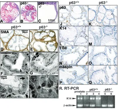
[image:3.612.48.384.73.515.2]appeared normal. Ducts of p63–/–prostate were surrounded by Fig. 1. Ontogeny of p63 in urogenital sinus (UGS) and phenotype of p63–/–UGS. (A-D) Ontogeny of p63 in embryonic UGS. CA, cloaca; HG, hind gut; MND,
mesonephric duct; NT, neural tube. Cross section (A) and saggital section (B-D) of mouse embryos. Scale bar: 50 µm. (E-O) Marker expression was examined in male p63+/+UGS (E-H,M) and male
(I-L,N) and female (O) p63–/–UGSs at E17.5. MD, Müllerian duct; WD, Wolffian duct. Gross morphology of the p63–/–UGS was normal and showed normal sexual dimorphism at E17.5 (compare O [female] with I-L [male]). p63–/–urogenital sinus epithelium (UGE) expressed differentiation markers identical to that of p63+/+UGE
[uroplakin (G,K), involucrin (H,L) and androgen receptor (AR) (M,N)] with the exception that p63 (E,I) and K14 (F,J) were absent in p63–/–UGE. In both male p63+/+
smooth muscle cells as in normal prostate (Fig. 2D,E). Although its gross morphology was similar to p63+/+prostate, p63–/– prostate lacked morphologically definable basal
epithelial cells (Fig. 2F-I). Instead, p63–/– prostate contained
goblet cell-like cells (Fig. 1G,I, white arrows). Basal cell markers [p63 (Fig. 2J,K), K14 (Fig. 2L,M), K19 (not shown), transglutaminase II (Friedrichs et al., 1995) (Fig. 2N,O)] were undetectable in p63–/– prostate epithelium. Maspin, which is
expressed in the basal cells of normal prostate (Pierson et al., 2002) (Fig. 2P), was also absent in p63–/–prostatic epithelium
(Fig. 1Q). Therefore, p63 is essential for development of prostatic basal cells. The result of IHC was confirmed by RT-PCR for K14 (Fig. 2R). K14 mRNA was detected in host prostate and p63+/+prostate, but not in p63–/–prostate (Fig. 2R).
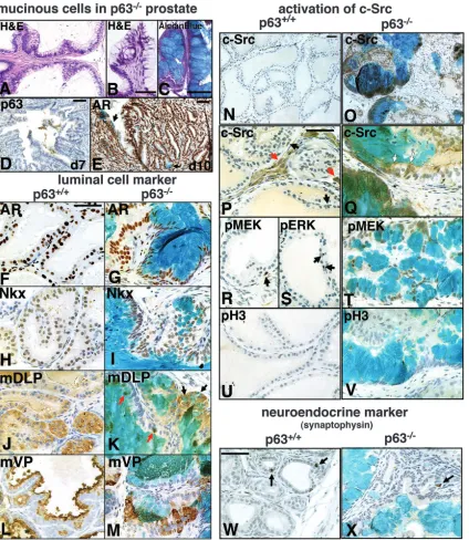
Ducts of p63–/–prostate were lined by columnar luminal and
atypical mucin-producing cells (Fig. 3A-C). These mucinous cells stained for PAS (not shown) and Alcian blue (pH 2.8)
(Fig. 3C). In time-course experiments the mucinous (Alcian blue-positive) cells were undetectable in p63–/– UGS seven
days after grafting (an early stage of prostatic development) (Fig. 3D). Ten to 14 days after grafting, a small number of Alcian blue-positive cells were detected in the prostatic ductal epithelium of p63–/– UGS grafts (Fig. 3E). This observation
suggests that differentiation of mucinous cells is secondary to the development of prostatic ducts.
Expression of luminal cell markers was examined in p63+/+ (Fig. 3F,H,J,L) and p63–/–(Fig. 3G,I,K,M) prostates. AR (Fig.
3F,G), Nkx3.1 (Fig. 3H,I), and the secretory proteins mDLP (Fig. 3J,K) and mVP (Fig. 3L,M) were expressed in the luminal cells in p63+/+ and p63–/– prostates. AR and Nkx3.1 were
expressed in some mucinous cells of p63–/–prostate.
[image:4.612.99.499.72.444.2]mDLP-positive (Fig. 3K) and mVP-mDLP-positive (Fig. 3M) cells were found among Alcian blue-positive cells. In some mucinous cells, mDLP was co-expressed with mucin (Fig. 3K, black Fig. 2. p63–/–prostate lacks basal cells. Both p63+/+(A) and p63–/–(B) urogenital sinus (UGS) formed prostate when grafted into intact male hosts. When p63–/–UGS was grafted into castrated male hosts, the UGS developed into a large cyst without ducts (C). The prostatic ducts were surrounded by smooth muscle cells expressing smooth muscle actin (SMA) (D,E). Morphologically definable basal cells (black arrow in F,H) were not observed in p63–/–prostate (G,I). p63–/–prostate contained goblet cells (G,I, white arrows) (see also Fig. 3). Elongated/flat nuclei of stromal cells should not be confused with basal cells. Expression of basal cell markers was examined in p63+/+(J,L,N,P) and p63–/–(K,M,O,Q) prostates. p63 (J,K), K14 (L,M), transglutaminase II (TGII,N,O) and maspin (P,Q) were detected only in p63+/+prostate. All basal cell markers
Fig. 3. Epithelial differentiation in p63–/–prostate. Scale bars: 50 µm. Goblet mucinous cells were detected in only p63–/–prostate (A,C). (C) The mucinous secretion stained for Alcian blue (pH 2.8). (D) Double staining for p63 and Alcian blue; one week after grafting the mucinous (Alcian blue-positive) cells were undetectable in p63–/–urogenital sinus (UGS). Double staining for androgen receptor (AR) and Alcian blue (E) in p63–/–UGS grafts; a small number of Alcian blue-positive cells was detected in epithelium of proximal ducts at 10 days after grafting (E, arrows). Expression of luminal cell markers was examined in p63+/+(F,H,J,L) and p63–/–(G,I,K,M) prostates. AR (F,G), Nkx3.1 (Nkx, H,I), and secretory proteins, mouse dorsolateral prostate (mDLP) (J,K) and mouse ventral prostate (mVP) (L,M) were expressed in the luminal cells in p63+/+and p63–/–prostates. mDLP-positive and mVP-positive cells were found among Alcian blue-positive cells (K,M). mDLP was co-expressed with mucin (K, red and black arrows). Secretory granules contained both mucin and mDLP in some mucinous cells (K, red arrows). Activity of Src was examined in p63+/+and p63–/–prostates (N-Q). In the p63+/+prostate,
activation of Src was detected in neurons (P, red arrows) and a subset of basal cells (P, black arrows) but not in luminal cells (N,P). In
p63–/–prostate, Src activity was detected mainly in Alcian blue-positive cells, but some non-mucinous luminal cells were also strongly positive for active Src (Q, white arrows). Downstream effecters in the Src-Ras-MAP kinase signal transduction pathway (MEK1/2 and ERK1/2) were also activated (phosphorylated) in neurons (not shown) and basal cells (R,S, arrows) in p63+/+prostate and in (mucinous
and non-mucinous) luminal cells in p63–/–prostate (T). Even though MAP kinase signaling was active, p63–/–prostate epithelium was proliferation-quiescent one month after grafting. Phosphorylation of histone H3 (pH3, U,V) was not detected in areas where
activated Src was detected in neurons (Fig. 3P, red arrows) and in a subset of basal cells (Fig. 3P, back arrows) but not in luminal cells (Fig. 3N,P) after one month of growth in intact male hosts. In contrast, in p63–/– prostate, Src activity was
detected in luminal epithelium (Fig. 3O,Q). Active Src was detected mainly in Alcian blue-positive cells, but some non-mucinous luminal cells were also strongly positive for active Src (Fig. 3Q, white arrows). In p63–/– prostate, focal
upregulation of Ras was also detected in the luminal cells which appeared to be in the process of trans-differentiation into mucinous cells (not shown). In the p63+/+prostate, downstream effecters in the Src-Ras-MAPK signal transduction (MEK1/2 and ERK1/2) were activated (phosphorylated) in the same cell types as Src activation; in neurons (not shown) and a subset of basal cells (Fig. 3R,S). In p63–/– prostate, MEK1/2 was
phosphorylated and translocated into nucleus in both mucinous and non-mucinous luminal cells (Fig. 3T). Even though MAP kinase signaling was active, p63–/– prostate was quiescent
in regard to proliferation one month after grafting. Phosphorylation of histone H3 was equally low in p63+/+and p63–/–prostate in intact male hosts (Fig. 3U,V).
Neuroendocrine cells developed independent of basal cells
Both p63+/+and p63–/–prostates contained rare neuroendocrine
cells as assessed by expression of synaptophysin (Fig. 3W,X). There was no distinctive difference between p63+/+ and p63–/– prostates in the distribution and concentration of
synaptophysin-positive cells.
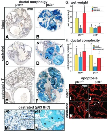
Effect of castration and testosterone-treatment One month after the grafting, p63+/+ and p63–/– UGSs
developed prostate with complex ductal structure (Fig. 4A,B). The p63+/+ prostate was negative for Alcian blue (Fig. 4A), whereas ducts of p63–/–prostate were filled with Alcian
blue-positive mucin (Fig. 4B). The wet weight of prostate is determined mostly by the water content. Therefore, p63+/+ prostate, which had more dilated ducts, was significantly heavier than p63–/– prostatic gland (Fig. 4G). In the intact
hosts, apoptotic cells were almost undetectable in both p63+/+ and p63–/– prostate (Fig. 4I,J). In response to androgen
deprivation epithelial apoptosis was detected in both the p63+/+ and p63–/–prostates three days after the castration of the host
(Fig. 4J,K). One month after the castration, p63+/+ prostatic regression was complete with marked reduction in luminal
in all p63 prostate grafts subjected to androgen depletion by castration (18/18). The mucinous pools in the stroma were connected to residual ducts or cysts lined at least in part by epithelial cells (not shown). Dead cells were also observed in the mucinous pools (Fig. 4N, black arrows), suggesting that the mucin was originally circumscribed by epithelium. In the surviving ducts of p63–/– prostate, both luminal and atypical
mucinous cells were present. Mucinous cells were stained for Alcian blue in the castrated host (Fig. 4D,N).
One month after castration, a 25 mg testosterone (T)-pellet was implanted into some of the hosts. After one month of continuous T-treatment, both p63+/+ and p63–/– prostate
regenerated (Fig. 4E,F). In p63+/+ prostate, ducts were filled with secretions (Fig. 4E) and the wet weight significantly (P=0.05) exceeded the original weight (Fig. 4G). Ducts of p63–/– prostate also regenerated and exhibited increased
complexity of ductal structure (Fig. 4F,H), but the wet weight remained the same (Fig. 4G).
The expression of proliferation makers clearly demonstrated the ability of p63+/+and p63–/–prostate to regenerate. In the
castrated hosts, pH3, phospho-MEK1/2 (pMEK) and Cdk4 were very low to undetectable in both p63+/+ (Fig. 5A) and p63–/–(Fig. 5B) prostates. Therefore, phosphorylation of MEK
in the epithelium of p63–/–prostate in the intact hosts (Fig. 3T)
is androgen-dependent. Five days after 25 mg T-pellet implantation, all three proliferation markers were upregulated in both p63+/+ (Fig. 5C) and p63–/– (Fig. 5D) prostates. In
p63+/+prostate, the lumen of existing ducts became enlarged, and the luminal epithelial cells increased in height as expected (compare Fig. 5A with Fig. 5C). In the p63–/– prostate, most
ducts disappeared after the castration. Therefore, regeneration of p63–/–prostate started as outgrowths of tightly packed ducts
from the large cysts, similar to the early prostatic development (Fig. 5D). Epithelial labeling indices for pH3 clearly showed regeneration of prostatic tissue in both p63+/+ and p63–/–
prostates. Epithelial pH3 labeling index was low (<1%) in the intact and castrated host in both p63+/+and p63–/–prostates.
T-pellet treatment significantly increased pH3 labeling index in both p63+/+and p63–/– prostates (Fig. 5E,*). One month after
T-pellet treatment to the castrated hosts, p63+/+ and p63–/–
prostates were fully regenerated and the pH3 labeling index returned to the basal level (Fig. 5E, +T one month).
all-hormone treatment groups. The regenerated ducts of p63–/–
prostates contained luminal cells positive for mucin, AR (Fig. 5F) and mDLP (Fig. 5G) or mVP (Fig. 5H), but not for mSV or mBUG (not shown), indicating prostatic differentiation.
Discussion
Studies with p63–/– mice have shown that p63 is essential for
differentiation of epidermis (Mills et al., 1999; Yang et al., 1999). In the Müllerian (paramesonephric) duct, loss of p63 transformed stratified squamous cervicovaginal epithelium
into simple columnar uterine epithelium (Kurita et al., 2004). Unlike the case of Müllerian duct, p63–/– UGE expressed
[image:7.612.192.564.209.665.2]proper differentiation markers for normal embryonic UGE (uroplakin, involucrin, K19 and AR) even though it failed to undergo squamous differentiation. Development of UGE into prostatic epithelia is regulated by androgens through AR in stromal cells (Cunha and Lung, 1978). In response to stromal signal(s), wild-type UGE proliferates, develops ducts and differentiates into luminal and basal prostatic epithelial cells. Our data clearly demonstrated that p63 is essential for development of prostatic basal cells. The unique phenotype of
Fig. 4. Effect of castration and androgen treatment. Double staining for androgen receptor (AR) and Alcian blue on p63+/+(A,C,E)
and p63–/–(B,D,F) prostates. After one month of growth in intact male hosts, p63+/+and p63–/–urogenital sinus (UGS) developed prostate having a complex ductal structure (intact, A,B). The ducts of p63+/+
prostates (A) were more dilated and had larger lumens than that of
p63–/–prostate (B). In grafts grown in intact hosts for one month followed by one month of castration, the lumen of ducts in
p63+/+prostate became smaller and
the entire graft was reduced in size (C). In p63–/–prostate, castration reduced the number of ducts, and the entire graft became cystic (D, arrows; large cystic ducts). Pools of secretion devoid of epithelium were always observed (D,N,*). One month after the T-pellet implantation to castrated hosts, ducts in p63+/+prostate became
dilated and filled with secretion (E). Ducts of p63–/–prostate also regenerated and exhibited increased complexity of ductal structure (F). (G) Wet weight of prostatic ducts. Data was analyzed with Fisher’s PSLD and ANOVA factorial tests. Data are expressed as mean+s.d. Lowercase letters indicate groups statistically distinguished. The data was significantly higher in the following order, c>a>b (P<0.05). (H) Ductal complexity. Total length of epithelial basement membrane (epithelial length, µm) in prostatic tissue was measured in sections of UGS grafts. The epithelial length was divided by the area of prostatic tissue (µm2). Data was analyzed
with Fisher’s PSLD and ANOVA factorial tests. Data are expressed as mean+s.d. Lowercase letters indicate groups statistically distinguished. There is no significant difference between a versus b, or a versus c. c is significantly higher than b. a, b and c are significantly higher than d (P<0.05). Detection of apoptosis in p63+/+(I,K) and p63–/–(J,L) prostate. In the intact hosts, apoptotic cells were rarely detected (I,J). Three days after the castration of the host, apoptotic epithelial cells were detected in both p63+/+and p63–/–prostates (K,L, white arrows). Double staining for p63 and Alcian blue (M,N). The concentration of p63-positive basal cells increased in the shrunken duct of p63+/+prostate (M).
p63–/– prostate lacking basal cells provides insights into the
function of basal cells in prostate in vivo. It is evident that development of prostatic ducts and differentiation of luminal cells do not require positive basal cells. Likewise, p63-positive basal cells are not required for differentiation of neuroendocrine cells. Moreover, basal cells are not required for smooth muscle differentiation of UGS mesenchyme, which is regulated by prostatic epithelium (Cunha et al., 1996).
Tsujimura et al. have suggested that luminal epithelium contains a self-renewing population of putative stem cells that are presumably required for prostatic regeneration (Tsujimura et al., 2002). Our results also demonstrated regenerative capacity of prostate lacking basal cells. Therefore, stem cell function (at least regenerative capacity) is not necessarily associated with ‘basal cells’. However, our results do not exclude possible trans-differentiation of basal to luminal cells in the normal adult prostate as suggested previously (Collins et al., 2001; Foster et al., 2002; Hudson et al., 2000; Uzgare et
al., 2004; van Leenders and Schalken, 2001; Wang et al., 2001). Moreover, our results do not exclude the presence of prostatic stem cells in the basal cells of normal wild-type prostate.
Although basal cells are not required for the differentiation of luminal cells, basal cells appear to be essential to maintain normal luminal cell differentiation. Luminal cells of p63–/–
prostate transformed into mucinous cells. Activation of Src, Ras and MAPK signaling can induce mucinous differentiation or overproduction of mucin in epithelial tissues (Lee et al., 2002; Li et al., 1998; Meerzaman et al., 2001; Perrais et al., 2002), and in the p63–/–prostate Src and MEK were activated
in the luminal cells. The activation of Src and MAPK signaling almost certainly plays a key role in mucinous transformation of luminal cells in p63–/– prostate. Src can be activated by
signals via adhesion molecules, cytokines or growth factors (Abram and Courtneidge, 2000; Thomas and Brugge, 1997). It is notable that in human prostate laminin 5 (α3β3γ2) is produced exclusively by basal cells (Calaluce et al., 2001) and
p63+/+(A) and p63–/–(B) prostates. Five days after implantation of a 25 mg T-pellet (castrated+T; C,D), all three proliferation markers were upregulated in both p63+/+(C) and p63–/–(D) prostates. In the p63–/–prostatic grafts, small bud-like outgrowths were visible (D, red arrows). (E) Labeling index for pH3. Data was analyzed with Fisher’s PSLD and ANOVA factorial test. Data was indicated as mean+s.d. The bars with asterisk are significantly higher than others (P<0.05). There’s no significant difference among bars without asterisk. Both p63+/+and p63–/– prostates were proliferation-quiescent in the intact and castrated hosts. T-treatment of castrated hosts increased pH3-positive cells equally in luminal cells of p63+/+and p63–/– prostates (+T 5 days). However, luminal cells in both p63+/+and p63–/–prostates became proliferation-quiescent one month after T-pellet implantation (+T one month). One month after T-pellet implantation, regenerated
[image:8.612.44.358.71.526.2]downregulation of laminin 5 affects membrane stability of integrin α6β4 and gene expression in the prostatic luminal cells (Hao et al., 2001). These observations clearly demonstrate that loss of basal cells and/or change in cell adhesion molecule indeed affect gene expression and phenotype of luminal cells in the prostate. Moreover, in the adult prostate, epithelial growth and functional differentiation are regulated by androgen via stromal paracrine factors. In the normal prostate, p63-positive basal cells reside on the basement membrane, and may mediate or modulate interactions between stromal cells and luminal cells. Therefore, it is likely that loss of basal cells disturbs the interaction between stromal cells and epithelial cells, and causes activation of Src in luminal cells. In the normal prostate, Src was active in a subset of basal cells in the proliferation-quiescent adult state, indicating the existence of Src-activating signals in the normal prostatic epithelium. The factors activating Src in luminal cells of p63–/–prostate are yet
to be identified, even though a wide spectrum of autocrine and paracrine growth factors have been described in the prostate (Cunha et al., 1998).
p63–/–prostate expressed proper secretory proteins specific
for mouse prostate. Therefore, expression of these androgen-regulated genes in luminal cells does not require basal cells. However, ducts in 63–/–prostate were less dilated than ducts in the p63+/+prostate, and the ducts in p63–/–prostate were unable
to reduce luminal secretory products in response to castration. This phenotype may be secondary to the mucinous differentiation of luminal cells, otherwise, basal cells may play a role in regulating secretory activity of luminal cells.
Basal cells also play key roles in maintaining ductal structure of the prostate in the absence of androgen. In response to castration, normal prostatic ducts lose luminal secretion content, and a substantial portion of luminal cells die via apoptosis. The entire process is highly coordinated, and therefore, normal prostatic ducts are able to shrink while maintaining their morphologic integrity. In contrast, in p63–/–
prostate complete loss of ducts occurred leaving only residual cystic structures or pools of mucinous secretion in the stroma following castration. In normal prostate, a substantial portion of luminal cells dies in response to castration, but always a subset of luminal cells survive to maintain ductal structure and to permit regeneration when androgens are restored. The mechanism controlling death or survival of luminal cells following castration is not known. Because castration selectively eliminates luminal (versus basal) cells, the ratio of basal cells to luminal cells increases (Rouleau et al., 1990), and after castration most surviving luminal cells are in direct contact with basal cells. Taken together, the phenotypes of the p63–/– prostate strongly suggest that basal cells play an
important role in the control of luminal cell differentiation and survival/apoptosis.
Adult prostate is a proliferation-quiescent organ in the mouse. Although androgen is a potent mitogen for developing and regenerating prostatic epithelia, androgen maintains prostatic tissue but does not stimulate epithelial proliferation in adult prostate. Luminal epithelial cells in developing or regenerating p63+/+ and p63–/– prostatic grafts became
proliferation-quiescent once the grafts reached a certain size. Therefore, the mode switch for androgen function, growth to maintenance, appears to be intact in p63–/–prostate. Moreover,
the labeling index for pH3 was identical between p63+/+and
p63–/– prostates in response to hormonal manipulation,
suggesting that p63 and basal cells are not essential for regulation of luminal epithelial proliferation.
Mucinous metaplasia of prostate has been suggested to be a pre-neoplastic condition (Scherl et al., 2004). Loss of basal cell is also a common characteristic in prostatic carcinogenesis. These observations suggest that changes caused by loss of basal cells may have an impact on carcinogenesis of the prostate. The effect of the loss of basal cells in carcinogenesis of the prostate is currently under investigation utilizing p63–/–
UGS and the hormonal carcinogenesis model with T+E2 -treatment (Wang et al., 2000).
In conclusion, p63 is essential for differentiation of prostatic basal cells, and basal cells are essential in maintaining normal differentiation of luminal cells and integrity of prostatic ducts. However, basal cells (therefore p63) are not required for development and regeneration of prostate. Further experimentation is required to define the role of p63 in basal cell differentiation. p63 isoforms are functionally distinct in regard to cell fate commitment, particularly in epidermal differentiation (Koster et al., 2004; McKeon, 2004). The differentiation of epidermis appears to be regulated by the balance between isoforms containing and lacking the transactivation domain. To understand the function of p63 in basal cell differentiation in prostate may require detailed analysis of isoform expression in the developing UGS and the adult prostate.
We thank Suzana S. Couto and Annemia Donjacour at UCSF for valuable advice, E. Birgit Lane for the anti-K14 LE001 and anti-K8 LE041 mouse monoclonal IgGs, and Hisaaki Kawakatsu at UCSF for anti-active-Src mouse monoclonal IgG. This work was supported by NIH grants CA84294 and CA89520.
References
Abram, C. L. and Courtneidge, S. A. (2000). Src family tyrosine kinases
and growth factor signaling. Exp. Cell Res. 254, 1-13.
Aumuller, G., Leonhardt, M., Renneberg, H., von Rahden, B., Bjartell, A. and Abrahamsson, P. A. (2001). Semiquantitative morphology of human
prostatic development and regional distribution of prostatic neuroendocrine cells. Prostate 46, 108-115.
Bhatia-Gaur, R., Donjacour, A. A., Sciavolino, P. J., Kim, M., Desai, N., Young, P., Norton, C. R., Gridley, T., Cardiff, R. D., Cunha, G. R. et al.
(1999). Roles for Nkx3.1 in prostate development and cancer. Genes Dev.
13, 966-977.
Calaluce, R., Kunkel, M. W., Watts, G. S., Schmelz, M., Hao, J., Barrera, J., Gleason-Guzman, M., Isett, R., Fitchmun, M., Bowden, G. T. et al.
(2001). Laminin-5-mediated gene expression in human prostate carcinoma cells. Mol. Carcinog. 30, 119-129.
Collins, A. T., Habib, F. K., Maitland, N. J. and Neal, D. E. (2001).
Identification and isolation of human prostate epithelial stem cells on alpha(2)beta(1)-integrin expression. J. Cell Sci. 114, 3865-3872.
Cooke, P. S., Young, P. and Cunha, G. R. (1991). Androgen receptor
expression in developing male reproductive organs. Endocrinology 128, 2867-2873.
Cunha, G. R. and Lung, B. (1978). The possible influences of temporal
factors in androgenic responsiveness of urogenital tissue recombinants from wild-type and androgen-insensitive (Tfm) mice. J. Exp. Zool. 205, 181-194.
Cunha, G. R. and Lung, B. (1979). The importance of stroma in
morphogenesis and function of urogenital epithelium. In Vitro 15, 50-71.
Cunha, G. R., Hayward, S. W., Dahiya, R. and Foster, B. A. (1996). Smooth
muscle-epithelial interactions in normal and neoplastic prostatic development. Acta Anat. 155, 63-72.
Cunha, G. R., Hayward, S. W., Hom, Y. K., Donjacour, A. A., Kurita, T., Cooke, P. S. and Lubahn, D. B. (1998). Growth factors as mediators of
at basal epithelial cells: three-dimensional views of the rat prostate, mammary gland and salivary gland. Differentiation 60, 219-227.
Hudson, D. L., O’Hare, M., Watt, F. M. and Masters, J. R. (2000).
Proliferative heterogeneity in the human prostate: evidence for epithelial stem cells. Lab. Invest. 80, 1243-1250.
Kawakatsu, H., Sakai, T., Takagaki, Y., Shinoda, Y., Saito, M., Owada, M. K. and Yano, J. (1996). A new monoclonal antibody which selectively
recognizes the active form of Src tyrosine kinase. J. Biol. Chem. 271, 5680-5685.
Koster, M. I., Kim, S., Mills, A. A., DeMayo, F. J. and Roop, D. R. (2004).
p63 is the molecular switch for initiation of an epithelial stratification program. Genes Dev. 18, 126-131.
Kurita, T., Mills, A. A. and Cunha, G. R. (2004). Roles of p63 in the
diethylstilbestrol-induced cervicovaginal adenosis. Development 131, 1639-1649.
Kurita, T., Wang, Y. Z., Donjacour, A. A., Zhao, C., Lydon, J. P., O’Malley, B. W., Isaacs, J. T., Dahiya, R. and Cunha, G. R. (2001). Paracrine
regulation of apoptosis by steroid hormones in the male and female reproductive system. Cell Death Differ. 8, 192-200.
Kurita, T., Young, P., Brody, J. R., Lydon, J. P., O’Malley, B. W. and Cunha, G. R. (1998). Stromal progesterone receptors mediate the inhibitory
effects of progesterone on estrogen-induced uterine epithelial cell deoxyribonucleic acid synthesis. Endocrinology 139, 4708-4713.
Lee, H. W., Ahn, D. H., Crawley, S. C., Li, J. D., Gum, J. R., Jr, Basbaum, C. B., Fan, N. Q., Szymkowski, D. E., Han, S. Y., Lee, B. H. et al. (2002).
Phorbol 12-myristate 13-acetate up-regulates the transcription of MUC2 intestinal mucin via Ras, ERK, and NF-kappa B. J. Biol. Chem. 277, 32624-32631.
Li, J. D., Feng, W., Gallup, M., Kim, J. H., Gum, J., Kim, Y. and Basbaum, C. (1998). Activation of NF-kappaB via a Src-dependent
Ras-MAPK-pp90rsk pathway is required for Pseudomonas aeruginosa-induced mucin overproduction in epithelial cells. Proc. Natl. Acad. Sci. USA 95, 5718-5723.
McKeon, F. (2004). p63 and the epithelial stem cell: more than status quo?
Genes Dev. 18, 465-469.
Meerzaman, D., Shapiro, P. S. and Kim, K. C. (2001). Involvement of the
MAP kinase ERK2 in MUC1 mucin signaling. Am. J. Physiol. Lung Cell Mol. Physiol. 281, L86-91.
Mills, A. A., Zheng, B., Wang, X. J., Vogel, H., Roop, D. R. and Bradley, A. (1999). p63 is a p53 homologue required for limb and epidermal
morphogenesis. Nature 398, 708-713.
prostate basal cell marker and is required for prostate development. Am. J. Pathol. 157, 1769-1775.
Sugimura, Y., Cunha, G. R. and Bigsby, R. M. (1986). Androgenic
induction of deoxyribonucleic acid synthesis in prostatic glands induced in the urothelium of testicular feminized (Tfm/y) mice. Prostate 9, 217-225.
Thomas, S. M. and Brugge, J. S. (1997). Cellular functions regulated by Src
family kinases. Annu. Rev. Cell Dev. Biol. 13, 513-609.
Tsujimura, A., Koikawa, Y., Salm, S., Takao, T., Coetzee, S., Moscatelli, D., Shapiro, E., Lepor, H., Sun, T. T. and Wilson, E. L. (2002). Proximal
location of mouse prostate epithelial stem cells: a model of prostatic homeostasis. J. Cell Biol. 157, 1257-1265.
Uzgare, A. R., Xu, Y. and Isaacs, J. T. (2004). In vitro culturing and
characteristics of transit amplifying epithelial cells from human prostate tissue. J. Cell. Biochem. 91, 196-205.
van Leenders, G. J. and Schalken, J. A. (2001). Stem cell differentiation
within the human prostate epithelium: implications for prostate carcinogenesis. BJU Int. 88 Suppl. 2, 35-42; discussion 49-50.
Walts, A. E., Said, J. W., Siegel, M. B. and Banks-Schlegel, S. (1985).
Involucrin, a marker of squamous and urothelial differentiation. An immunohistochemical study on its distribution in normal and neoplastic tissues. J. Pathol. 145, 329-340.
Wang, Y., Hayward, S., Cao, M., Thayer, K. and Cunha, G. (2001). Cell
differentiation lineage in the prostate. Differentiation 68, 270-279.
Wang, Y., Hayward, S. W., Donjacour, A. A., Young, P., Jacks, T., Sage, J., Dahiya, R., Cardiff, R. D., Day, M. L. and Cunha, G. R. (2000). Sex
hormone-induced carcinogenesis in Rb-deficient prostate tissue. Cancer Res. 60, 6008-6017.
Wu, X. R., Lin, J. H., Walz, T., Haner, M., Yu, J., Aebi, U. and Sun, T. T.
(1994). Mammalian uroplakins. A group of highly conserved urothelial differentiation-related membrane proteins. J. Biol. Chem. 269, 13716-13724.
Yang, A., Kaghad, M., Wang, Y., Gillett, E., Fleming, M. D., Dotsch, V., Andrews, N. C., Caput, D. and McKeon, F. (1998). p63, a p53 homolog
at 3q27-29, encodes multiple products with transactivating, death-inducing, and dominant-negative activities. Mol. Cell. 2, 305-316.
Yang, A., Schweitzer, R., Sun, D., Kaghad, M., Walker, N., Bronson, R. T., Tabin, C., Sharpe, A., Caput, D., Crum, C. et al. (1999). p63 is essential