INTRODUCTION
The emergence of function in sensory circuits requires accurate synaptic connectivity between peripheral sensory neurons and their central targets. A large body of literature has shown that this depends on a complex interplay of molecular mechanisms for proliferation control, neuronal fate specification, neuronal pathfinding and target recognition. An important contribution to the generation of neuronal circuits between sensory neurons in the periphery and their target neurons in the CNS is made by ensembles of transcription factors that operate as cell-intrinsic determinants in the regulation of temporal and spatial gene expression in the developing neurons (Chen et al., 2003; Komiyama and Luo, 2006; Shirasaki and Pfaff, 2002; Skeath and Thor, 2003).
An excellent model for the analysis of the molecular mechanisms that control sensory neuron connectivity is the developing olfactory system. In both insects and mammals, precise neuronal circuitry is established by the ordered axonal projection of olfactory sensory neurons (OSNs) to specific target neurons in the olfactory glomeruli (Axel, 1995; Jefferis and Hummel, 2006; Mombaerts et al., 1996; Rodrigues and Hummel, 2008). In Drosophila, OSNs are housed in ~500 hair-like sense organs called sensilla, located on two peripheral structures: the third segment of the antenna and the maxillary palps. Morphologically, three major
types of olfactory sensilla can be identified on the third antennal segment, namely the sensilla basiconica, trichoidea, and coeloconica; each of these antennal sense organs contains between one and four sensory neurons (Shanbhag et al., 2000). Each OSN expresses a single odorant receptor (OR) molecule and sends its axon to a specific glomerulus in the antennal lobe where it forms connections with postsynaptic target neurons, the projection neurons (PNs) and local interneurons (LNs) (Fishilevich and Vosshall, 2005).
Sense organs on the antenna derive from progenitor cells (sense organ precursors; SOPs) in the eye-antennal disc. Initial disc development begins during embryogenesis when a set of cells acquire antennal identity through the combinatorial action of the homeodomain transcription factors Homothorax, Extradenticle and Distalless, as well as the basic helix-loop-helix (bHLH) protein Spineless (Haynie and Bryant, 1986). The interaction of these transcription factors with the EGF signaling pathway generates a prepattern upon which neurogenic and proneural genes act to generate SOPs. The molecular mechanisms that control the selection of a single progenitor from the undifferentiated field of epidermal cells in the antennal disc are thought to be similar to those involved in the development of other sense organs in the fly PNS, where proneural domains are refined to single SOPs through Notch signaling (Rhyu et al., 1994). Two proneural genes are involved in the specification of SOPs for the different sensillar types on the antennal surface (Goulding et al., 2000; Gupta et al., 1998; zur Lage et al., 2003). atonal (ato), a bHLH transcription factor-encoding gene, specifies the progenitors that give rise to coeloconic sensilla, while amos, a second bHLH transcription factor-encoding gene specifies the basiconic and trichoid SOPs. The choice between basiconic and trichoid sensillar development appears to be controlled by the dosage of the Runx family Development 137, 3687-3695 (2010) doi:10.1242/dev.052407
© 2010. Published by The Company of Biologists Ltd
1National Centre for Biological Sciences, Tata Institute of Fundamental Research, GKVK Campus, Bellary Road, Bangalore 560065, India. 2Biozentrum, University of Basel, Basel CH4056, Switzerland.
*Authors for correspondence ([email protected]; [email protected])
Accepted 3 September 2010 SUMMARY
In Drosophila, the cephalic gap gene empty spiraclesplays key roles in embryonic patterning of the peripheral and central nervous system. During postembryonic development, it is involved in the development of central olfactory circuitry in the antennal lobe of the adult. However, its possible role in the postembryonic development of peripheral olfactory sense organs has not been investigated. Here, we show that empty spiracles acts in a subset of precursors that generate the olfactory sense organs of the adult antenna. All empty spiracles-expressing precursor cells co-express the proneural gene amosand the early patterning gene lozenge. Moreover, the expression of empty spiracles in these precursor cells is dependent on both amos and lozenge. Functional analysis reveals two distinct roles of empty spiraclesin the development of olfactory sense organs. Genetic interaction studies in a lozenge-sensitized background uncover a requirement of empty spiracles in the formation of trichoid and basiconic olfactory sensilla. MARCM-based clonal mutant analysis reveals an additional role during axonal targeting of olfactory sensory neurons to glomeruli within the antennal lobe. Our findings on empty spiraclesaction in olfactory sense organ development complement previous studies that demonstrate its requirement in olfactory interneurons and, taken together with studies on the murine homologs of empty spiracles, suggest that conserved molecular genetic programs might be responsible for the formation of both peripheral and central olfactory circuitry in insects and mammals.
KEY WORDS: Olfactory sense organs, empty spiracles, amos, lozenge, Axonal targeting
Expression and function of the
empty spiracles
gene in
olfactory sense organ development of
Drosophila
melanogaster
Sonia Sen1, Beate Hartmann2, Heinrich Reichert2,* and Veronica Rodrigues1,*
D
E
V
E
LO
P
M
E
N
transcription factor Lozenge (Lz), which regulates amos expression; high levels of Lz produce basiconic sensilla, while lower levels produce trichoidea (Gupta et al., 1998).
After their generation, OSNs initiate axogenesis and target specific glomeruli in the antennal lobe. Considerable progress has been made in understanding the mechanisms that control this wiring specificity, and a number of molecules involved in signaling, cell adhesion and axonal guidance have been identified (Rodrigues and Hummel, 2008). However, the role of transcription factors in the control of OSN pathfinding and connectivity remains far less well understood; so far, only two POU domain transcription factors, Acj6 and Pdm3 (Bai et al., 2009; Komiyama et al., 2004; Tichy et al., 2008), and mastermind (mam), a nuclear factor required for Notch signaling, have been implicated (Sakurai et al., 2009). This paucity of information on transcription factors involved in OSN axonal wiring specificity contrasts with the large number of transcription factors known to control the wiring of the PNs into the antennal glomeruli (Komiyama and Luo, 2006).
The cephalic gap gene empty spiracles (ems) encodes a homeodomain-containing transcription factor that is required during embryogenesis for the development of the antennal head segment from which the larval olfactory sense organs derive. ems loss-of-function mutations result in a gap-like phenotype in the embryonic head and brain, and an absence of peripheral sensory structures in the antennal cephalic segment (Cohen and Jurgens, 1990; Dalton et al., 1989; Schmidt-Ott et al., 1994; Walldorf and Gehring, 1992). During postembryonic brain development, emsis expressed in two of the deutocerebral neuroblast lineages that give rise to the antennal lobe PNs and LNs; emsfunction is necessary for the specification of these olfactory interneurons, as well as for targeting of their neurites in the antennal lobe (Das et al., 2008; Lichtneckert et al., 2008). By contrast, virtually nothing is known about the function of ems in antennal olfactory sense organ development in Drosophila.
This lack of information on emsaction in the development of the adult olfactory sense organs contrasts with the large amount of information on the role of the emsorthologs Emx1and Emx2 in the formation of the mammalian olfactory system. In the mouse, both genes are expressed in the developing olfactory epithelium, as well as in the developing olfactory bulb, and mutant analysis indicates that they play important roles in proliferation and axonal wiring (Bishop et al., 2003; Mallamaci et al., 1998; Matsuo et al., 1997; Nedelec et al., 2004; Shinozaki et al., 2004; Simeone et al., 1992a; Simeone et al., 1992b). Given the importance of Emx1/2 in the development of axonal projections of murine olfactory receptor neurons, we set out to investigate whether the fly emsgene might also be required for targeting of OSNs in Drosophila.
Here, we show that emsis expressed post-embryonically in a subset of precursors of olfactory sense organs, and acts in their development as well as in the correct targeting of the OSN axons that derive from them. emsis expressed transiently during early pupal life in a subset of progenitors that co-express amosand lz, and genetic analysis using loss-of-function mutations suggests that ems expression in the developing olfactory sense organs is dependent on both lz and amos. Although ems-null clones did not reveal any obvious defects in sense organ development, analysis of heterozygotes in a lzmutant background uncovers a functional requirement in the formation of trichoid and basiconic sensilla. MARCM-based mutant analysis using OR-specific Gal4 drivers reveals a further functional requirement of ems in OSN axonal
pathfinding. Our findings on the role of emsin the development of olfactory sense organs in the PNS complement previous reports of a requirement for emsin the development of olfactory interneurons in the CNS. Taken together with studies on the murine homologs of ems, these results suggest that conserved molecular genetic programs might be responsible for the formation of peripheral and central olfactory circuit elements in insects and mammals.
MATERIALS AND METHODS Fly strains and genetics
Fly stocks were obtained from the Bloomington Stock Centre (IN, USA) and, unless otherwise stated, were grown on cornmeal media, at 25°C.
amos1, amos2, amos3and the amos-Gal4 stock were kindly provided by
Andrew Jarman (University of Edinburgh, UK). For staging, white prepupae (0 hours after puparium formation; APF) were collected on a moist filter paper and aged under humid conditions at 25°C.
MARCM experiments
For tubulin marked MARCM clones (Lee and Luo, 2001), females of genotype yhsFLP; Tubulin-Gal4,UAS-mCD8::GFP,UAS-LacZ/CyO-GFP; FRT82B GAL80/TM6Bwere crossed to males of either UAS-LacZ,UAS-mCD8::GFP/CyO; FRT82B/TM6Bor UAS-LacZ,UAS-mCD8::GFP/CyO; FRT82B ems3/TM6B.
In MARCM experiments where OR-Gal4 lines were used to mark specific OSNs, females of genotype yhsFLP; UAS-LacZ,UAS-mCD8::GFP/CyO; FRT82B/TM6B or yhsFLP; UAS-LacZ,UAS-mCD8::GFP/CyO; FRT82B ems3/TM6Bwere crossed to males of the
following genotypes:
wt1118,OR59b- Gal4/FM7a;;FRT82B Tub-Gal80/MKRS,
wt1118,OR85f-Gal4/FM7a;;FRT82B Tub-Gal80/MKRS,
w1118;OR10a-Gal4/CyO; FRT82B Tub-Gal80/TM6B,
wt1118,OR67d- Gal4/FM7a;;FRT82B Tub-Gal80/MKRS,
w1118;OR43a-Gal4/CyO-GFP; FRT82B Tub Gal80/MKRS,
w1118; OR88a-Gal4/CyO-GFP; FRT82B Tub Gal80/MKRS,
w1118; OR83c-Gal4/CyO-GFP; FRT82B Tub Gal80/MKRS,
w1118; OR47b-Gal4/CyO-GFP; FRT82B Tub-Gal80/MKRS,
w1118; OR23a-Gal4/CyO; FRT82B Tub Gal80/MKRSor
w1118;OR65a-Gal4/CyO-GFP; FRT82B Tub-Gal80/MKRS.
For generating large clones in the antenna, the Minute technique was used. Here, yhsFLP;UAS-LacZ,UAS-mCD8::GFP/CyO; FRT82B/TM6B
or yhsFLP; UAS-LacZ,UAS-mCD8::GFP/CyO; FRT82B ems3/TM6B
females were crossed to w1118; FRT82B, Ubi::GFPnls, 3R
P{A92}RpS3[Plac92]/TM6Bmales. Progeny of all the clonal crosses were heat shocked for 1 hour at 37°C at the late second instar stage. Results were observed in the adult, except for experiments shown in Fig. 3E-L, where 5 hour APF animals were dissected.
Immunolabeling
Pupal antennal discs and adult brains were dissected and stained as described previously (Jhaveri and Rodrigues, 2002; Wu and Luo, 2006). Primary antibodies used were: rabbit anti-GFP (1:10,000; Molecular Probes, Invitrogen, Delhi, India); mouse anti-Bruchpilot (mAbnc82, 1:20; DSHB, Iowa, USA); rabbit anti-Ems (1:500) and rat anti-Ems (1:100; U. Walldorf, University of Saarland, Hamburg, Germany); mouse anti-Lz (1:10; DSHB Iowa, USA); sheep Atonal (1:5000) and rabbit anti-Amos (1:500; A. Jarman, University of Edinburgh, UK); and guinea-pig anti-Senseless (1:1000; H. Bellen, Baylor College of Medicine, USA). The secondary antibodies used were Alexa Fluor-488-, Alexa Fluor-568- and Alexa Fluor-647-coupled antibodies generated in goat (1:400; Molecular Probes).
Cuticle preparation and sensillar counting
Adult antennae were dissected in PBS, placed on a glass slide with Faure’s solution, covered with a coverslip and allowed to clear at 75oC overnight
(Gupta et al., 1998). Cuticle mounts were imaged using Nomarski optics in a Nikon E-1000 microscope. Static images of the antenna taken at different focal planes were imported into Image J and sensilla were counted using the ‘cell counter’ plug-in (http://rsbweb.nih.gov/ij/).
D
E
V
E
LO
P
M
E
N
Microscopy and image processing
Fluorescent preparations were imaged on an Olympus Fluoview (FV1000) or Leica TCS SP5 scanning confocal microscope. Optical sections were taken at 1 m intervals with a picture size of 512⫻512 pixels and digitally processed using Image J and Adobe Photoshop CS3 (Adobe Systems, San Jose, CA, USA).
RESULTS
emsis co-expressed in a subset of amos
-expressing cells in the developing antennal disc The progenitors of the olfactory sense organs are generated in the third segment of the antennal disc and are specified by two bHLH transcription factor-encoding genes, amosand ato. For a precise characterization of the spatial and temporal pattern of ems expression during this process, we first studied emsin the context of amosand ato using immunocytochemistry.
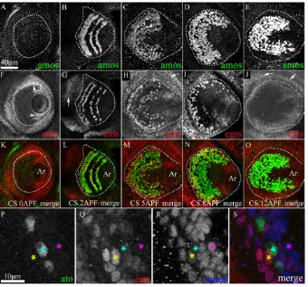
Fig. 1A-E shows the spatiotemporal expression of amosin the third antennal segment from 0-12 hours APF. At 0 hours APF, amosis not yet expressed (Fig. 1A) and first appears at 1 hour APF (not shown). At 2 hours APF, Amos appears in three to four semi-circular domains in the third antennal segment (Fig. 1B), which broaden by 8 hours APF and 12 hours APF to merge into a single large ‘C-shaped’ band of cells (Fig. 1C-E). The time course of ems expression is comparable with that of amos, although it is sparser and terminates earlier (Fig. 1F-J). At 0 hours APF, emsis detected in superficial cells located along the periphery of the disc and in the second antennal segment (arrow in Fig. 1F) and at the base of the presumptive arista (arrowhead in Fig. 1F). This staining is probably within the epidermal cells of the disc, including the peripodial membrane, and was not analyzed further in this study. No ems-expressing cells are seen within the third antennal segment
proper at this time. At 1 hour APF, initial expression is observed in the third antennal segment (not shown), and by 2 hours APF this increases to include cells in four semi-circular domains (Fig. 1G). Expression in these domains broadens by 5 hours APF (Fig. 1H). By 8 hours APF, ems levels begin to diminish (Fig. 1I) and are undetectable by 12 hours APF (Fig. 1J).
A comparison of merged spatiotemporal expression patterns of amosand emsshows that the onset of emsexpression is temporally coincident with that of amos, but is downregulated earlier (Fig. 1K-O). emsis present in a subset of amos-expressing but in none of the ato-expressing cells (Fig. 1P-S). This was confirmed at all pupal stages in the third antennal segment (data not shown). We conclude that although all ems-expressing cells co-express amos(and none co-express ato), not all amos-expressing cells co-express ems.
As lzis known to activate amos (zur Lage et al., 2003), both genes should be expressed together in precursors of the trichoid and basiconic (but not coeloconic) sensilla. As expected, we find that all ems-expressing cells also co-express lz, and that emsexpression is restricted to a subset of lz-expressing cells (see Fig. S1 in the supplementary material).
The emsgene is expressed in a subset of olfactory sense organ precursors
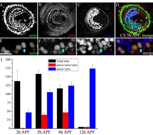
[image:3.612.53.393.60.377.2]Although amosis expressed in the proneural cell clusters from which the trichoid and basiconic SOPs derive, the senseless (sens) gene is highly expressed in the SOPs themselves and is, thus, a marker for these progenitor cells (Nolo et al., 2000). Fig. 2A,E shows SOPs marked by Sens in the antennal disc 5 hours APF. Only a small proportion of the emsand amos-expressing cells co-express sens(Fig. 2A-H). We conclude from this that emsis expressed (with amos) in a subset of the proneural domains from which SOPs are selected.
Fig. 1. emsis co-expressed in a subset of amos-expressing cells in the developing antennal disc. (A-O)The third antennal segment in the antennal disc is highlighted within broken lines. Ar, arista. (A,F,K) 0 hours APF; (B,G,L) 2 hours APF; (C,H,M) 5 hours APF; (D,I,N) 8 hours APF; (E,J,O) 12 hours APF. Merged images of discs immunolabeled with anti-Amos (A-E) and anti-Ems (F-J) shown in K-O. Both Amos (A) and Ems (F) are absent from sensory cells of the disc at 0 hours APF. At 2 hours APF, expression of both Amos and Ems is seen (B,G); at 12 hours APF, Ems expression has decayed (J), while Amos expression is still strong (E). In addition to sensory cells, Ems can be detected in the superficial layer of the disc (arrows in F-J) and in the epidermal cells at the periphery of the presumptive arista (arrowhead in F).
(P-S)Magnified images of the third antennal
segment stained with anti-Ato (P), anti-Ems (Q) and anti-Amos (R). The merged image (S) shows that Ems is always co-expressed with Amos (yellow star), but never with Atonal (cyan star). However, not all Amos-positive
cells express Ems (pink star). Scale bars: 40m
for A-O, 10m for P-S.
D
E
V
E
LO
P
M
E
N
Initial observations indicate that the number of ems-expressing SOPs increases transiently during early pupal development. In order to quantify this, we counted the number of cells co-expressing ems, sens and amos within the anlage of the third antennal segment at different time points during early pupal development (Fig. 2I). At 5 hours APF, ~25% of ems- and amos -expressing cells also co-express sens; this number increases to ~40% at 8 hours APF. At 12 hours APF, emsexpression declines markedly and is found in only a few cells.
Taken together, these findings indicate that ems is expressed, together with amos, in a subset of proneural clusters within the third segment of the antennal disc. Moreover, its expression is transient within SOPs, which are selected from these clusters. This suggests that emscould be involved in the development of a subset of the trichoid and/or basiconic olfactory sense organs.
amosregulates emsexpression in the developing third antennal segment
Given the central role of amosin the specification of basiconic and trichoid SOPs, we reasoned that expression of ems in the third antennal segment might require amos. We characterized two mutant alleles of amos, amos1and amos3, which show a strong reduction in basiconic and trichoid antennal sensilla (zur Lage et al., 2003). In both mutant alleles, emsexpression was almost totally absent in the third antennal segment except for a few cells seen in the periphery of the third antennal segment (white arrowhead in Fig. 3B). Fig. 3A-D demonstrates that emsexpression is absent in proneural clusters and SOPs (ems is detected only in non-sensory/non-progenitor cells; compare with control discs, Fig. 1H). As expected, atoexpression is unaffected (not shown), whereas the number of sens-expressing SOPs was reduced as compared with
controls (Fig. 3C; Fig. 2A). The ~250 (252.3±38.8; n3) Sens-positive cells in wild-type antennal discs at 5 hours APF are reduced to ~110 (110.33±6.8; n3) in amos3mutants.
To determine whether ems might in turn regulate amos expression, we generated marked clones of a loss-of-function allele of ems(which is embryonic lethal) using MARCM (mosaic analysis with a repressible cell marker) (Lee and Luo, 2001). In these experiments, ubiquitously expressed tub-Gal4was used to drive UAS-mCD8::GFPin clones induced at the second larval instar stage and recovered at 5 hours APF. In control clones, as expected, all ems-expressing cells co-expressed amos, and the overall expression domains of the two genes were comparable with wild type (Fig. 3E-H). In ems mutant clones, the overall expression domains of amosalso remained normal and all cells that expressed the mutant emsgene invariably co-expressed amos (Fig. 3I-L). [The emsmutant allele used encodes a truncated non-Fig. 2. emsis expressed in a subset of olfactory sense organ
progenitors. (A-D)5 hours APF antennal disc co-stained with
anti-Senseless (A), anti-Ems (B) and anti-Amos (C). (D)Merge.
(E-H)Magnified images from a 5 hour APF antennal disc. Scale bar:
10m. The cell marked with arrows expresses all three markers and are
identified as an Ems-positive, Amos-positive SOP. Ems-positive cells that express Amos but not Sens are likely to be cells of the proneural
domains. (I)Quantification of Ems-positive and Amos-positive cells,
[image:4.612.52.297.58.274.2]which also express Sens. Histograms represent the mean ± s.d.; n4.
Fig. 3. Lz and Amos regulate emsexpression in the developing third antennal segment.(A-D)Antennal discs from 5 hour APF animals. Anlage of the third antennal segment (and arista) is
highlighted with broken lines. (A)amos3completely lacks Amos
immunoreactivity. (B)Ems expression is present in the non-sensory cells
in the superficial layers of the disc (arrows) and in a few cells within the
third segment (arrowheads). (C)Sens-positive cells are reduced in
number when compared with wild type (Fig. 2A). (D)Merge of A-C.
(E-L)MARCM clones labeled with tub-Gal4>UAS-GFP. In each case, a
region of the clone (boxed) is magnified in the inset. (E-H)Control
clones show normal expression of Ems (F, arrow indicates the
non-sensory staining) and Amos (G). (H)Merge of GFP, Ems and Amos.
Arrowheads in E-H indicate cells that co-expresses Amos and Ems.
(I-L)ems-null clones. Anti-Ems recognizes the non-functional truncated
Ems in the mutant (arrowhead in J). In these clones, cells mutant for
Ems express Amos (arrowheads in K,L) and the overall domains of amos
expression remain normal (K). (M-P)Antennal discs of 5 hours APF lz3
animals. Expression of Amos is reduced, leaving a small domain of cells (M). Ems staining within sensory progenitors is absent, leaving only the staining in superficial cells (N, arrows). The number of Sens-positive
(SOPs) (O) is greatly reduced. (P)Merge. Scale bar: 50m.
D
E
V
E
LO
P
M
E
N
[image:4.612.315.563.61.318.2]functional protein that is still detected in the cytoplasm by the anti-Ems antibody (see Lichtneckert et al., 2007).] We conclude that ems does not regulate amos in the developing third antennal segment.
As lzis known to regulate amos(zur Lage et al., 2003), loss-of-function lzalleles might be expected to affect emsexpression in the third antennal segment. To investigate this, we characterized the developing antennal disc of two viable strong hypomorphic lz alleles, lz3and lz34. In both alleles, the number of amos-expressing cells was strongly reduced (Fig. 3M) and ems expression was completely absent in the anlage of the third antennal segment, leaving only the epidermal staining (Fig. 3N). This is shown for lz3 in Fig. 3M-P, which also documents the fact that the overall number of SOPs as visualized by sens-expression was reduced as expected (wild-type 252.3±38.8, n3; lz392.7±0.6, n3). These results are in accordance with the notion that lz, by acting through amos, is also involved in regulation of emsin the anlage of the developing third antennal segment.
emsis involved in the development of olfactory sensilla
As the two genes known to specify trichoid and basiconic sensilla, lzand amos, also regulate emsexpression, we reasoned that ems might itself play a role in olfactory sensillar development. In order to test this, we generated large ems–/– mutant clones using the Minute method (see Materials and methods) in the antenna and examined sensillar types in cuticular whole mounts. This analysis failed to reveal any change in the number of any of the three sensillar types in mutant versus wild-type antennae (see Fig. S2 in the supplementary material).
This negative result could mean that emsis not necessary for olfactory sensillar development. Alternatively, it could reflect a functional redundancy of ems with other genes that control the development of the olfactory sensilla. Ectopic expression of emsin the sense organ precursors using lz-Gal4 (lz-Gal4/+;UAS-ems/+) leads to a decrease in the number of basiconic and trichoid sensilla (P<0.001; see Fig. S3 in the supplementary material). When ems is expressed using a later driver amos-Gal4, only the trichoid sensilla are affected (P<0.01; see Fig. S3 in the supplementary material). These results suggest that the dynamic expression of Ems in the sensory precursors is important for determination of sensillar types.
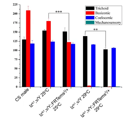
Because lz(acting through amos) is a key player in sense organ development, a possible functional redundancy of emsmight be uncovered by testing for genetic interactions with lz alleles. To investigate this, we examined the sensillar numbers of flies heterozygous for emsin the background of a temperature-sensitive allele of lz(lzts1). When reared at a permissive temperature (25°C), expression patterns of amosand emswere only mildly affected (see Fig. S4A-C in the supplementary material) and the number of different sensillar types was comparable with that of the wild type (Fig. 4). However, when these hemizygous mutants also carried a heterozygous ems-null allele (lzts1/Y; ems3/+), they showed a marked reduction in the number of basiconic sensilla (Fig. 4; P<0.001). The trichoid and coeloconic sensilla were unaffected (P<0.3). As expected, lzts1 animals reared at non-permissive temperature (29°C) showed a severe reduction in basiconic sensilla (Fig. 4). Staining of 5 hours APF antennal discs from these animals revealed a strong decrease in emsas well as in amosexpression (see Fig. S4D-F in the supplementary material). Significantly, however, in the background of an ems heterozygote at 29°C, these animals showed an additional reduction in the number of trichoid
sensillae (Fig. 4; P<0.001). The coeloconic sensilla were not affected in any of these experiments. A small number of mechanosensory bristles appeared on the antennae of the lzts1 mutants at 29°C and in lzts1/Y; ems3/+at 29°C; these were never observed in controls (data not shown).
In view of the observation that ems-null mutations by themselves do not affect sensillar development, we hypothesize that emsis involved in a functionally redundant manner in the development of a subset of trichoid and basiconic (but not coeloconic) sense organs. It therefore seems likely that ems acts in a redundant pathway together with other gene(s) regulated by lz.
emsis required for olfactory receptor neuron wiring
[image:5.612.327.534.59.257.2]The experiments described above were assayed at the level of external sensillar morphology and uncovered a redundant role of emsin sense organ development. However, in these experiments, the development of the internal cells comprising the sense organs – neurons and support cells – was not analyzed. Could emshave an additional non-redundant role in the development of the OSNs that are generated from the SOPs? To investigate this, we carried out a MARCM-based mutant analysis using OR-specific Gal4 drivers and assayed for axonal wiring defects in the adult antennal lobe. Three Gal4 drivers specific for ORs expressed in neurons of basiconic sense organs (OR10a, OR59b, OR85f) and seven Gal4 drivers specific for ORs expressed in neurons of trichoid sense organs (OR23a, OR47b, OR67d, OR88a, OR43a, OR83c, OR65a) were used. Clones were induced at the late second larval instar stage and recovered in the adult (see Materials and methods). This protocol allowed us to avoid large ems–/–neuroblast clones in the antennal lobe, which are rarely generated 48 hours after larval Fig. 4. emsinteracts genetically with lzmutants to produce phenotypes that affect the antennal sense organs.Numbers of trichoid, basiconic and coeloconic sensilla, as well as ectopic mechanosensory bristles are indicated. Bars represent mean ± s.e.m.
[n3 for wild type (Canton special, CS); n5 in all other genotypes].
Numbers of sensilla are marginally affected in lzts1/Y reared at 25°C.
When one mutant copy of the emsmutant is introduced (lzts1/Y;
ems3/+), the number of basiconica is reduced (P<0.001). lzts1/Y animals
reared at 29°C show almost no basiconica, leaving trichoidea
unaffected. In lzts1/Y; ems3/+ animals, the numbers of trichoidea are
also reduced (P<0.001).
D
E
V
E
LO
P
M
E
N
hatching; nevertheless, we excluded brains that showed anatomical defects visible upon immunostaining with mAbnc82. Previous investigators have shown that in most cases clones including only one or two interneurons are likely to be generated at these time points (Das et al., 2008).
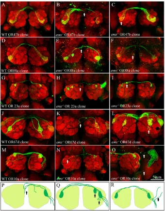
OSNs deficient for emsshowed a variety of connectivity defects exemplified in Fig. 5 and summarized in Table S1 in the supplementary material. In control clones, labeled receptor neuron axons enter the antennal lobe, converge onto the appropriate ipsilateral glomerulus, and also innervate the corresponding contralateral glomerulus via a commissural projection (Fig. 5A,D,G,J,M). By contrast, marked defects in projections across the midline were observed in mutant clones induced with four out of seven trichoid-specific driver lines (OR47b, OR88a, OR23a and OR67d; arrow in Fig. 5B,E,H,K). In these cases, labeled mutant axons often failed to enter the contralateral lobe and either stalled or mis-projected back to the ipsilateral lobe (schematic in Fig. 5P). Moreover, when multiple receptor axons were labeled a marked reduction in the intensity of contralateral versus ipsilateral glomerular innervations was seen (arrow in Fig. 5C). We also observed targeting defects in which OSNs innervated ectopic
glomeruli, in addition to the cognate glomerulus in the antennal lobe (e.g. OR23a; arrow in Fig. 5I). In some cases, OSN terminals ‘spilled out’ of the boundaries of the cognate glomerulus into neighboring regions in antennal lobe (OR67d; arrow in Fig. 5L; schematic in Fig. 5Q). Only a subset of the clones obtained (~25%) showed the defects described above (see Table S1 in the supplementary material for details). This could imply that the penetrance of the phenotype is low. However, because only a subset of the basiconic/trichoid SOPs express ems, it is possible that low frequencies of phenotypes are to be expected even if penetrance is high.
[image:6.612.51.391.59.495.2]Targeting defects were also observed in one of the three basiconic specific OR lines (OR10a). Mutant axons that projected across the midline often failed to target to the appropriate contralateral glomerulus and meandered across the lobe, whereas in other cases, they failed to form ipsilateral projections and innervated only the contralateral glomerulus (Fig. 5N). In one preparation, we observed mutant axons projecting beyond the antennal lobe into adjacent ‘non-olfactory’ neuropile (arrowheads in Fig. 5O; schematic in Fig. 5R). The remaining basiconic-specific OR lines (OR59b and OR85f) yielded a significantly smaller
Fig. 5. Ems is necessary for wiring of OSNs to the antennal glomeruli. Adult antennal lobes immunostained with mAbnc82 to visualize the glomeruli. Clonal OSNs are marked by expression of GFP
induced by MARCM. (A-O)OSNs expressing
OR-47b (A-C), OR-88a (D-F), OR-23a (G-I), OR-67d (J-L) and OR-10a (M-O) are labeled. Wild-type OSNs (A,D,G,J,M) target to their cognate glomeruli and send collaterals to the contralateral lobe via the antennal
commissure. (P-R)The connectivity defects
seen in ems–/–OSNs that lack emsfunction
are summarized. (1) Midline crossover defects, marked by arrows in B,E,H,K, where
projection across the commissure is compromised. In C, OSNs innervate the contralateral glomerulus less than the ipsilateral glomerulus, indicating a possible crossover defect (arrow). (2) Target recognition defects where OSNs target ectopic glomeruli (F,I, arrows; compare with controls in D,G,Q). In some cases, OSNs fail to innervate the ipsilateral glomerulus (N, arrow). (3) OSNs spill-over defects, where terminals extend beyond their target glomeruli to a neighboring glomerulus (arrow, L). (4) Misrouting defects, where OSNs project outside the antennal lobe to non-olfactory neuropile (arrows in O,R). The green staining demarcated with broken lines in E,H,O is a
staining artifact. Scale bar: 50m.
D
E
V
E
LO
P
M
E
N
number of labeled clones in the antennal lobe than in their wild-type controls (see Table S1 in the supplementary material). This observation could have two possible explanations. (1) ems could regulate the expression of some ORs; hence the promoter would be unable to drive Gal4 expression in mutant clones. In the mouse, Emx2 is known to initiate transcription of several OR genes (McIntyre et al., 2008). (2) Some OSNs lacking emsfunction are unable to target to the antennal glomeruli. These possibilities require further investigation.
Taken together, these findings show that emsplays an important role in axonal pathfinding and targeting of OSNs, which is seen most prominently among neurons of the trichoid sensilla, but is also observed in basiconic OSNs. We conclude that emsis not only involved in the formation of external sensillar structures but also plays an important, non-redundant role in OSN axonal wiring in the antennal lobe.
DISCUSSION
emsis expressed in a subset of proneural clusters and SOPs specified by amos
In this report, we studied the expression and function of emsin the developing third antennal segment of Drosophila. Our analysis of emsexpression combined with data on co-expression of other key genes involved in antennal sense organ development allowed the identification of the ems-positive cell types. Moreover, it demonstrated that ems is expressed in a restricted spatial and temporal pattern in these developing sense organs.
In antennal development, SOPs derived from proneural domains specified by amosform trichoid and basiconic sensilla and those specified by atoform coeloconic sensilla (Gupta et al., 1998; zur Lage et al., 2003). We find that ems-expressing cells comprise a subset of the cells in the amos-expressing proneural domains and SOPs, and argue that emsconfers specific properties on this subset of SOPs. Although the precise identity of sensilla that are influenced by emsexpression has not been determined, it is unlikely that these are confined to a specific region of the antennal surface. Furthermore, it is unlikely that ems-expressing SOPs develop into a distinct sensillar type because the sensillar phenotypes we observed upon interaction with lz alleles affect both trichoid and basiconic lineages.
Previous work has established that the pre-patterning gene lzfirst appears early in the antennal disc (at late third instar larva) and regulates the expression of amosin proneural clusters (Goulding et al., 2000; Gupta et al., 1998). Our immunocytochemical studies show that emsexpression is coincident with the appearance of amos, arguing that lz could regulate both genes in proneural clusters.
Genetic analysis demonstrates that emsexpression is lost in amos mutants, favoring the idea of a hierarchy of gene function with lz controlling amos, which in turn regulates ems (model in Fig. 6).
What is the mechanism that selects emsexpression in a spatially defined pattern of cells in the antennal disc? It seems unlikely that expression could be determined solely through the action of lz and amos, as emsis expressed in a small subset of these lineages. We propose that additional genes interact in a genetic cascade, together with lzand amosto select specific ems-expressing proneural clusters and SOPs. This idea is supported by our observation of a haplo-insufficient interaction of ems with lzmutations. The identity of these unknown genes needs to be deciphered by further genetic studies.
Function of emsin adult olfactory sense organ development
Two mutant phenotypes in olfactory sense organ development were observed in ems loss-of-function experiments: deficits in the number of external olfactory sensilla and axonal path finding/ targeting defects of mutant OSNs in the antennal lobe.
Although ems loss-of-function mutations did not provide evidence for a requirement of emsin sensillar development, we did observe a clear haplo-insufficient interaction between emsmutations and lzalleles that affected the formation of trichoid and basiconic sense organs. This leads us to propose that emsplays a redundant role, acting with gene(s) regulated by lz, in the development of some of the basiconic and trichoid sensilla. Genes that act together with lz to regulate formation of trichoid or basiconic sensilla have not yet been identified. Ha and his colleagues (Ha and Smith, 2006) identified two genes, tod1and tot1, that affect trichoid sensilla alone without affecting basiconica. However, the nature of these genes has not been characterized, and it will be interesting to study whether they impinge on emsexpression.
A marked non-redundant role of emswas observed in OSN axon pathfinding and targeting in MARCM-based analyses. Several different types of axonal projection defects were seen in the ems mutant OSNs of trichoid sensilla (schematized in Fig. 5P-R). These include defects in commissural projections, mistargeting to inappropriate glomeruli, spillover of axon terminals and unequal innervation of ipsilateral versus contralateral glomeruli. Comparable defects were seen in the ems mutant OSNs of basiconic sensilla, albeit at lower frequency.
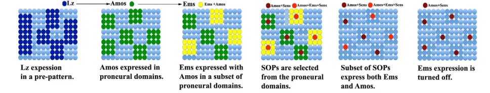
[image:7.612.62.546.63.155.2]As emsis expressed transiently in the SOPs and their proneural domains during early pupal development, it is likely that emsacts as an early intrinsic determinant in sense organ progenitors to influence cell fate decisions, which indirectly result in appropriate axonal projections of OSNs later in postembryonic development. Fig. 6. Summary diagram of the development of SOPs in the antennal disc.Lz is expressed in a zone of epidermal cells defining a prepattern
in the third antennal disc. amos is regulated by lz and is turned on within a set of cells called the proneural domain. Although amos and ems are
expressed simultaneously within the resolution of our experiments, genetic experiments have shown that amos turns on ems in a subset of
proneural domains. Lateral inhibition acts upon these cells, leading to the selection of a single SOP that expresses higher levels of Amos and/or Ems
and Sens. Expression of emsis turned off prior to amos. The gene(s) that negatively regulate emshave not been identified.
D
E
V
E
LO
P
M
E
N
Thus, OSN axonal targeting is likely to be mediated by other factors that are themselves regulated by ems and subsequently affect components of the wiring machinery.
Targeting of the OSNs to cognate glomeruli has been studied in several laboratories and the roles of several cell adhesion and signaling molecules have been identified. Roundabout proteins (Robo1, Robo2 and Robo3) (Jhaveri et al., 2004), semaphorins (Komiyama et al., 2007; Sweeney et al., 2007), N-cadherin (Hummel and Zipursky, 2004), DSCAM (Hummel et al., 2003), Wnt5 (Sakurai et al., 2009; Yao et al., 2007) and the small GTPases Pak and Dock (Ang et al., 2003) have been implicated. However, to date, the only transcription factors other than Ems that have been shown to affect OSN targeting are the POU domain molecules Acj6 and Pdm3 (Komiyama et al., 2004; Tichy et al., 2008), and the Notch signaling pathway acting through Mastermind (Sakurai et al., 2009). Although Ems, along with other transcription factors, has also been shown to be required for the precise targeting of PNs and LNs, it remains to be determined how these transcription factors can regulate cell surface and signaling molecules in developing OSN axons.
emsis required for the development of both larval and adult olfactory sense organs and olfactory interneurons
During embryonic development, emsis first expressed at the early cellular blastoderm stage in a single circumferential stripe at the anterior end of the embryo that subsequently becomes regionalized to discrete ectodermal patches of the labral, antennal and intercalary segment of the anterior head (Dalton et al., 1989; Walldorf and Gehring, 1992). The large ems domain in the ectoderm/neuroectoderm of the antennal segment gives rise to a set of peripheral cephalic sense organs and to the anlage of the antennal brain neuromere (Hartmann et al., 2000; Lichtneckert and Reichert, 2008; Urbach and Technau, 2004).
Mutation of ems leads to a gap-like phenotype in the embryonic head, which includes deletions of cephalic sense organs in the antennal (and intercalary) segments (Cohen and Jurgens, 1990; Dalton et al., 1989; Jürgens et al., 1984; Walldorf and Gehring, 1992). The major olfactory sense organ of the larva – the dorsal organ – is also lacking in emsmutants (Schmidt-Ott et al., 1994; Stocker, 2008). In addition, lack of ems function also results in defects in the embryonic brain, including a deletion of the deutocerebral brain neuromere, which contains the larval olfactory lobe (Hirth et al., 1995). This phenotype is due to defective specification of the neuroectoderm and correlates with the absence of the proneural gene lethal of scute, which is thought to be required for neuroectodermal cells to adopt the competence to become neuroblasts (Younossi-Hartenstein et al., 1997).
Remarkably, emsplays important roles in both peripheral and central olfactory system development of both the larval and adult. This is despite the fact that the olfactory sense organs of the adult antenna (and maxillary palps) have a distinct origin from those of the larvae. Most of the olfactory interneurons of the adult are generated postembryonically from a set of deutocerebral brain neuroblasts, and previous reports showed that emsfunction is necessary in at least two of these neuroblast lineages (Jefferis and Hummel, 2006; Rodrigues and Hummel, 2008). In the anterodorsal neuroblast lineage, ems plays a role in the appropriate dendritic targeting of PNs to the olfactory glomeruli, whereas in the lateral neuroblast lineage it determines the correct number of PNs and LNs. It is interesting that ems acts in the development of both the peripheral and central olfactory system; the possibility that this transcription factor plays a role in matching these classes of neurons is intriguing.
Evolutionary conservation of ems/Emxroles in olfactory system development?
The organization of the olfactory system is strikingly similar in insects and mammals (Hildebrand and Shepherd, 1997; Komiyama and Luo, 2006). OSNs expressing a given OR project to the same glomerulus in the antennal lobe of insects and the olfactory bulb of mammals. In the glomeruli of both animal groups, OSNs make specific synaptic connections with olfactory interneurons: PNs and LNs in insects; and the mitral, tufted cells, the periglomerular cells and granule cells in mammals. In Drosophila, emsplays important roles in the development of both peripheral sense organs and central olfactory interneurons at both larval and adult stages. Remarkably, the vertebrate homologs of ems, Emx1/2, has comparable functions in the development of the olfactory system. In the mouse, genes of the ems/Emx family have been shown to be important both for the development of the OSNs and the olfactory interneurons. The two murine ems gene homologs, Emx1 and Emx2, are expressed peripherally in the developing olfactory epithelium and centrally in cells in the developing olfactory bulb, notably the mitral cells, which are the vertebrate counterpart of the insect olfactory PNs (Mallamaci et al., 1998; Simeone et al., 1992a; Simeone et al., 1992b). Mutational loss of the Emxgenes leads to marked defects in both nasal epithelium and olfactory bulb (Bishop et al., 2003; Cecchi and Boncinelli, 2000; Shinozaki et al., 2004). In Emx2 mutants, the olfactory nerve is present; however, no connection is formed between the nerve and the bulb, implying that most of the olfactory sensory neurons fail to project to the brain. In Emx1/2 double mutants the olfactory bulbs are reduced and severely disorganized, and the olfactory tract is deficient.
The comparable expression and function ofems/Emxgenes in the development of olfactory sensory neurons and olfactory interneurons in insects and mammals, argue for evolutionarily conserved roles of the ems/Emxgenes in olfactory system development. Thus, although the similarity in anatomical organization of the peripheral and central olfactory system in insects and mammals may be due to functional convergence, it might also reflect a remarkable conservation in the molecular mechanisms for central and peripheral olfactory system development in both animal groups.
Acknowledgements
This work was supported by grants from TIFR, SNSF and the Indo-Swiss Joint Research Program. We are grateful to Hugo Bellen, Andrew Jarman and Uwe Walldorf; the Bloomington DrosophilaStock Centre DSHB for fly stocks and reagents; and K. VijayRaghavan, Abhijit Das and Ajeet Pratap Singh for many useful discussions and comments on the manuscript. We thank the Department of Science and Technology, Government of India (Centre for Nanotechnology No. SR/S5/NM-36/2005) for the Olympus FV1000 confocal microscopes in the Central Imaging and Flow Cytometry Facility (CIFF) and NOMIC, NCBS. We acknowledge Tripti Gupta, Melanie Gentner, Mario Metzler, Susanne Flister and Bruno Bello for technical help. S.S. was funded by a short term fellowship from EMBO.
Competing interests statement
The authors declare no competing financial interests.
Supplementary material
Supplementary material for this article is available at
http://dev.biologists.org/lookup/suppl/doi:10.1242/dev.052407/-/DC1
References
Ang, L. H., Kim, J., Stepensky, V. and Hing, H.(2003). Dock and Pak regulate olfactory axon pathfinding in Drosophila. Development130, 1307-1316. Axel, R.(1995). The molecular logic of smell. Sci. Am.273, 154-159. Bai, L., Goldman, A. L. and Carlson, J. R.(2009). Positive and negative
regulation of odor receptor gene choice in Drosophila by acj6. J. Neurosci. 29, 12940-12947.
Bishop, K. M., Garel, S., Nakagawa, Y., Rubenstein, J. L. and O’Leary, D. D. (2003). Emx1 and Emx2 cooperate to regulate cortical size, lamination, neuronal
D
E
V
E
LO
P
M
E
N
differentiation, development of cortical efferents, and thalamocortical pathfinding. J. Comp. Neurol. 457, 345-360.
Cecchi, C. and Boncinelli, E.(2000). Emx homeogenes and mouse brain development. Trends Neurosci.23, 347-352.
Chen, H. H., Hippenmeyer, S., Arber, S. and Frank, E.(2003). Development of the monosynaptic stretch reflex circuit.Curr. Opin. Neurobiol.13, 96-102. Cohen, S. M. and Jurgens, G.(1990). Mediation of Drosophila head
development by gap-like segmentation genes. Nature346, 482-485. Dalton, D., Chadwick, R. and McGinnis, W.(1989). Expression and embryonic
function of empty spiracles: a Drosophila homeo box gene with two patterning functions on the anterior-posterior axis of the embryo. Genes Dev.3, 1940-1956. Das, A., Sen, S., Lichtneckert, R., Okada, R., Ito, K., Rodrigues, V. and
Reichert, H.(2008). Drosophila olfactory local interneurons and projection neurons derive from a common neuroblast lineage specified by the empty spiracles gene. Neural Dev.3, 33.
Fishilevich, E. and Vosshall, L. B.(2005). Genetic and functional subdivision of the Drosophila antennal lobe.Curr. Biol. 15, 1548-1553.
Goulding, S. E., zur Lage, P. and Jarman, A. P.(2000). amos, a proneural gene for Drosophila olfactory sense organs that is regulated by lozenge. Neuron25, 69-78.
Gupta, B. P., Flores, G. V., Banerjee, U. and Rodrigues, V.(1998). Patterning an epidermal field: Drosophila lozenge, a member of the AML-1/Runt family of transcription factors, specifies olfactory sense organ type in a dose-dependent manner. Dev. Biol. 203, 400-411.
Ha, T. S. and Smith, D. P.(2006). A pheromone receptor mediates 11-cis-Vaccenyl acetate-induced responses in Drosophila. J. Neurosci. 26, 8727-8733. Hartmann, B., Hirth, F., Walldorf, U. and Reichert, H.(2000). Expression,
regulation and function of the homeobox gene empty spiracles in brain and ventral nerve cord development of Drosophila. Mech. Dev.90, 143-153. Haynie, J. L. and Bryant, P. J.(1986). Development of the eye-antenna imaginal
disc and morphogenesis of the adult head in Drosophila melanogaster. J. Exp. Zool. 237, 293-308.
Hildebrand, J. G. and Shepherd, G. M.(1997). Mechanisms of olfactory discrimination: converging evidence for common principles across phyla. Annu. Rev. Neurosci. 20, 595-631.
Hirth, F., Therianos, S., Loop, T., Gehring, W. J., Reichert, H. and Furukubo-Tokunaga, K.(1995). Developmental defects in brain segmentation caused by mutations of the homeobox genes orthodenticle and empty spiracles in Drosophila. Neuron15, 769-778.
Hummel, T. and Zipursky, S. L.(2004). Afferent induction of olfactory glomeruli requires N-cadherin. Neuron42, 77-88.
Hummel, T., Vasconcelos, M. L., Clemens, J. C., Fishilevich, Y., Vosshall, L. B. and Zipursky, S. L.(2003). Axonal targeting of olfactory receptor neurons in Drosophila is controlled by Dscam. Neuron37, 221-231.
Jefferis, G. S. and Hummel, T.(2006). Wiring specificity in the olfactory system.
Semin. Cell Dev. Biol. 17, 50-65.
Jhaveri, D. and Rodrigues, V.(2002). Sensory neurons of the Atonal lineage pioneer the formation of glomeruli within the adult Drosophila olfactory lobe.
Development129, 1251-1260.
Jhaveri, D., Saharan, S., Sen, A. and Rodrigues, V.(2004). Positioning sensory terminals in the olfactory lobe of Drosophila by Robo signaling. Development
131, 1903-1912.
Jürgens, G., Wieschaus, E., Nüsslein-Volhard, C. and Kluding, H.(1984). Mutations affecting the pattern of the larval cuticle inDrosophila melanogaster.
Dev. Genes Evol.193, 283-295.
Komiyama, T. and Luo, L.(2006). Development of wiring specificity in the olfactory system.Curr. Opin. Neurobiol.16, 67-73.
Komiyama, T., Carlson, J. R. and Luo, L.(2004). Olfactory receptor neuron axon targeting: intrinsic transcriptional control and hierarchical interactions.Nat. Neurosci.7, 819-825.
Komiyama, T., Sweeney, L. B., Schuldiner, O., Garcia, K. C. and Luo, L.(2007). Graded expression of semaphorin-1a cell-autonomously directs dendritic targeting of olfactory projection neurons. Cell128, 399-410.
Lee, T. and Luo, L.(2001). Mosaic analysis with a repressible cell marker (MARCM) for Drosophila neural development. Trends Neurosci.24, 251-254. Lichtneckert, R. and Reichert, H.(2008). Anteroposterior regionalization of the
brain: genetic and comparative aspects. Adv. Exp. Med. Biol. 628, 32-41. Lichtneckert, R., Bello, B. and Reichert, H.(2007). Cell lineage-specific
expression and function of the empty spiracles gene in adult brain development of Drosophila melanogaster. Development134, 1291-1300.
Lichtneckert, R., Nobs, L. and Reichert, H.(2008). Empty spiracles is required for the development of olfactory projection neuron circuitry in Drosophila.
Development135, 2415-2424.
Mallamaci, A., Iannone, R., Briata, P., Pintonello, L., Mercurio, S., Boncinelli, E. and Corte, G.(1998). EMX2 protein in the developing mouse brain and olfactory area. Mech. Dev.77, 165-172.
Matsuo, I., Suda, Y., Yoshida, M., Ueki, T., Kimura, C., Kuratani, S. and Aizawa, S.(1997). Otx and Emx functions in patterning of the vertebrate rostral head. Cold Spring Harbor Symp. Quant. Biol. 62, 545-553.
McIntyre, J. C., Bose, S. C., Stromberg, A. J. and McClintock, T. S.(2008). Emx2 stimulates odorant receptor gene expression. Chem. Senses33, 825-837. Mombaerts, P., Wang, F., Dulac, C., Vassar, R., Chao, S. K., Nemes, A.,
Mendelsohn, M., Edmondson, J. and Axel, R.(1996). The molecular biology of olfactory perception. Cold Spring Harbor Symp. Quant. Biol. 61, 135-145. Nedelec, S., Foucher, I., Brunet, I., Bouillot, C., Prochiantz, A. and Trembleau,
A.(2004). Emx2 homeodomain transcription factor interacts with eukaryotic translation initiation factor 4E (eIF4E) in the axons of olfactory sensory neurons.
Proc. Natl. Acad. Sci. USA101, 10815-10820.
Nolo, R., Abbott, L. A. and Bellen, H. J.(2000). Senseless, a Zn finger transcription factor, is necessary and sufficient for sensory organ development in Drosophila. Cell102, 349-362.
Rhyu, M. S., Jan, L. Y. and Jan, Y. N.(1994). Asymmetric distribution of numb protein during division of the sensory organ precursor cell confers distinct fates to daughter cells. Cell76, 477-491.
Rodrigues, V. and Hummel, T.(2008). Development of the Drosophila olfactory system. Adv. Exp. Med. Biol. 628, 82-101.
Sakurai, M., Aoki, T., Yoshikawa, S., Santschi, L. A., Saito, H., Endo, K., Ishikawa, K., Kimura, K., Ito, K., Thomas, J. B. et al.(2009). Differentially expressed Drl and Drl-2 play opposing roles in Wnt5 signaling during Drosophila olfactory system development. J. Neurosci. 29, 4972-4980.
Schmidt-Ott, U., Gonzalez-Gaitan, M., Jackle, H. and Technau, G. M.(1994). Number, identity, and sequence of the Drosophila head segments as revealed by neural elements and their deletion patterns in mutants. Proc. Natl. Acad. Sci. USA91, 8363-8367.
Shanbhag, S. R., Muller, B. and Steinbrecht, R. A.(2000). Atlas of olfactory organs of Drosophila melanogaster 2. Internal organization and cellular architecture of olfactory sensilla. Arthropod. Struct. Dev. 29, 211-229. Shinozaki, K., Yoshida, M., Nakamura, M., Aizawa, S. and Suda, Y.(2004).
Emx1 and Emx2 cooperate in initial phase of archipallium development. Mech. Dev.121, 475-489.
Shirasaki, R. and Pfaff, S. L.(2002). Transcriptional codes and the control of neuronal identity. Annu. Rev. Neurosci.25, 251-281.
Simeone, A., Acampora, D., Gulisano, M., Stornaiuolo, A. and Boncinelli, E. (1992a). Nested expression domains of four homeobox genes in developing rostral brain. Nature358, 687-690.
Simeone, A., Gulisano, M., Acampora, D., Stornaiuolo, A., Rambaldi, M. and Boncinelli, E.(1992b). Two vertebrate homeobox genes related to the Drosophila empty spiracles gene are expressed in the embryonic cerebral cortex.
EMBO J.11, 2541-2550.
Skeath, J. B. and Thor, S.(2003). Genetic control of Drosophila nerve cord development.Curr. Opin. Neurobiol.13, 8-15.
Stocker, R. F.(2008). Design of the larval chemosensory system. Adv. Exp. Med. Biol. 628, 69-81.
Sweeney, L. B., Couto, A., Chou, Y. H., Berdnik, D., Dickson, B. J., Luo, L. and Komiyama, T.(2007). Temporal target restriction of olfactory receptor neurons by Semaphorin-1a/PlexinA-mediated axon-axon interactions. Neuron53, 185-200.
Tichy, A. L., Ray, A. and Carlson, J. R.(2008). A new Drosophila POU gene, pdm3, acts in odor receptor expression and axon targeting of olfactory neurons.
J. Neurosci. 28, 7121-7129.
Urbach, R. and Technau, G. M.(2004). Neuroblast formation and patterning during early brain development in Drosophila. BioEssays 26, 739-751. Walldorf, U. and Gehring, W. J.(1992). Empty spiracles, a gap gene containing a
homeobox involved in Drosophila head development. EMBO J.11, 2247-2259. Wu, J. S. and Luo, L.(2006). A protocol for dissecting Drosophila melanogaster
brains for live imaging or immunostaining.Nat. Protoc.1, 2110-2115. Yao, Y., Wu, Y., Yin, C., Ozawa, R., Aigaki, T., Wouda, R. R., Noordermeer, J.
N., Fradkin, L. G. and Hing, H.(2007). Antagonistic roles of Wnt5 and the Drl receptor in patterning the Drosophila antennal lobe.Nat. Neurosci.10, 1423-1432.
Younossi-Hartenstein, A., Green, P., Liaw, G. J., Rudolph, K., Lengyel, J. and Hartenstein, V.(1997). Control of early neurogenesis of the Drosophila brain by the head gap genes tll, otd, ems, and btd. Dev. Biol. 182, 270-283.
zur Lage, P. I., Prentice, D. R., Holohan, E. E. and Jarman, A. P.(2003). The Drosophila proneural gene amos promotes olfactory sensillum formation and suppresses bristle formation. Development130, 4683-4693.