ORIGINAL RESEARCH
PEDIATRICS
Early Diagnosis of Spastic Cerebral Palsy in Infants with
Periventricular White Matter Injury Using Diffusion
Tensor Imaging
XH. Jiang,XX. Li,XC. Jin,XM. Wang,XC. Liu,XK.C. Chan, andXJ. Yang
ABSTRACT
BACKGROUND AND PURPOSE: Periventricular white matter injury is the common cause of spastic cerebral palsy. However, the early diagnosis of spastic cerebral palsy still remains a challenge. Our aim was to investigate whether infants with periventricular white matter injury with bilateral spastic cerebral palsy have unique lesions different from those in infants without cerebral palsy and to evaluate the efficiency of DTI in the early diagnosis of spastic cerebral palsy.
MATERIALS AND METHODS: Infants with periventricular white matter injury and controls underwent MR imaging at 6 –18 months of age. Fractional anisotropy was calculated from DTI. Cerebral palsy was diagnosed by 24 –30 months of age. Subjects were divided into 3 groups: infants with periventricular white matter injury with bilateral spastic cerebral palsy, infants with periventricular white matter injury without cerebral palsy, and controls. Tract-Based Spatial Statistics and Automated Fiber Quantification were used to investigate intergroup differences. Receiver operating characteristic curves were used to assess the diagnostic accuracy of spastic cerebral palsy. Correlations between motor function scores and fractional anisotropy were evaluated along white matter tracts.
RESULTS:There were 20, 19, and 33 subjects in periventricular white matter injury with spastic cerebral palsy, periventricular white matter injury without cerebral palsy, and control groups, respectively. Decreased fractional anisotropy in the corticospinal tract was only observed in infants with periventricular white matter injury with spastic cerebral palsy, whereas decreased fractional anisotropy in the posterior thalamic radiation and genu and splenium of the corpus callosum was seen in both periventricular white matter injury subgroups. Fractional anisotropy in the corticospinal tract at the internal capsule level was effective in differentiating infants with periventricular white matter injury with spastic cerebral palsy from those without cerebral palsy by a threshold of 0.53, and it had strong correlations with motor function scores.
CONCLUSIONS: Corticospinal tract lesions play a crucial role in motor impairment related to spastic cerebral palsy in infants with periventricular white matter injury. Fractional anisotropy in the corticospinal tract at the internal capsule level could aid in the early diagnosis of spastic cerebral palsy with high diagnostic accuracy.
ABBREVIATIONS:CP⫽cerebral palsy; CST⫽corticospinal tract; CST-CP⫽CST at the cerebral peduncle level; CST-CR⫽CST at the corona radiata level; CST-IC⫽ CST at the internal capsule level; FA⫽fractional anisotropy; GCC⫽genu of the corpus callosum; GMFCS⫽Gross Motor Function Classification System; PTR⫽ posterior thalamic radiation; PWMI⫽periventricular white matter injury; SCC⫽splenium of the corpus callosum; SCP⫽spastic cerebral palsy
P
eriventricular white matter injury (PWMI) is a major form of white matter injury in both preterm and term infants and is the common cause of spastic cerebral palsy (SCP).1-3MR imaging iseffective in identifying PWMI and demonstrates periventricular white matter signal abnormality and/or volume loss, enlargement of the lateral ventricles, and thinning of the corpus callosum.3On the basis of signs on MR imaging, PWMI can be classified into mild,
Received June 6, 2018; accepted October 30.
From the Department of Radiology (H.J., X.L., C.J., M.W., C.L., J.Y.), First Affiliated Hospital, and Department of Biomedical Engineering (H.J., J.Y.), Key Laboratory of Biomedical Information Engineering of the Ministry of Education, School of Life Science and Technology, Xi’an Jiaotong University, Xi’an, China; and Department of Ophthalmology and Radiology (K.C.C.), School of Medicine, New York University, New York, New York.
Haoxiang Jiang and Xianjun Li contributed equally to this work.
This study was supported by the National Key Research and Development Pro-gram of China (2016YFC0100300); the National Natural Science Foundation of China (81471631, 81771810 and 81171317); the 2011 New Century Excellent Talent Sup-port Plan of the Ministry of Education, China (NCET-11-0438); the Fundamental Re-search Funds for the Central Universities (xjj2018265); and the Fundamental
Research Funds of the First Affiliated Hospital of Xi’an Jiaotong University (2017QN-09).
Preliminary data from this research were previously presented at: Annual Meeting of the International Society for Magnetic Resonance in Medicine, April 22–27, 2017; Honolulu, Hawaii.
Please address correspondence to Jian Yang, PhD, Department of Radiology, First Affiliated Hospital, Xi’an Jiaotong University, Xi’an, Shaanxi, China; e-mail: cjr.yangjian@vip.163.com
Indicates open access to non-subscribers at www.ajnr.org
Indicates article with supplemental on-line tables.
Indicates article with supplemental on-line photos.
moderate, and severe grades.4Severe PWMI develops frequently into cerebral palsy (CP).4,5However, the early diagnosis of SCP in infants with mild and moderate PWMI still remains a major challenge.
MR imaging and neurologic examinations are predictive tools for detecting the risk of CP.6-8Compared with neurologic exam-inations, MR imaging is preferred for revealing the anatomic po-sition and severity of brain lesions.6However, conventional MR imaging has limitations in delineating white matter tracts pre-cisely and has failed to differentiate individual tracts specifically.9 DTI is effective in overcoming the above limitations and improv-ing the diagnostic accuracy.6Patients with SCP typically present with motor deficits. DTI studies have revealed that the damage in the corticospinal tract (CST) is related to motor impairment.10-12 Meanwhile, the sensory pathways or commissural tract lesions or both are also involved in the motor deficits in children with SCP.13-15 However, the prerequisite white matter lesions ac-countable for SCP remain unclear.16The exploration of the dif-ferences in white matter alterations between infants with PWMI with and without CP may provide clues for the identification of the responsible white matter tracts related to motor impairment. In this study, the analyses of DTI data with Tract-Based Spatial Statistics (TBSS; http://fsl.fmrib.ox.ac.uk/fsl/fslwiki/TBSS) and Automated Fiber Quantification (AFQ; https://pypi.org/project/ AFQ-Browser/) were used to compare the differences among groups of infants with PWMI with bilateral SCP, infants with PWMI without CP, and controls. The aim was to investigate whether infants with PWMI with bilateral SCP have unique le-sions different from lele-sions in those without CP and to evaluate the early diagnostic efficiency of DTI for differentiating infants with SCP from those with PWMI without CP.
MATERIALS AND METHODS
This was a retrospective cohort study approved by the institu-tional review board of the First Affiliated Hospital of Xi’an Jiao-tong University.
Participants
Parents of the infants were informed of the potential risks of MR imaging. Written consent was obtained before the brain MR im-aging examination.
Brain MR imaging was performed in infants 6 –18 months of age, from July 2011 to January 2014. The reasons for MR imaging examination included suspected developmental delay, seizures, or other risks of cerebral disorders. The MR imaging scanner and sequences did not change throughout the study period. Next, these infants underwent follow-up neurologic examinations be-tween 24 and 30 months of age, performed by a pediatric neurol-ogist using several measurements, including motor milestones, primitive reflexes, postural reactions, muscle strength, and cogni-tive and psychomotor development assessment using the second edition of the Bayley Scales of Infant Development and the Gross Motor Function Classification System (GMFCS; https://research. cerebralpalsy.org.au/what-is-cerebral-palsy/severity-of-cerebral-palsy/gross-motor-function-classification-system/) assessment. Developmental delay was defined by a score of the Mental or Psychomotor Development Index of⬍85 according to the Bayley Scales of Infant Development assessment.
CP was diagnosed in infants on the basis of the follow-up examination results using the definition provided by the Interna-tional Executive Committee in the United States.17The infants with CP were classified into GMFCS levels I–V, representing mild-to-severe motor dysfunction.10The GMFCS level zero rep-resented infants without motor dysfunction in this study.
The inclusion criteria of infants with PWMI were as follows: 1) MR imaging performed between 6 and 18 months of age and 2) PWMI diagnosed by MR imaging. The severity of PWMI was graded as mild, moderate, or severe on the basis of the MR imag-ing signs as follows: 1) mild, abnormally high signal in the periventricular white matter on T2-weighted images with mild white matter reduction limited to the peritrigonal region; 2) mod-erate, abnormally high white matter signal on T2-weighted im-ages, with moderate periventricular white matter decrease and irregular enlargement of the ventricles; 3) severe, large, or exten-sive cystic changes of periventricular white matter, with marked reduction of white matter and severe irregular enlargement of the ventricles (On-line Fig 1).4The exclusion criteria included severe PWMI, intrauterine congenital infection, and unsuccessful fol-low-up neurologic examinations. According to the folfol-low-up outcomes, infants with mild and moderate PWMI were divided into those with PWMI with bilateral SCP and those without CP after the exclusion of unilateral and nonspastic CP.
The infants with MR imaging performed between 6 and 18 months of age were selected as controls after excluding the following conditions: 1) abnormalities on MR imaging, in-cluding PWMI; dilated perivascular spaces; arachnoid cyst; hy-drocephalus; congenital malformations; hematencephalon; subarachnoid space enlargement; encephalomalacia and so forth; 2) history of nervous system infection; preterm birth; hypoxic-ischemic encephalopathy and congenital heart dis-ease; 3) seizures; developmental delay; and hypertonia; and 4) lack of follow-up neurologic examinations.
MR Imaging Protocols
Image Postprocessing and Data Analysis
Fractional anisotropy (FA) maps were obtained after brain extrac-tion and eddy current correcextrac-tion using the FMRIB Software Li-brary (FSL; http://www.fmrib.ox.ac.uk/fsl).19Linear and nonlin-ear image registrations were used for alignment of the FA maps of all subjects to a selected FA map. An averaged image of the coreg-istered FA maps was created as the target map. Then, FA maps of all subjects were registered to the target map. A mean FA map and mean FA skeleton were created. The aligned FA map of each sub-ject was prosub-jected onto the mean FA skeleton (threshold⫽0.2). Voxelwise statistical analysis was performed to assess the inter-group differences in FA.
AFQ software was used to quantitatively analyze white matter tracts.20Automatic fiber tract segmentation and cleaning were performed first. Then, FA values along the tracts were calculated by clipping each fiber to 100 equally spaced nodes. The CST, pos-terior thalamic radiation (PTR), genu of the corpus callosum (GCC), and splenium of corpus callosum (SCC) were evaluated. To characterize the properties of the CST in detail, we divided the CST into 3 parts according to the brain atlas21: CST at the cerebral peduncle level (CST-CP; nodes: 1–37), CST at the internal capsule level (CST-IC; nodes: 38 – 81), and CST at the corona radiata level (CST-CR; nodes: 82–100).
Statistical Analysis
Categoric variables were analyzed using the2test in SPSS soft-ware (Version 17.0; IBM, Armonk, New York). Continuous vari-ables were analyzed across groups using analysis of variance with
the least significant difference test. Sex ratios were compared across tracts by using the 2 test. Interrater reliability analyses for GMFCS were performed with thetest. The variables of gesta-tional age, birth weight, and age at MR imaging were adjusted during the inter-group comparisons and correlation analyses to remove their potential effects on the variations in DTI metrics. Inter-group comparisons in FA were per-formed by TBSS with the threshold-free cluster enhancement and family-wise error correction. In the AFQ analysis, a Mann-WhitneyUtest with a Bonferroni correction was used to assess the inter-group differences in FA. Receiver oper-ating characteristic curves were used to assess the diagnostic performances of FA in different regions for differentiating infants with PWMI with bilateral SCP from those without CP. Correlations be-tween FA values of the white matter tracts and motor function scores (GMFCS levels and Psychomotor Devel-opment Index) in infants with PWMI were analyzed by partial correlation analysis.Pvalues⬍.05 were considered statistically significant for all tests.
RESULTS
Demographic and Clinical Information
Thirty-nine infants with PWMI and 33 controls were finally en-rolled on the basis of the inclusion and exclusion criteria (Fig 1). According to the follow-up diagnosis, the infants with PWMI were divided into infants with bilateral SCP (n⫽20) and those without CP (n⫽19) groups. The time between the MR imaging examination and diagnosis of SCP was 14.8⫾3.2 months.
Among infants with PWMI with bilateral SCP, 7 (35%), 6 (30%), 4 (20%), and 3 (15%) infants were classified into GMFCS levels I, II, III, and IV, respectively. Thevalue for the interrater reliability of the GMFCS assessment was 0.83. There was no sig-nificant difference in gestational age and birth weight between infants with PWMI with bilateral SCP and those without CP. The gestational age and birth weight in infants with PWMI with bilat-eral SCP and without CP were less than those in controls. More-over, there was no significant difference across groups in the sex constituent ratio or age at MR imaging (Table 1). More details about the perinatal characteristics of the infants with PWMI are listed in On-line Table 1.
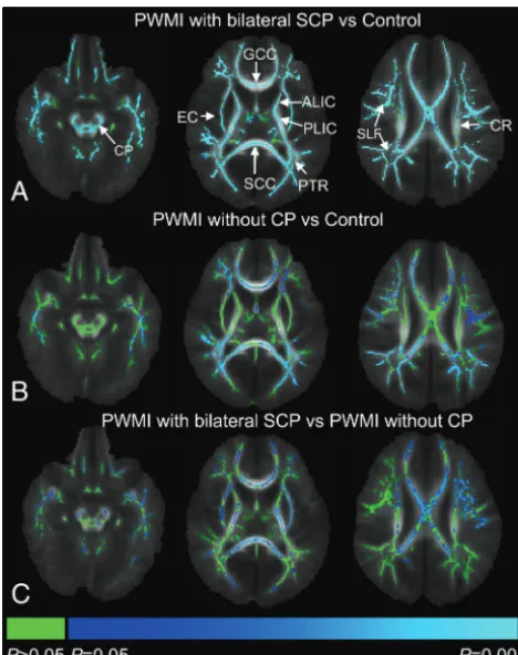
TBSS Analysis of Intergroup Differences
[image:3.594.55.375.49.366.2]showed significantly lower FA values than infants with PWMI without CP in the bilateral CST-CP, CST-IC, CST-CR, external capsule, GCC, and SCC (Fig 2).
AFQ Analysis of Intergroup Differences along Tracts
The tract profiles of infants with PWMI with bilateral SCP, infants with PWMI without CP, and controls are presented inFig 3. Ob-vious deceases in FA values in infants with PWMI with bilateral SCP were found along the CST, mainly located at the internal capsule level, while these changes in FA were not found in infants with PWMI without CP. The PTR, GCC, and SCC showed signif-icantly decreased FA values in both groups.
The receiver operating characteristic curves for the diagnostic performances of FA in different regions for differentiation of
in-fants with PWMI with bilateral SCP from those without CP are depicted in On-line Fig 2. The area under the curve was larger in the CST than that in the PTR, GCC, or SCC (Table 2). Among the 3 parts of the CST, an FA threshold of 0.53 at the internal capsule level had the highest area under the curve value, which demon-strated high specificity and sensitivity.
Correlation between Motor Function Scores and FA
The correlations between motor function scores and FA values along white matter tracts are presented in On-line Table 2. There were more significant correlations along the CST than along the PTR, GCC, and SCC (P⬍.05); and GMFCS levels were negatively correlated with FA values within the CST at the level of the inter-nal capsule (leftr⫽ ⫺0.80; rightr⫽ ⫺0.79). The Psychomotor Development Index scores showed a positive correlation with FA values within the CST at the level of internal capsule (leftr⫽0.69; rightr⫽0.53).
DISCUSSION
This study found that the spatial distribution of white matter al-terations was different between the 2 PWMI groups. Structural integrity was more severely damaged in more widespread white matter areas in infants with PWMI with bilateral SCP than in those without CP. Furthermore, injured CST was found only in infants with PWMI with bilateral SCP. The FA threshold of 0.53 in the CST at the internal capsule level was useful for the differ-entiation of infants with PWMI with SCP from those without CP, and it had significant correlations with motor functions.
DTI with TBSS analysis is a powerful approach that effectively reveals alterations in the main white matter tracts of the whole brain. Several studies have reported the presence of FA reduction within widespread white matter areas, especially in the central and posterior white matter, in infants with PWMI with SCP.10,12,22 However, knowledge of the spatial distribution characteristics of white matter lesions in infants with PWMI with and without CP is limited. In this study, the infants with PWMI with SCP had lower FA and more widespread distribution than those without CP. Additionally, the age distribution of patients here was different from that in previous studies.10,12,15,22We focused on 6- to 18-month-old infants with PWMI, which is the period before the definite diagnosis. Identification of the differences between in-fants with PWMI with SCP and those without CP during this time interval is beneficial for determining candidates at risk of SCP.
[image:4.594.54.531.66.142.2]In this study, the white matter tracts potentially involved in SCP were further analyzed using the AFQ. Previously, Hoon et al13had suggested that PTR injury altered the sensorimotor con-nections to the motor cortex and attenuated descending CST. However, our study showed that the PTR, SCC, and GCC were the Table 1: Demographics of infants with PWMI with bilateral spastic cerebral palsy, infants with PWMI without cerebral palsy, and controlsa
Infants with PWMI with SCP (n= 20)
Infants with PWMI
without CP (n= 19) Controls (n= 33)
PValues PWMI with SCP
vs PWMI without CP
PWMI with SCP vs Control
PWMI without CP vs Control
Sex (female/male) 8:12 6:13 11:22 .58 .62 .90
Gestational age (wk) 37.38⫾1.90 (34.57–41.29) 38.07⫾2.45 (34.00–41.43) 39.16⫾1.14 (37.00–41.00) .23 ⬍.01 .04 Age at MRI (mo) 11.74⫾2.13 (8.00–17.66) 11.56⫾2.99 (6.40–16.76) 11.06⫾2.38 (6.10–17.33) .83 .34 .49 Birth weight (kg) 2.95⫾0.47 (2.20–3.70) 3.06⫾0.60 (1.80–3.85) 3.38⫾0.50 (2.50–4.38) .50 .01 .04
a
Data in columns 2– 4 are mean and standard deviation (range).
[image:4.594.53.287.162.458.2]injured regions in both PWMI subgroups, whereas decreased FA in CST was observed only in infants with PWMI with SCP. These results indicate that PTR injury may not be directly responsible for SCP. The CST, as the major projectional motor tract situated in close proximity to the periventricular white matter region, is vulnerable in patients with PWMI.23CST lesions can interrupt the corticomotor circuit in executing movement and have also been shown to be involved in spasticity.24These findings suggest that CST lesions are a prerequisite for bilateral SCP in infants with
PWMI and provide clues for determining whether patients with PWMI will develop SCP.
[image:5.594.56.530.69.580.2]previous MR imaging studies for detecting the risk of CP.8,26The results obtained in this study suggest that CST at the internal capsule level is more suitable than the whole tract for the early diagnosis of SCP. Furthermore, the multiple regression results revealed that FA values of infants with PWMI were not correlated to age (On-line Table 3). This finding suggests that the FA thresh-old is applicable to the individual infants with PWMI between 6 and 18 months of age.
Apart from the determination of the risk of CP, the evaluation of the degree of motor impairment in CP can further facilitate the targeting of interventional strategies.27,28Consistent with previ-ous findings,23,24FA in the CST was significantly correlated with motor function scores. A strong correlation was found along the CST at the internal capsule level, which may be associated with the vulnerability of white matter in the internal capsule, where the descending motor axons are densely concentrated.29,30 More-over, there were more significant correlations along the CST than along the PTR, GCC, and SCC. It has been suggested that PTR lesions are responsible for the weakness in motor function in pa-tients with CP because of the role of PTR in visuospatial perfor-mance.24 The corpus callosum may play an important role in dexterity and bimanual motor coordination.31Overall, the results suggest that FA of the CST at the internal capsule level is suitable for assessing the motor function in infants with PWMI with SCP. Nevertheless, this study has several limitations. First, it ex-cluded severe PWMI because these cases accounted for a minority (3 subjects) of all the infants with PWMI. Moreover, it was rela-tively easy to determine the risk of CP in these infants. Second, unilateral SCP and nonspastic CP were not included due to the small sample sizes (2 with unilateral SCP and 1 with athetoid CP) and divergent MR imaging signs, such as asymmetric brain lesions in unilateral SCP and concurrent basal ganglia lesions in athetoid CP. Third, the FA threshold in the internal capsule of the CST was objectively extracted from an Automated Fiber Quantification. This threshold may be different from those obtained by other analysis methods, such as the ROI analysis. Fourth, this work tried to investigate the characteristics of DTI metric changes associated with SCP before the definite diagnosis. However, DTI was per-formed just several months ahead of the diagnosis of SCP. Whether SCP can be predicted by DTI in earlier periods needs further research. Finally, this was a retrospective study.
Prospec-tive research is required to test the accuracy of the early diagnosis of SCP.
CONCLUSIONS
CST lesions play a crucial role in motor impairment associated with SCP in infants with PWMI. FA in the CST at the internal capsule level could aid in the early diagnosis of SCP with high diagnostic accuracy.
ACKNOWLEDGMENTS
The authors are grateful to Drs Liming Liu and Jie Yue in the Department of Child Health Care for the diagnosis of CP.
REFERENCES
1. Bax M, Tydeman C, Flodmark O.Clinical and MRI correlates of cerebral palsy: the European Cerebral Palsy Study.JAMA2006;296: 1602– 08CrossRef Medline
2. Back SA, Rivkees SA.Emerging concepts in periventricular white matter injury.Semin Perinatol2004;28:405–14CrossRef Medline
3. Jauhari P, Singhi P, Sankhyan N, et al.A comparison of spastic di-plegia in term and preterm-born children.J Child Neurol2018;33: 333–39CrossRef Medline
4. Imamura T, Ariga H, Kaneko M, et al.Neurodevelopmental out-comes of children with periventricular leukomalacia.Pediatr Neo-natol2013;54:367–72CrossRef Medline
5. Woodward LJ, Anderson PJ, Austin NC, et al.Neonatal MRI to pre-dict neurodevelopmental outcomes in preterm infants.N Engl J Med2006;355:685–94CrossRef Medline
6. Hadders-Algra M.Early diagnosis and early intervention in cerebral palsy.Front Neurol2014;5:185CrossRef Medline
7. Morgan C, Crowle C, Goyen TA, et al.Sensitivity and specificity of General Movements Assessment for diagnostic accuracy of detect-ing cerebral palsy early in an Australian context.J Paediatr Child Health2016;52:54 –59CrossRef Medline
8. Novak I, Morgan C, Adde L, et al.Early, accurate diagnosis and early intervention in cerebral palsy: advances in diagnosis and treat-ment.JAMA Pediatr2017;171:897–907CrossRef Medline
9. Son SM, Park SH, Moon HK, et al.Diffusion tensor tractography can predict hemiparesis in infants with high risk factors.Neurosci Lett2009;451:94 –97CrossRef Medline
10. Lee JD, Park HJ, Park ES, et al.Motor pathway injury in patients with periventricular leucomalacia and spastic diplegia.Brain2011;134: 1199 –210CrossRef Medline
11. Skranes J, Vangberg T, Kulseng S, et al.Clinical findings and white matter abnormalities seen on diffusion tensor imaging in adoles-Table 2: Diagnostic performance of fractional anisotropy in different regions for differentiation of infants with PWMI with bilateral spastic cerebral palsy and those without cerebral palsy
PWMI with SCP (Mean and standard deviation)
PWMI without CP (Mean and standard deviation)
Area under the
Curve (95% CI) Threshold Sensitivity Specificity
CST-L 0.42⫾0.06 0.50⫾0.04 0.90 (0.75–0.98) 0.48 94% 72%
CST-R 0.43⫾0.06 0.50⫾0.04 0.85 (0.68–0.95) 0.49 93% 67%
PTR-L 0.34⫾0.06 0.35⫾0.05 0.55 (0.37–0.73) 0.27 27% 100%
PTR-R 0.33⫾0.07 0.34⫾0.05 0.54 (0.35–0.72) 0.36 67% 50%
GCC 0.41⫾0.05 0.45⫾0.05 0.72 (0.59–0.82) 0.46 87% 50%
SCC 0.43⫾0.11 0.49⫾0.09 0.62 (0.49–0.74) 0.40 40% 88%
CST-CP-L 0.36⫾0.08 0.42⫾0.04 0.74 (0.57–0.88) 0.40 69% 83%
CST-CP-R 0.36⫾0.06 0.42⫾0.06 0.77 (0.60–0.90) 0.40 73% 72%
CST-IC-L 0.48⫾0.06 0.59⫾0.05 0.95 (0.82–0.99) 0.53 94% 89%
CST-IC-R 0.48⫾0.07 0.58⫾0.05 0.88 (0.72–0.97) 0.53 93% 78%
CST-CR-L 0.43⫾0.09 0.46⫾0.09 0.60 (0.42–0.77) 0.49 81% 50%
CST-CR-R 0.47⫾0.10 0.48⫾0.11 0.52 (0.34–0.70) 0.59 100% 17%
[image:6.594.55.532.65.209.2]cents with very low birth weight.Brain2007;130:654 – 66CrossRef Medline
12. Ceschin R, Lee VK, Schmithorst V, et al.Regional vulnerability of longitudinal cortical association connectivity: associated with structural network topology alterations in preterm children with cerebral palsy.Neuroimage Clin2015;9:322–37CrossRef Medline
13. Hoon AH Jr, Stashinko EE, Nagae LM, et al.Sensory and motor deficits in children with cerebral palsy born preterm correlate with diffusion tensor imaging abnormalities in thalamocortical path-ways.Dev Med Child Neurol2009;51:697–704CrossRef Medline
14. Trivedi R, Agarwal S, Shah V, et al.Correlation of quantitative sen-sorimotor tractography with clinical grade of cerebral palsy. Neu-roradiology2010;52:759 – 65CrossRef Medline
15. Nagae LM, Hoon AH Jr, Stashinko EE, et al.Diffusion tensor imag-ing in children with periventricular leukomalacia: variability of in-juries to white matter tracts.AJNR Am J Neuroradiol 2007;28: 1213–22CrossRef Medline
16. Colver A, Fairhurst C, Pharoah PO.Cerebral palsy.Lancet2014;383: 1240 – 49CrossRef Medline
17. Rosenbaum P, Paneth N, Leviton A, et al.A report: the definition and classification of cerebral palsy April 2006.Dev Med Child Neurol
2007;109:8 –14Medline
18. Cote´ CJ, Wilson S; American Academy of Pediatrics; American Acad-emy of Pediatric Dentistry.Guidelines for monitoring and manage-ment of pediatric patients during and after sedation for diagnostic and therapeutic procedures: update 2016.Pediatrics2016;138. pii: e20161212CrossRef Medline
19. Smith SM, Jenkinson M, Woolrich MW, et al.Advances in functional and structural MR image analysis and implementation as FSL. Neu-roimage2004;23:S208 –19CrossRef Medline
20. Yeatman JD, Dougherty RF, Myall NJ, et al.Tract profiles of white matter properties: automating fiber-tract quantification.PLoS One
2012;7:e49790CrossRef Medline
21. Oishi K, Faria AV, Yoshida S, et al.Quantitative evaluation of brain development using anatomical MRI and diffusion tensor imaging. Int J Dev Neurosci2013;31:512–24CrossRef Medline
22. Arrigoni F, Peruzzo D, Gagliardi C, et al.Whole-brain DTI
assess-ment of white matter damage in children with bilateral cerebral palsy: evidence of involvement beyond the primary target of the anoxic insult.AJNR Am J Neuroradiol2016;37:1347–53CrossRef Medline
23. Resˇic´ B, Tomasovic´ M, Kuzmanic´-Sˇamija R, et al. Neurodevelop-mental outcome in children with periventricular leukomalacia. Coll Antropol008;32(Suppl 1):143– 47Medline
24. Scheck SM, Boyd RN, Rose SE.New insights into the pathology of white matter tracts in cerebral palsy from diffusion magnetic reso-nance imaging: a systematic review.Dev Med Child Neurol2012;54: 684 –96CrossRef Medline
25. Murakami A, Morimoto M, Yamada K, et al.Fiber-tracking tech-niques can predict the degree of neurologic impairment for periventricular leukomalacia.Pediatrics2008;122:500 – 06CrossRef Medline
26. Thayyil S, Chandrasekaran M, Taylor A, et al.Cerebral magnetic resonance biomarkers in neonatal encephalopathy: a meta-analy-sis.Pediatrics2010;125:e382–95CrossRef Medline
27. Kendall GS, Melbourne A, Johnson S, et al.White matter NAA/Cho and Cho/Cr ratios at MR spectroscopy are predictive of motor out-come in preterm infants. Radiology 2014;271:230 –38 CrossRef Medline
28. Melhem ER, Hoon AH Jr, Ferrucci JT Jr, et al.Periventricular leukomalacia: relationship between lateral ventricular volume on brain MR images and severity of cognitive and motor impairment. Radiology2000;214:199 –204CrossRef Medline
29. Jaspers E, Byblow WD, Feys H, et al.The corticospinal tract: a bio-marker to categorize upper limb functional potential in unilateral cerebral palsy.Front Pediatr2016;3:112CrossRef Medline
30. Rose J, Mirmiran M, Butler EE, et al.Neonatal microstructural de-velopment of the internal capsule on diffusion tensor imaging cor-relates with severity of gait and motor deficits.Dev Med Child Neu-rol2007;49:745–50CrossRef Medline