ORIGINAL RESEARCH
ADULT BRAIN
Lateral Posterior Choroidal Collateral Anastomosis Predicts
Recurrent Ipsilateral Hemorrhage in Adult Patients with
Moyamoya Disease
J. Wang, Y. Yang, X. Li, F. Zhou, Z. Wu, Q. Liang, Y. Liu, Y. Wang, S. Na, X. Chen, X. Zhang, and
B. Zhang
ABSTRACT
BACKGROUND AND PURPOSE: Choroidal collateral anastomosis is associated with hemorrhage recurrence in patients with Moyamoya disease. However, the relationship between recurrent ipsilateral hemorrhage and choroidal collateral anastomosis sub-types (anterior choroidal artery anastomosis, lateral posterior choroidal artery anastomosis, and medial posterior choroidal artery anastomosis) is unclear. This study aimed to assess this potential association in adult patients with Moyamoya disease.
MATERIALS AND METHODS:Patients angiographically diagnosed with Moyamoya disease who underwent conservative treatment between January 2008 and December 2018 were included in this retrospective study. Two readers assessed the angiographic images to identify choroidal collateral anastomosis subtypes, and Cox proportional hazard regression models were used to estimate the risk of recurrent hemorrhage associated with each subtype.
RESULTS:Thirty-nine patients (mean age ¼45.2 years) were included in this study. During 52.4 637.0 months of follow-up, recur-rent ipsilateral hemorrhage occurred in 48.7% (19/39) of patients. Patients with recurrecur-rent hemorrhage had a higher prevalence of choroidal collateral (94.8% versus 60.0%; P¼.02) and lateral posterior choroidal artery (78.9% versus 25.0%;P,.01) anastomoses than those without recurrent hemorrhage. Lateral posterior choroidal artery anastomosis was associated with recurrent hemor-rhage before (hazard ratio ¼6.66; 95% CI, 2.18–20.39; P,.01) and after (hazard ratio ¼5.78; 95% CI, 1.58–21.13;P,.01) adjust-ments were made for age, sex, and other confounding factors.
CONCLUSIONS:Choroidal collateral anastomosis is responsible for most cases of recurrent hemorrhage in adult patients with Moyamoya disease; lateral posterior choroidal artery anastomosis is a significant risk factor for these recurrent events.
ABBREVIATIONS:AChA ¼anterior choroidal artery; ChCA¼choroidal collateral anastomosis; HR¼hazard ratio; LPChA¼lateral posterior choroidal artery; MMD¼Moyamoya disease; MPChA¼medial posterior choroidal artery
M
oyamoya disease (MMD) is an uncommon but potentially catastrophic cerebrovascular disorder characterized by progressive occlusion in the terminal portion of the internal ca-rotid artery and its main branches within the circle of Willis.1Intracranial hemorrhage accounts for half of initial manifesta-tions of MMD in adult patients,2,3 and the recurrence rate of hemorrhage can be as high as 17.1% per year during the natural course of the disease.4,5 Patients with hemorrhage recurrence generally have poor outcomes such as life-long disabilities or even death.4Identifying risk factors for recurrent hemorrhage is therefore useful for clinical decision-making in terms of manage-ment and follow-up.
The occurrence of periventricular hemorrhage is a major clinical concern in adult patients with MMD who experience recurrent hemorrhage.6 In brain hemispheres with recurrent hemorrhage, the hemorrhage is more likely to occur around the posterior territory, particularly in the periventricular area around the atrium or the posterior portion of the body of the lateral ven-tricle.5,7 A previous postmortem study demonstrated that the subependymal collateral arteries supplying the above territories originate mainly from the lateral posterior choroidal arteries (LPChAs) but rarely from the anterior choroidal arteries
Received April 29, 2019; accepted after revision July 29.
From the Department of Neurosurgery (J.W., Z.W.), The Affiliated Changzhou No. 2 People's Hospital of Nanjing Medical University, Changzhou, China; Departments of Neurosurgery (J.W., Y.Y., Q.L., Y.W., S.N., X.C.) and Radiology (X.L., F.Z., X.Z., B.Z.), The Affiliated Drum Tower Hospital of Nanjing University Medical School, Nanjing, China; and Department of Neurosurgery (Y.L.), West China Hospital, Sichuan University, Chengdu, China.
J. Wang, Y. Yang, X. Li, F. Zhou, and B. Zhang contributed equally to this study. This work was supported by the Natural Science Foundation of China
(81720108022, B.Z.) and the Social Development Project of Science and Technology in Jiangsu Province (BE2016605, BE201707, B.Z.).
Please address correspondence to Bing Zhang, MD, PhD, Department of Radiology, The Affiliated Drum Tower Hospital of Nanjing University Medical School, Nanjing, China, 210008; e-mail: [email protected]
Indicates open access to non-subscribers at www.ajnr.org
http://dx.doi.org/10.3174/ajnr.A6208
(AChAs) or medial posterior choroidal arteries (MPChAs).8 Therefore, we suspect that the LPChAs may play a key role in the occurrence of recurrent hemorrhage. Choroidal collateral anastomosis (ChCA) has previously been shown to be associ-ated with recurrent hemorrhage in this patient population.5,9,10 However, the relationship between the subtypes of ChCA and recurrent ipsilateral hemorrhage is still unclear.
In this retrospective study, we therefore sought to assess the association between the subtypes of ChCA and recurrent ipsilat-eral hemorrhage in adult patients with MMD.
MATERIALS AND METHODS
Study PopulationThis study was approved by The Affiliated Drum Tower Hospital of Nanjing University Medical School with a waiver of informed consent. Consecutive patients with MMD who had experienced initial intracranial hemorrhage and received conservative treatment (no surgical intervention) and clinical follow-up between January 2008 and December 2018 were recruited for this retrospective study. We enrolled as many patients as possible with.5 years of clinical follow-up for nonrecurrent hemorrhage controls. Considering that the main purpose of our study was to evaluate the association between the ChCA subtypes and recurrent ipsilateral rhage, the factors associated with non-MMD-related hemor-rhage and influencing the target vessel evaluation were excluded in the present study. All cases of MMD had bilateral involvement and were diagnosed via angiography according to the guidelines proposed by the Ministry of Health, Labor, and Welfare of Japan.11 Patients with autoimmune disease, meningitis, brain tumor, Down syndrome, neurofibromatosis type 1, or a history of head irradiation were excluded from the study. We also excluded patients who met any of the fol-lowing 3 conditions: 1) age older than 65 years12; 2) the pres-ence of factors influencing the evaluation of recurrent hemorrhage such as bleeding diathesis, uncontrolled diabetes mellitus (fasting blood glucose level of .300 mg/dL), or uncontrolled hypertension (systolic pressure of.180 mm Hg and/or diastolic pressure of.110 mm Hg); or 3) a history of intracranial hemorrhage. The occurrence of recurrent hemor-rhage was recorded if there was an acute neurologic symptom during follow-up with a corresponding new intracranial hem-orrhage on brain imaging. Demographic and clinical charac-teristics for all patients were collected from the medical records.
Imaging Techniques
For the initial hemorrhagic event, a cerebrovascular DSA exami-nation was performed on an x-ray scanner (Allura Xper FD20/ 2D; Philips Healthcare, Best, the Netherlands); this examina-tion included selective common, internal, and external carotid artery arteriography on both sides and vertebral arteriography on at least 1 side. The imaging parameters for this examination were as follows: 6 frames/s, injection pressure =300 psi/kg, and contrast medium administered at a rate of 3 mL/s. When a recurrent hemorrhage occurred, non-contrast-enhanced CT of the brain was performed to identify the intracranial
hemorrhagic site. When diffuse ventricular hemorrhage occurred, SWI examinations were conducted within 2 months of the hemorrhagic event, with all scans obtained on a 3T MR imaging scanner (uMR 770; United Imaging Healthcare; Shanghai) with a 24-channel phased array head and neck coil. The MR images were acquired using the following parameters: TR/TE¼24.2/15 ms, flip angle¼15°, FOV¼190190 mm2, section thickness¼ 1.2 mm, voxel size¼0.60.61.2 mm3, and acquisition time¼7 minutes 35 seconds.
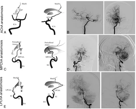
Definitions and Analysis of Angiographic Variables The definition of ChCA provided by Funaki et al9was adapted to the present study. For this study, the subtypes of ChCA were categorized into AChA anastomosis, LPChA anastomosis, and MPChA anastomosis.9AChA anastomosis is defined as anasto-mosis between the extreme dilation and extension of the AChA with sudden deviation from the shape of the lateral ventricle at its peripheral portion to connect the medial end of the medullary ar-tery. LPChA anastomosis refers to the extreme extension of the LPChA beyond the atrium of the lateral ventricle to reach the body of the lateral ventricle. MPChA anastomosis is defined as the extreme extension of the MPChA beyond the level of the peri-callosal artery to the corpus callosum. Schematic illustrations and representative examples of each subtype are shown in Fig 1. Other variables such as Suzuki stages1(stages I–VI), involvement of the posterior cerebral arteries (Mugikura stage II–IV),13 tha-lamic collateral anastomosis,9the presence of an intracranial an-eurysm, and the presence of a fetal posterior communicating artery at the time of initial hemorrhage were also evaluated. The initial angiographic images were reviewed in consensus by 2 neu-roradiologists, both with.5 years’experience in neurovascular imaging and both blinded to the brain images. A third investiga-tor with.10 years’experience in neurovascular imaging resolved any discrepancies.
Follow-Up
After the initial hemorrhagic event was investigated and conserv-ative treatment was initiated, all patients underwent follow-up in the outpatient clinic every 3–6 months so that cases of hemor-rhage recurrence could be identified. When hemorhemor-rhage reoc-curred, the site of the hemorrhage was determined on CT within 1 week after the onset of the event. When diffuse ventricular hem-orrhage occurred, the presumed origin of the recurrent hemor-rhage was identified by SWI within 2 months of the event. The interval between the initial event and hemorrhage recurrence was also recorded.
Reproducibility
All angiographic data were interpreted by the 2 raters twice, with a 1-month time interval between sessions to minimize memory bias. Interrater and intrarater agreement values for the subtypes of ChCA were calculated.
Statistical Analysis
hemorrhage and hemispheres with and without recurrent hemor-rhage in terms of baseline characteristics. Univariate and multi-variate Cox proportional hazard regression models were used to estimate the risk of recurrent ipsilateral events for each ChCA subtype. Interrater and intrarater agreement values for ChCA subtype evaluation were calculated using the unweighted Cohenk. AP value,.05 was considered sig-nificant. All statistical analyses were performed using SPSS 22.0 (IBM, Armonk, New York).
RESULTS
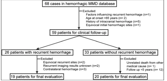
From January 2008 to December 2018, a total of 68 patients pre-sented with hemorrhagic MMD and underwent conservative treatment. Of these 68 patients, 9 (13.2%) were excluded for the following reasons: uncontrolled hypertension and diabetes melli-tus (n¼1), age older than 65 years (n¼2), history of intracranial
hemorrhage (n¼5), and equivocal initial hemorrhagic sites (n¼1). During the follow-up period, another 20 (29.4%) patients were excluded for the following reasons: equivocal recurrent hemorrhagic sites (n¼2), follow-up images of recurrent hemor-rhage unknown (n¼2), presenting with recurrent hemorrhage in the contralateral hemispheres (n¼3), unrelated death from other medical causes (liver tumor, n¼1), and follow-up period of ,5 years (patients without recurrent hemorrhage,n¼12). The remaining 39 (57.4%) patients (29 women; mean age at diag-nosis¼45.269.1 years) met the eligibility criteria for the final analysis (Fig 2). Of these 39 patients, 2 (5.1%) were smokers, 13 (33.3%) had hypertension, 8 (20.5%) had dyslipidemia, 3 (7.7%) had diabetes mellitus, and 3 (7.7%) had a history of is-chemic stroke (defined as symptomatic infarctions confirmed on DWI). None of the study patients were treated with antipla-telets or anticoagulants. During 52.4 6 37.0 months (range, 1–114 months) of follow-up, 48.7% (19/39) of patients
FIG 1. Schematic illustrations and angiographicfindings from representative cases of each subtype of choroidal collateral anastomosis.AandB,
Anterior-posterior and lateral right carotid artery angiograms show a dilated anterior choroidal artery extending beyond the lateral ventricle to the cortex (arrows).CandD, Anterior-posterior and lateral right vertebral artery angiograms show a medial posterior choroidal artery extending beyond the level of the pericallosal artery (arrows) to the corpus callosum.EandF, Anterior-posterior and lateral left vertebral artery angio-grams show a lateral posterior choroidal artery extending beyond the body of the lateral ventricle to the cortex (arrows). MedA indicates the medullary artery; P1 and P2, the proximal portion of posterior cerebral artery; BA, the basilar artery.
[image:3.585.56.534.47.430.2]experienced recurrent ipsilateral hemorrhage. Of the baseline clinical risk factors assessed, only the number of subarachnoid hemorrhages in patients with recurrent events was signifi-cantly higher than in those without recurrent events (21.1% versus 0.0%;P¼.05;Table 1).
Intracranial Hemorrhage
In 39 hemispheres, the initial hemorrhagic sites were in the sub-ependymal area of the lateral ventricle in 21 hemispheres (53.8%), the insular lobe in 5 hemispheres (12.8%), the temporal lobe in 3 hemispheres (7.7%), the basal ganglia in 2 hemispheres (5.1%), the occipital lobe in 2 hemispheres (5.1%), the corpus cal-losum in 1 hemisphere (2.6%), the frontal lobe in 1 hemisphere (2.6%), and the subarachnoid in 4 hemispheres (10.2%). Of the 39 hemispheres with intracranial hemorrhage, 11 (28.2%) dem-onstrated recurrent hemorrhage at the initial hemorrhage site and 8 (20.5%) demonstrated recurrent hemorrhage at a different site in the same hemisphere. Comparisons of imaging features in hemispheres with and without recurrent hemorrhage are shown inFig 3.
Analysis of Angiographic Variables
Of the 39 cases in the study, none demonstrated intracranial lesions at Suzuki stage I, one (2.6%) demon-strated lesions at stage II, nineteen (48.7%) demonstrated lesions at stage III, five (12.8%) demonstrated lesions at stage IV, twelve (30.8%) demon-strated lesions at stage V, and 2 (5.1%) demonstrated lesions at stage VI. In addition, 11 (28.2%) intracra-nial arteries were found to have ste-notic-occlusive lesions involving the posterior cerebral arteries. The fetal posterior communicating artery and thalamic anastomosis were identi-fied in 33.3% (13/39) and 15.4% (6/39) of cases, respectively. An in-tracranial aneurysm was identified in those 4 patients presenting with subarachnoid hemorrhage, includ-ing the posterior cerebral artery in 2 patients, the lenticulostriate ar-tery in 1 patient, and the middle meningeal artery in 1 patient.
A ChCA was identified in 76.9% (30/39) of patients, including AChA in 13 patients, LPChA in 20 patients, and MPChA in 14 patients. Com-pared with hemispheres without hemorrhage recurrence, hemispheres with hemorrhage recurrence demon-strated a higher prevalence of ChCA (94.8% versus 60.0%; P ¼ .02), LPChA anastomosis (78.9% versus 25.0%; P , .01), and intracranial aneurysms (21.1% versus 0.0%;P¼.03). A representative case demonstrating LPChA anastomosis and recurrent ipsilateral hemorrhage is shown inFig 4. No other significant differences were noted in baseline angiographic characteristics between hemispheres with and those without recurrent hemorrhage (Table 2).
Association between Subtypes of ChCA and Recurrent Ipsilateral Hemorrhage
Table 3 summarizes the radiographic characteristics of the 19 hemispheres in which recurrent hemorrhage was seen. In 14 hemispheres, ChCA was considered responsible for the recur-rence because the recurrent hemorrhage occurred in the hemi-sphere containing a ChCA and corresponded to the distribution of the choroidal arteries: Five stemmed from the AChA, 7 stemmed from the LPChA, and 2 stemmed from the MPChA.
In univariate Cox regression analysis, a significant association was demonstrated between LPChA anastomosis and hemorrhage recurrence (hazard ratio [HR]¼6.66; 95% CI, 2.18–20.39;P, .01). In multivariate Cox regression analysis, after adjustments
[image:4.585.54.381.51.211.2]FIG 2. Flowchart of patient recruitment.
Table 1: Baseline characteristics in patients with MMD with and without recurrent ipsilateral hemorrhage
Characteristic
Patients without Recurrent Hemorrhage
(n¼20)
Patients with Recurrent Hemorrhage
(n¼19) PValue
Women (No.) (%) 15 (75.0) 14 (73.7) .93
Age (mean) (yr) 43.7610.6 46.967.0 .27
Smokers (No.) (%) 1 (5.0) 1 (5.3) ..99
Concurrent disease (No.) (%)
Hypertension 8 (40.0) 5 (26.3) .37
Dyslipidemia 4 (20.0) 4 (21.1) ..99
Diabetes mellitus 2 (10.0) 1 (5.3) ..99
History of ischemia (No.) (%) 1 (5.0) 2 (10.5) .61
Hemorrhagic type (No.) (%)
IVH 13 (65.0) 13 (68.4) .82
Only IVH 6 (30.0) 10 (52.6) .20
ICHþIVH 7 (35.0) 3 (15.8) .46
SAH 0 (0.0) 4 (21.1) .05
Only SAH 0 (0.0) 1 (5.3) .49
SAHþIVH 0 (0.0) 3 (15.8) .27
ICH 7 (35.0) 2 (10.5) .13
[image:4.585.57.383.271.466.2]were made for age, sex, a history of ischemia, Suzuki stage.III, involvement of the posterior cerebral arteries, thalamic anasto-mosis, the fetal posterior communicating artery, and intracranial aneurysm, the association between LPChA anastomosis and hemorrhage recurrence remained significant (HR¼ 5.78; 95% CI, 1.58–21.13;P,. 01) (Table 4).
Reproducibility
Thekvalues for intrarater agreement in the identification of AChA, MPChA, and LPChA were 0.84, 0.71, and 0.69, respectively. Thek values for interrater agreement in the identification of AChA, MPChA, and LPChA were 0.76, 0.66, and 0.61, respectively.
DISCUSSION
In this study, we found that recurrent ipsilateral hemorrhage was common among adult patients with MMD, and ChCA was re-sponsible for most cases of hemorrhage recurrence in these patients. In addition, LPChA anastomosis was found to be an in-dependent predictor of recurrent ipsilateral hemorrhage. Our findings suggest that serial imaging follow-up of LPChA anasto-mosis may provide additional information for risk stratification in patients with MMD.
Previous research has suggested that MMD-related intracra-nial hemorrhage may differ from primary intracraintracra-nial hemor-rhage in terms of location: MMD-related hemorhemor-rhage is more likely to present as intraventricular hemorrhage with or without intrace-rebral hemorrhage.6 One study showed that the presence of intra-ventricular hemorrhage was signifi-cantly correlated with the occurrence of recurrent hemorrhage in patients with MMD.14 Similarly, our study demonstrated that .50% of hemor-rhage recurrence events presented as intraventricular hemorrhages; how-ever, this result did not reach statisti-cal significance. These differences in results between studies may be par-tially due to the different exclusion criteria used in different studies.
In the current study, ChCA was found to be more prevalent in hemi-spheres with recurrent hemorrhage among patients with MMD, findings consistent with previous results.5,9We also found that the subtypes of ChCA were more frequently observed in the hemispheres with recurrent hemor-rhage, though this difference was not significant for AChA or MPChA anas-tomosis. The recurrent hemorrhagic sites were distributed mainly in the
FIG 3. Topographic analysis showing the distribution of initial and recurrent hemorrhagic sites.A,
Topographic analysis of initial hemorrhagic sites for those with (black dots) and those without (white dots) recurrent hemorrhage.B, Another topographic analysis shows the distribution of recurrent hemorrhagic sites (black dots). Four patients with recurrent hemorrhage attributable to intracranial aneurysm rupture are not shown in this analysis.
FIG 4. A 50-year-old man experienced a recurrent hemorrhage in the ipsilateral hemisphere.A, CT image indicates the initial hemorrhage in the
posterior portion of the body of the left lateral ventricle.B, CT image obtained 49 months later reveals a recurrent hemorrhage in the initial hemorrhagic site.C. Left anterior-posterior carotid artery angiogram obtained at baseline demonstrates no obvious anterior choroidal anasto-mosis from the internal carotid artery. Anterior-posterior (D) and lateral (E) views of the left vertebral angiogram obtained at baseline reveal the typicalfinding of lateral posterior choroidal anastomosis responsible for recurrent hemorrhage (black arrows).
[image:5.585.57.374.210.466.2] [image:5.585.54.532.550.673.2]periventricular area around the atrium or the posterior portion of the body of the lateral ventricle, suggesting that fragile choroidal collateral vessels around the periventricular area are mainly derived from the posterior circulation, particularly for LPChA.
LPChA anastomosis was shown to be significantly correlated with recurrent ipsilateral hemorrhage in this study, suggesting that LPChA anastomosis plays a key role in the occurrence of recurrent hemorrhage in patients with MMD. However, the mechanism of this correlation is not fully understood. We pro-pose 2 possible mechanisms to explain this relationship.
The first potential mechanism involves persistent hemody-namic stress of the collateral vessels in different periventricular
regions. Yamamoto et al15demonstrated that the Moyamoya col-lateral vessels longitudinally shift from the anterior to posterior circulation during disease progression. In the current study, we found that recurrent hemorrhage was rarely observed in the insular lobe and basal ganglia but was more prevalent in the atrium and the posterior portion of the body of the lateral ventricle. The collat-eral anastomoses around the insular lobe and basal ganglia are mainly derived from the single blood supply of the anterior circula-tion, whereas the collateral anastomoses around the atrium and the posterior portion of the body of the lateral ventricle derive from the multiple blood supplies of both the anterior and posterior circulations. Persistent hemodynamic stress is thus added from both the anterior and posterior circula-tions, a potential trigger of recurrent hemorrhage.
[image:6.585.55.377.221.417.2]The second potential mechanism is related to the development of ventricular microaneurysms in the LPChA. A previous study using 7T TOF MRA found that a high number of patients with MMD had ventricular microaneurysms in the periventricular region.16In the current study, excluding cases of intracranial aneurysm rupture, recurrent hemorrhage occurred in the initial hemorrhagic sites in 46.7% (7/15) of hemispheres. The coexisting anterior and posterior choroidal anastomoses in MMD indicate that ventricular micro-aneurysms may be present in choroidal collateral vessels, particularly in the LPChA. Future studies using high-reso-lution MR imaging to characterize the
Table 2: Baseline variables in hemispheres with and without recurrent hemorrhage in adult patients with MMD
Variables
Hemispheres without Recurrent Hemorrhage
(n= 20)
Hemispheres with Recurrent Hemorrhage
(n= 19) PValue Suzuki stage (No.) (%)
I 0 (0.0) 0 (0.0)
II 1 (5.0) 0 (0.0)
III 10 (50.0) 9 (47.4)
IV 3 (15.0) 2 (10.5)
V 5 (25.0) 7 (36.8)
VI 1 (5.0) 1 (5.3)
Suzuki stage.III (No.) (%) 9 (45.0) 10 (52.6) .63
Involved PCA (No.) (%) 4 (20.0) 7 (36.8) .30
Fetal PcomA (No.) (%) 7 (35.0) 6 (31.6) .82
IA (No.) (%) 0 (0.0) 4 (21.1) .03
TC anastomosis (No.) (%) 2 (10.0) 4 (21.1) .41
ChCA subtype (No.) (%) 12 (60.0) 18 (94.8) .02
AChA anastomosis 5 (25.0) 8 (42.1) .26
LPChA anastomosis 5 (25.0) 15 (78.9) ,.01
MPChA anastomosis 5 (25.0) 9 (47.4) .15
Note:—PCA indicates posterior cerebral artery; PcomA, posterior communicating artery; IA, intracranial aneurysm; TC, thalamic collateral.
Table 3: Clinical features of adult patients with MMD who experienced recurrent ipsilateral hemorrhage
Case (yr)/SexAge
Hemorrhagic Site Interval
(mo)
Target ChCA Site of Initial Hemorrhage Site of Recurrent Hemorrhage AChA LPChA MPChA
1 46/M Right body of lateral ventricle Right body of lateral ventricle 5 þ
2 43/M Left atrium Left atrium 10 þ
3 44/F Right anterior body of lateral ventricle Right temporal horn of lateral ventricle 2 þ 4 42/F Right posterior body of lateral ventricle Right posterior body of lateral ventricle 10 þ 5 50/M Left posterior body of lateral ventricle Left posterior body of lateral ventricle 49 þ 6 63/F Left occipital horn of lateral ventricle Left occipital horn of lateral ventricle 1 þ 7 46/M Right frontal horn of lateral ventricle Right splenium of corpus callosum 4 þ
8 51/F Left posterior horn of lateral ventricle Left atrium 12 þ
9 51/F Left atrium Left atrium 5 þ
10 45/F Left temporal horn of lateral ventricle Left posterior body of lateral ventricle 58 þ
11 50/F Left insular lobe Left insular lobe 47 þ
12 58/F Right anterior body of lateral ventricle Right body of corpus callosum 14 þ
13 36/F Left body of lateral ventricle Left atrium 59 þ
14 32/F Right posterior horn of lateral ventricle Right temporal horn of lateral ventricle 1 þ
15a 42/F Left temporal-occipital lobe Left putamen 25
16b 56/M Left perimesencephalic subarachnoid Left perimesencephalic subarachnoid 39 17b 45/F Right insular lobe and subarachnoid Right insular lobe and subarachnoid 1 18b 57/F Left quadrigeminal cistern Left quadrigeminal cistern 57 19b 42/F Left temporal lobe and subarachnoid Left temporal lobe and subarachnoid 2 Note:—þindicates present.
[image:6.585.53.535.483.703.2]arteriopathy of the LPChA in patients with MMD are warranted. This study is limited by its retrospective nature. In addition, because of the rarity of nonsurgical intervention in patients with hemorrhagic MMD and the large number of patients excluded, this study is limited by its small sample size. Furthermore, inter-rater and intrainter-rater agreement values for the identification of LPChA were low due to technical flaws in angiographic imaging of some unilateral vertebral arteries and the use of a weak contrast agent for angiography in some cases. The relationship between subtypes of ChCA and recurrent hemorrhage was determined only via analysis of the initial angiographic images, with no follow-up angiographic validation. Therefore, the development of ChCA sub-types was unclear in our study. Nevertheless, this study remains one of the first to analyze the association between ChCA subtypes and recurrent ipsilateral hemorrhage in patients with MMD. Considering the unique collateral anastomosis seen in patients with MMD, multicenter prospective studies are needed to deter-mine the natural course of these collateral vessels. MR imaging may be able to be used for serial imaging follow-up of LPChA anastomosis in patients with MMD. In a recent study, sliding thin-slab maximum-intensity-projection coronal MRA images were found to provide reliable follow-up of periventricular anastomosis.17 Future studies are warranted to assess surgical revascularization in the area of the LPChA in nonsurgical patients and serial imaging fol-low-up of LPChA changes in surgical patients with MMD.
CONCLUSIONS
Recurrent ipsilateral hemorrhage is common in the natural course of hemorrhagic MMD, and LPChA anastomosis is associated with recurrent hemorrhage in this patient population. In light of these findings, LPChA anastomosis may serve as a marker of the risk of recurrent ipsilateral hemorrhage in patients with MMD.
ACKNOWLEDGMENTS
We thank Zhongzhi Jia, MD, The Affiliated Changzhou No. 2 People’s Hospital of Nanjing Medical University, Changzhou, China, and Megan Griffiths, scientific writer, Cleveland, Ohio, for help with revising the manuscript.
REFERENCES
1. Suzuki J, Takaku A.Cerebrovascular“Moyamoya”disease: disease showing abnormal net-like vessels in base of brain.Arch Neurol 1969;20:288–99CrossRef Medline
2. Kuroda S, Houkin K. Moyamoya disease: current concepts and future perspectives.Lancet Neurol2008;7:1056–56CrossRef Medline 3. Takahashi JC, Miyamoto S.Moyamoya disease: recent progress and outlook.Neurol Med Chir (Tokyo)2010;50:824–32CrossRef Medline
4. Kobayashi E, Saeki N, Oishi H, et al.Long-term natural history of hemorrhagic Moyamoya disease in 42 patients. J Neurosurg 2000;93:976–80CrossRef Medline
5. Funaki T, Takahashi JC, Houkin K, et al.High rebleeding risk asso-ciated with choroidal collateral vessels in hemorrhagic Moyamoya disease: analysis of a nonsurgical cohort in the Japan Adult Moyamoya trial.J Neurosurg2018 Feb 1:1–8 [Epub ahead of print] CrossRef Medline
6. Nah HW, Kwon SU, Kang DW, et al.Moyamoya disease-related versus primary intracerebral hemorrhage: [corrected] location and outcomes are different [erratum in Stroke 2013;44:e119].Stroke 2012;43:1947–50CrossRef Medline
7. Takahashi J, Funaki T, Houkin K, et al; JAM Trial Investigators. Significance of the hemorrhagic site for recurrent bleeding: pre-specified analysis in the Japan Adult Moyamoya Trial. Stroke 2016;47:37–43CrossRef Medline
8. MarinkovićS, Gibo H, FilipovićB, et al.Microanatomy of the sub-ependymal arteries of the lateral ventricle. Surg Neurol 2005; 63:451–58; discussion 458Medline
9. Funaki T, Takahashi JC, Houkin K, et al.Angiographic features of hemorrhagic moyamoya disease with high recurrence risk: a supplementary analysis of the Japan Adult Moyamoya Trial. J Neurosurg2018;128:777–84CrossRef Medline
10. Funaki T, Takahashi JC, Houkin K, et al.Effect of choroidal collat-eral vessels on de novo hemorrhage in Moyamoya disease: analysis of nonhemorrhagic hemispheres in the Japan Adult Moyamoya Trial.J Neurosurg2019 Feb 8:1–7 [Epub ahead of print]CrossRef Medline
11. Research Committee on the Pathology and Treatment of Spontaneous Occlusion of the Circle of Willis; Health Labour Sciences Research Grant for Research on Measures for Infractable Diseases.Guidelines for diagnosis and treatment of Moyamoya disease (spontaneous occlusion of the circle of Willis). Neurol Med Chir (Tokyo) 2012;52:245–66CrossRef Medline
12. Miyamoto S, Yoshimoto T, Hashimoto N, et al.Effects of extracra-nial-intracranial bypass for patients with hemorrhagic Moyamoya disease: results of the Japan Adult Moyamoya Trial. Stroke 2014;45:1415–21CrossRef Medline
13. Mugikura S, Takahashi S, Higano S, et al.Predominant involvement of ipsilateral anterior and posterior circulations in Moyamoya dis-ease.Stroke2002;33:1497–500CrossRef Medline
14. Kim KM, Kim JE, Cho WS, et al.Natural history and risk factor of recurrent hemorrhage in hemorrhagic adult Moyamoya disease.
Neurosurgery2017;81:289–96CrossRef Medline
15. Yamamoto S, Hori S, Kashiwazaki D, et al.Longitudinal anterior-to-posterior shift of collateral channels in patients with Moyamoya disease: an implication for its hemorrhagic onset. J Neurosurg 2019;130:884–90CrossRef Medline
16. Matsushige T, Kraemer M, Schlamann M, et al.Ventricular microaneur-ysms in Moyamoya angiopathy visualized with 7T MR angiography.
AJNR Am J Neuroradiol2016;37:1669–72CrossRef Medline
[image:7.585.49.533.68.138.2]17. Miyakoshi A, Funaki T, Takahashi JC, et al.Restoration of periven-tricular vasculature after direct bypass for Moyamoya disease: intra-individual comparison.Acta Neurochir (Wien)2019;161:947– 54CrossRef Medline
Table 4: Univariate and multivariate adjustedaanalyses of the association between ChCA subtypes and recurrent ipsilateral
hemorrhage in adult patients with MMD
ChCA Subtype
Presence of Recurrent Hemorrhage
Univariate Analysis Multivariate Analysis
HR 95% CI PValue HR 95% CI PValue
AChA anastomosis 1.77 0.71–4.41 .22 8.23 1.41–48.13 .02
MPChA anastomosis 2.14 0.86–5.29 .10 3.43 0.80–14.80 .10
LPChA anastomosis 6.66 2.18–20.39 ,.01 5.78 1.58–21.13 ,.01
a
Multivariate analysis was adjusted for confounding factors including age, sex, a history of ischemia, Suzuki stage.III, involvement of posterior cerebral arteries, thalamic anastomosis, fetal posterior communicating artery, and intracranial aneurysm.