Neuroendocrine Differentiation in the Progression of
Prostate Cancer: An Update on Recent Developments
Valérie Perrot
Laboratory M.E.R.C.I.-EA 3829, Institute of Research and Biomedical Innovation (IRIB), University of Rouen, Rouen, France Email: valerie.perrot@univ-rouen.fr
Received July 31, 2012; revised August 30, 2012; accepted September 10, 2012
ABSTRACT
Neuroendocrine (NE) differentiation, either benign or malignant, is the hallmark of prostate cancer (PCa). Clusters of malignant NE cells are found in most prostate cancer cases. NE differentiation is among the non-mutually exclusive theories proposed to explain the progression to androgen independence of PCa. NE differentiation is usually associated with an increased aggressivity and invasiveness of prostate tumors and a poor prognosis. This review aims to present an overview of current knowledge on neuroendocrine differentiation in PCa to improve our understanding of tumour pro-gression and androgen independence. The NE component represents an important therapeutic axis. Development of new generation of drugs that selectively target NE-like cells may lead to the development of new therapeutic modalities for advanced and hormone-refractory PCa.
Keywords: Prostate Cancer; Hormonal Therapy; Neuroendocrine; Neuroendocrine Differentiation; Neuropeptides; Chromogranin A; Tumorigenesis; Adenocarcinoma
1. Introduction
Prostate cancer (PCa) is a leading cause of cancer-related deaths among men in the Western countries. Its incidence is increasing annually worldwide due to better and earlier detection and also because of general aging of the world’s population [1]. PCa tumours are heterogenous, most men harbour slow-growing tumours while others have neoplasms rapidly progressing to metastatic disease [2]. When PCa metastasizes, the patient 5-year survival rate drops to near 30% from virtually 100% when disease remains localized (confined to primary site) or regional (spread to regional lymphnodes) [3]. PCa depends on
androgens in the early stages. Therefore, androgen abla-tion is the most common therapy, aiming to deprive the androgen-receptive cells of their growth stimulus. Al-though most patients respond initially to this treatment, the tumour eventually recurs and enters an androgen-ind- ependent stage for which treatment options are few and generally ineffective [4].
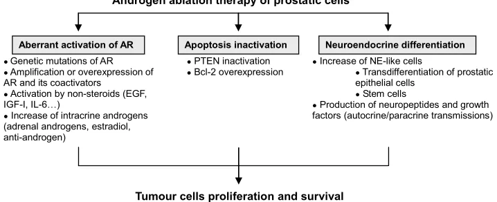
A lot of progress has been made in understanding the mechanisms which drive the development and the pro-gression of PCa, and in particular factors leading to the development of androgen independence (Figure 1). In this regard, evidence has emerged that androgen-resistant
Androgen ablation therapy of prostatic cells
Apoptosis inactivation Neuroendocrine differentiation Aberrant activation of AR
● PTEN inactivation
● Bcl-2 overexpression
● Genetic mutations of AR
● Amplification or overexpression of AR and its coactivators
● Activation by non-steroids (EGF, IGF-I, IL-6…)
● Increase of intracrine androgens (adrenal androgens, estradiol, anti-androgen)
● Increase of NE-like cells
● Transdifferentiation of prostatic epithelial cells
● Stem cells
● Production of neuropeptides and growth factors (autocrine/paracrine transmissions)
[image:1.595.119.478.563.709.2]Tumour cells proliferation and survival
PCa are often associated with tumour enrichment in neuro- endocrine (NE) cells. In the past years a growing body of literature showed increased interest in the NE differentia- tion phenomenon that occurs in PCa. In contrast, less information is available on the specific role that NE cells play in the pathophysiology and prognosis of PCa. This review aims to present an overview of the current know- ledge on NE cells and differentiation in PCa to improve our understanding of tumour progression and androgen independence. Better knowledge of PCa initiation and progression will help for developing new strategies for tumour prevention and treatment.
2. Neuroendocrine Cells of the Normal
Prostate: Origin, Localisation and
Function
Normal prostatic tissue is composed of stromal and epithelial compartments. The stroma not only acts as a supporting tissue, but also participates to the endocrine and paracrine microenvironment that controls prostatic epithelium growth and differentiation.
Three types of epithelial cells are found in adult pro- static gland: secretory, basal and neuroendocrine (NE). Luminal secretory cells are the most abundant and re- quire androgens for growth and survival. They synthesize and secrete products of the seminal plasma, including prostatic-specific antigen (PSA). Basal cells are the prin- cipal epithelial proliferating cell type and are androgen- insensitive. They give rise to pluripotent cells, which are responsive to androgens, and can differentiate into basal cells, differentiated luminal cells, and possibly also NE cells. NE cells are ubiquitously present throughout the body and constitute a minor epithelial cell population widely distributed in normal prostatic acini and ducts. NE cells do not express the proliferation associated Ki 67 and MIB-1 antigens [5]. These post-mitotic cells are highly specialized cells, which share structural, func- tional and metabolic properties with neurons [6]. NE cells do not express androgen receptors [7,8], suggesting that they are androgen-insensitive [9].
The origin of NE cells in normal prostate is still under debate. The fact that NE cells do not express cytokeratin, a basal cell layer marker, suggests that they originate differently from other prostatic epithelial cells. NE cells may be derived either from undifferentiated basal cells of the prostatic epithelium [10] or represent an independent cell lineage derived from a neurogenic origin [11]. Fur- ther investigations will be necessary to clearly establish the origin of NE prostatic cells.
Histological studies revealed that in normal prostate gland, NE cells exhibit two distinct morphologies: 1) open cells with extensions at their apex that connect with the lumen, and 2) closed cells with dendritic-like proc- esses that extend between adjacent cells and do not have
contact with the lumen [12]. Both subpopulations of NE cells participate to a communication network, in particu- lar with the prostatic stroma, through their various secre- tory products.
In normal prostate, NE cells regulate prostatic growth, differentiation and secretion in an androgen-independent manner. NE cells contain neurosecretory granules rich in various peptide hormones and biogenic amines such as calcitonin [13], parathyroid hormone-related protein (PTHrP) [14], NE markers like chromogranins (CgA, CgB) [15] and neuron-specific enolase (NSE), serotonin [16], bombesin [17] and somatostatin [18] (Table 1).
The majority of these products can be either released into the blood stream or act locally. Relatively high lev- els of peptides are also found in the seminal fluid, sug- gesting that they may regulate sperm function. Therefore, the secretory products of NE cells affect target cells by endocrine but also paracrine and/or autocrine transmis- sions in an androgen-independent fashion due to the lack of androgen receptor.
The prostatic epithelium contains cells expressing a continuum of biological properties, differentiation mark- ers, and variable degrees of androgen-dependence. It is assumed that the prostatic epithelium is under hormonal control of androgens for growth and survival. Conse- quently, androgen deprivation, which is the most com- mon PCa therapy, elicits massive loss (up to 90%) of prostatic epithelial cells and their androgen-dependent precursors [19], and a concomitant increase of androgen- independent cells, such as NE and basal cells.
3. Neuroendocrine Differentiation,
Hormone-Independence and Tumour
Progression in Pca
Androgens play an important role in the development of normal prostate as well as in the carcinogenesis of PCa through the activation and signaling of the nuclear andro- gen receptor (AR). During prostatic tumorigenesis, a pro- gresssion from normal prostate to prostatic intraepithelial neoplasia (PIN) to adenocarcinoma, and finally to small- cell carcinoma of the prostate is observed. It is generally admitted that NE differentiation is part of the oncogenic process.
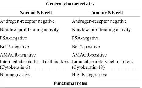
Table 1. General characteristics, functional roles, products and receptors of normal NE and NE tumour cells in pros-tate.
General characteristics
Normal NE cell Tumour NE cell
Androgen-receptor negative Androgen-receptor negative
Non/low-proliferating activity Non/low-proliferating activity
PSA-negative PSA-negative
Bcl-2-negative Bcl-2-positive AMACR-negative AMACR-positive Intermediate and basal cell markers Luminal secretory cell markers
(Cytokeratin-5) (Cytokeratin-18)
Non-aggressive Highly aggressive
Functional roles Regulation of cell growth and differentiation Regulation of homeostasis
Regulation of prostatic secretion Products Adrenomedullin
Bombesin/gastrin releasing peptide (GRP) Calcitonin, calcitonin gene-related peptide Cholecystokinin (CKK)
Chromogranins (CgA, CgB) Gastrin-releasing peptide Histamine
-human chorionic gonadotropin (HCG) Katacalcin
Neuron-specific enolase (NSE) Neuropeptide Y
Parathyroid hormone-related protein (PTHrP) Proadrenomedullin N-terminal peptide Serotonin
Somatostatin
Thyroid stimulating hormone (TSH)-like peptide Vascular endothelial growth factor (VEGF) Vasoactive intestinal peptide (VIP)
Receptors Bombesin/GRP (GRPR)
Calcitonin (hCTR-2) Cholecystokinin c-erbB-2
Gastrin releasing peptide (GRPR) Neuropeptide Y
Neurotensin
Serotonin (5HTR1A, B) Somatostatin (SSTR1-5) Vasoactive intestinal peptide PTHrP receptor
A recent report, from Hirano et al., [23] described dif- ferences in NE cell distribution in PCa. In low grade PCa, morphological features of NE differentiation can be ob- served in CgA-positive areas or occasionally in single CgA cells. In moderately differentiated PCa, NE cells are mostly organized in clusters of focal agglomerates, where- as in high grade PCa, diffuse areas of NE differentiation morphologically similar to surrounding carcinoma cells can be observed.
The origin of NE tumour cells in PCa
The origin of NE cells in prostate tumoral lesions and the underlying molecular mechanisms of enrichment remain controversial. As proposed by Bonkhoff et al. [5, 24], NE tumour cells can arise from the intermediate stem cells, under pathological conditions such as andro- gen deprivation, contributing to increase NE cell popula- tion beyond to normal. Alternatively, PCa cells can un- dergo a “transdifferentiation” process to become NE-like cells, which express NE markers and acquire a NE phe- notype. They begin synthesizing their own growth fac- tors, becoming more autonomous and less dependent on the tissue microenvironmental control. These cells still retain some epithelial characteristics [25], in particular cytokeratin K8/18 [26] and prostatic acid phosphatase [27] luminal cell markers, suggesting that they originate from cancerous luminal epithelial cells. Thus, the in- crease of NE tumour cells as a result of malignant trans- formation of epithelial/basal cells is a common charac- teristic of PCa progression. Further investigations will, however, be necessary to clearly establish the origin of NE tumour cells in prostate adenocarcinoma.
Despite controversies regarding their origin, it is be- coming increasingly clear that NE tumour cells are dis- tinct from NE cells in normal prostate (Table 1). Ac- cording to histological studies, NE tumour cells are mor- phologically similar to the surrounding carcinoma cells [28]. Moreover, a recent study deciphered some genetic features of NE tumour cells and concluded to their rela- tionship with carcinoma cells [29]. They also differ from normal NE cells in the overexpression of proteins, such as anti-apoptotic protein B cell lymphoma protein 2 (Bcl- 2) [30] as well as-methylacyl-CoA racemase (AMACR), an enzyme involved in the β-oxidation of fatty acids [27]. Additionally, NE tumour cells are posi- tive for cyto- keratin 18, a luminal cell-type cytokeratin, whereas nor- mal NE cells rather express cytokeratin 5, a basal cell marker [25,26].
Mechanisms of androgen deprivation therapy-induced NE differentiation
One major feature regarding prostatic cancer cells is their ability following androgen deprivation to undergo a transdifferentiation process, in which they acquire char- acteristics of NE cells and express NE markers, thus be- coming NE-like cells. As they grow independently of androgen, these subpopulations of NE-like cells give rise to clones that have growth advantage, due to the loss of checks and balances, over their counterparts that retain androgen dependence. They may additionally acquire other alterations at both genetic and epigenetic levels that could contribute to the progression to androgen inde- pendence. Thus, through the course of androgen depriva- tion therapy, NE-like cells gradually substitute the func- tion of stromal cells and allow continued proliferation of cancer cells, contributing to progression and aggressive- ness of PCa.
A growing body of literature confirms that NE differ- entiation is more abundant in PCa after androgen with- drawal, indicating that NE component of PCa is resistant to hormone therapy [32]. Moreover, the increase in NE differentiation also appears to be related to the duration of treatment, including hormone deprivation therapy or chemotherapy [33]. Indeed, a long-term androgen depri- vation therapy can induce downstream signaling path- ways that drive the acquisition of a NE aggressive phe- notype. Acquisition of NE characteristics could also oc- cur in response to the influence of prostatic environment, and in particular the synergistic functional network be- tween epithelial and NE prostatic system that may pro- mote internal cell abnormalities, thus triggering the in- duction, and maintenance of NE differentiation.
These observations initially made using clinical sam- ples have been confirmed and the underlying molecular mechanisms of androgen depletion-induced NE differen- tiation studied in vitro and inanimal models of PCa. In an effort to reproduce NE differentiation of prostatic epi- thelial cells in vitro, a number of human PCa cell lines (i.e. LNCaP, DU145, PC3, PC-82, LAPC-4) as well as stable clones of NE-like PCa cells and co-cultures of prostatic cells which summarize tumour characteristics in vivo have now been established. Several studies revealed that androgen deprivation induced NE transdifferentia- tion of both androgen-sensitive and androgen-insensitive PCa cell lines which acquire NE phenotype and express NE markers and peptides [34]. Androgen depletion in- duced NE differentiation through the increase of intra- cellular cyclic AMP (cAMP) levels that in turn induce the activation of protein kinase A (PKA) [35]. Conflict- ing results, however, have reported the involvement of Erk/Mitogen-Activated Protein Kinase (MAPK) pathway in cAMP/PKA-mediated NE differentiation.
The use of these androgen-sensitive and androgen-in- sensitive PCa cell lines has led to interesting data and, could still provide insights into molecular mechanisms of
NE differentiation, in addition to identifying novel thera- peutic targets. Transdifferentiation processes of these PCa cell lines by various stimuli are, however, transient and cells can fully revert to their original phenotype in the absence of inducers, thus making them different from terminally differentiated human PCa NE cells. A better characterization of expression profiles of NE markers and tumorigenic activity of these different PCa cell lines is still needed.
In this regards, stable clones of NE-like PCa cells have been established to elucidate the molecular mechanisms of NE differentiation and its role in PCa progression. These NE-like LNCaP subclone cells obtained by pro- longed culturing in androgen-reduced medium, termi- nally transdifferentiate into NE-like cells and grow to become independent cell lines which exhibit a high tu- morigenicity compared to LNCaP parental cells [36,37]. Importantly, they cannot be reverted to LNCaP phenol- type and they maintain the expression of various NE markers. Therefore, LNCaP subclone cells resemble to NE-like PCa cells found in adenocarcinomas and may have a better clinical relevance than PCa cell lines pre- viously mentioned.
In addition, co-culture models have been developed in order to reconstitute the complex interplay existing be- tween prostatic epithelial, stromal and extracellular ma- trix (ECM) components of the prostate gland [38]. Ef- forts need to be pursued to develop these cell culture models that are taking into account dynamic cellular in- teractions within the prostatic microenvironment which may influence or promote internal cell abnormalities in the progression towards the acquisition of NE phenotype and the hormone-independent PCa cell growth.
Studies on in vivo animal models of PCa (human tu- mour xenografts and transgenic mice (TRAMP and LADY mice)) validate in vitro observations, supporting the importance of NE differentiation in PCa. In these animal models, tumour progression is associated with the acquisition of NE characteristics by cancer cells. More- over, NE differentiation has been reported to markedly increase after castration in xenograft models of human PCa and to precede the emergence of tumour cell prolif- eration and progression to androgen independence [39].
Data from in vitro and in vivo studies collectively sup- port the notion that androgen deprivation induces trans- differentiation of androgen-sensitive adenocarcinoma cells to become NE-like cells in PCa lesions. It remains however to determine whether those transdifferentiated NE-like cells exhibit similar biochemical properties as that of NE-like PCa cells.
citonin, growth factors and cytokines as well as through the activation of various intracellular signaling pathways, including signal transducer and activator of transcription (STAT-3), MAPKs, cAMP/PKA and phosphatidylinosi- tol 3-kinase (PI3K) [40]. The diversity of the pathways that may promote NE cell transdifferentiation could ex- plain at least in part PCa heterogeneity. It remains yet to determine if NE differentiation induced by agents other than androgen depletion also occurs through inhibition of AR signalling. A better knowledge of these alternative signalling pathways will help to determine what drives acquisition of the NE process and may also allow the identification of new therapeutic targets in order to block or prevent PCa progression.
Paracrine to autocrine shift in tumour cell regulation during malignant transformation of epithelial cells
NE tumour cells do not express androgen receptors [7, 8]; they sustain their function in the androgen-deprived environment by establishing autocrine and paracrine networks to stimulate androgen-independent growth of prostate carcinoma cells. These cells produce a wide range of neuropeptides and neurotransmitters involved in the interactions between the different compartments of the prostate. An ever growing list of prostatic NE cell products have been identified and cell surface receptors for some of these NE products have been identified in non-NE tumour cells [25,41].
The ability of non steroids like growth factors (insu- lin-like growth factor I (IGF-I), keratinocyte growth fac- tor (KGF)) and cytokines (interleulin 6 (IL-6)) to mimic the effect of androgens through the mitogen-activated protein kinase (MAPK) signaling pathway [25] contrib- utes to the adaptation of NE tumour cells to an andro- gen-depleted environment, thus increasing their malig- nant potential. Therefore, the autocrine transmission may become more important in androgen-independent tu- mours and contribute to CaP progression. Furthermore, during the evolution from normal to malignant cancer cells, the shift to autocrine transmission may also be as- sociated with the acquired regulation of genes involved in proliferation and survival that are not normally ex- pressed in normal prostate epithelial cells.
Cell-cell interactions are extremely important in main- taining homeostasis between epithelial and stromal com- partments of the prostate. Through the course of andro- gen deprivation therapy, prostatic epithelial cell growth and differentiation can be induced by paracrine growth factors such as epidermal growth factor (EGF), fibroblast growth factor (FGF), KGF, nerve growth factor (NGF) and IGF-I [42,43] that will diffuse from stromal to epithelial compartments, leading to the activation of AR target genes in a ligand-independent manner. Growth factors produced by prostatic epithelial cells can also act in an autocrine manner to stimulate this process.
Aside from these observations, there is considerable evidence indicating that during the transformation pro- cess of prostatic epithelial cells some factors are shifted from paracrine to autocrine regulation, as documented for neurotropins. Classically, NGF, neurotropin-3 and brain-derived growth factor are involved in the NE regu- lation of prostatic function. NGF is produced by stromal cells and is not a survival factor for epithelial cells in the normal prostate. Interestingly, reports indicate that in PCa, NGF can be secreted by malignant prostatic epithet- lial cells and regulate an acquired survival pathway through an autocrine mechanism [44].
Therefore, identification of other growth and survival factors produced by NE tumour cells and a better knowledge of the molecular mechanisms underlying this acquired expression is critical to understand what causes NE cells to become malignant and to effectively treat the disease.
Role of NE tumour cells in PCa
NE tumour cell population is highly aggressive and exhibit tumorigenic activity. Several studies reported that the proportion of NE tumour cells is increased in high grade and high stage tumours [16,45], and in particular in androgen-deprived and androgen-independent tumours. In most cases, NE differentiation is correlated with a poor prognosis [46].
Tumour enrichment in NE-like cells and the conse- quent increase in neurosecretory products can contribute to the androgen-independent proliferation of PCa through their mitogenic effects on adjacent cancer cells, thus in- creasing malignancy and reducing responsiveness of can- cer cells to androgen ablation therapy. Secretory products of NE-like cells have been shown to increase the prolif- eration index (Ki-67-positive cells) of neighboring can- cer cells through paracrine mechanisms [5]. Some of the peptide products of NE cells in the prostate, like bombe- sin, calcitonin and PTHrP, can affect prostate adenocar- cinoma cell proliferation in vitro [9,47].
Moreover, neurosecretory factors produced by NE-like cells may also act by enhancing the sensitivity of prosta- tic cancer cells to lower circulating androgen levels, consequently allowing PCa to escape androgen ablation therapy. NE-like cells may regulate adjacent tumour cells by paracrine mechanisms, highlighting the strong po- tential of neurosecretory products [48].
member of the inhibitor of apoptosis (IAP) family with direct inhibitory action on caspase-3 and caspase-7 activ- ity. Its expression is observed during fetal development but not in normal, terminally differentiated adult human tissues [51]. Additionally, survivin overexpression has been reported in various adenocarcinomas including PCa. Interestingly, increased expression of survivin seems to be associated with PCa progression after radical prost- atectomy [52]. Clusterin is a glycoprotein, involved in diverse biologic processes including transport of lipo- proteins, modulation of cell-cell interactions, cell death and tissue remodeling [53].
In addition, overexpression of the anti-apoptotic Bcl-2 protein by NE tumour cells may also protect them from apoptotic stimuli, and thereby increase their resistance to androgen deprivation therapy and their ability to progress towards hormone-refractory prostate cancers (HRPC) [49]. Therefore, enhancing the pro-apoptotic potential of PCa malignant cells through targeting key players in- volved in apoptosis resistance is one strategy to treat an- drogen-independent PCa.
Furthermore, NE tumour cells may also block the apo- ptotic process of prostatic cells in their vicinity through their neurosecretory products, contributing to andro- gen-independent proliferation of PCa cells and cancer progression. Bombesin and calcitonin have been reported to decrease apoptosis of PCa cells in vitro [54].
Several reports indicated that NE tumour cells might also promote neovascularisation of PCa. They may mod- ulate angiogenesis through secreted angiogenic factors such as vascular endothelial growth factor (VEGF), pla- telet-derived growth factor (PDGF) and basic fibroblast growth factor (FGF), and thus influence prostatic cell growth and differentiation. This idea is supported by the fact that benign prostatic tissue contains low level of VEGF, whereas VEGF staining intensity correlates with Gleason grade. Complete androgen blockade for three months before surgery, decreased VEGF level and vas- cularisation, except in areas where cells are NE-like [55]. In radical prostatectomy samples, there is a correlation between NE differentiation and neovascularisation and both correlate with tumour grade and tumour stage.
Increasing knowledge of the role of NE-like cells throughout the course of prostatic carcinogenesis and tumour progression will pave the way to the development of new therapeutic modalities for advanced and hor- mone-refractory PCa.
4. Pronostic Significance of NE
Differentiation in PCa
Discrepancies in reported incidence (30% - 100%) of NE differentiation in PCa mainly occur from the limited number of NE cell biomarkers used to score NE features. Classically, NE differentiation can be assessed by im-
munoreactivity for neuroendocrine markers or bioactive hormones (somatostatin, 5-HT), measurement of serum levels of NE markers or electron microscopy (neurose- cretory granules).
Among the different NE markers, most of the attention has been given to the detection of CgA and NSE in PCa clinical specimens. CgA belongs to the granin family of acidic secretory glycoproteins that is found in secretory granules of the regulated pathway of a wide variety of endocrine cells and neuron, including NE tumour cells. Classically, CgA serves as a generic marker of the NE cell population. So far, detection of CgA in neoplastic tissue remains one of the most reliable methods to assess NE differentiation [25]. CgA is considered as one of the PCa progression marker after radical prostatectomy [56]. The use of CgA as a prognostic factor in androgen-sen- sitive PCa is still controversial.
Despite the fact that PSA is commonly used as a mark- er for the early detection of PCa as well as monitoring the therapeutic response and tumour recurrence, it is not enough specific and sensitive. A growing body of litera- ture suggests that, independently of PSA, serum CgA is a significant predictor of poor prognosis in patients with advanced and hormone-refractory prostate cancers [22]. Consistent with histological findings, levels of CgA are increased in PCa patients and correlate with tumour stage. A microarray study on PCa revealed that CgA gene is among the 5 genes strongly correlated with the Gleason score, which can predict the outcome following radical prostatectomy [56]. Therefore, serum measurements of NE markers in conjunction with PSA could help screen patients.
In addition to CgA, other serum markers such as CgB, secretoneurin, a proteolytic product of secretogranin II (also known as CgC), gastrin-releasing peptide/ProGRP and NSE, may serve as additional prognostic and/or di-agnostic markers [57,58]. Serum calcitonin level has been reported to be a more specific marker for prostatic small cell carcinoma [59].
The possibility to treat NE differentiation in androgen- independent PCa is related to the development of specific and sensitive markers, clinically detectable in patients with PCa. Further investigations will be necessary to strengthen the prognostic significance of NE tissue mark- er in PCa.
5. Neuroendocrine-Targeted Therapy
taxane-based chemotherapy (paclitaxel, docetaxel) which is of limited benefit represented the main therapeutic option in the occurrence of hormone-refractory PCa. Novel chemotherapeutics and targeted agents for patients with metastatic hormone-refractory PCa stages have been very recently introduced into clinical practice and include abiraterone acetate (a new androgen biosynthesis inhibi- tor), cabazitaxel (a novel microtubule inhibitor), MDV- 3100 (a novel androgen-receptor antagonist), the radio- isotope alpharadin (radium-223), sipuleucel-T (an immu- notherapeutic agent) and denosumab (a bone-targeting agent) [60].
Other novel approaches currently being tested in early clinical trials for advanced PCa include immunological therapies (PCa vaccines, PCa antibodies), angiogenesis inhibitors (targeting VEGF, PDGF, PDGF receptor), epi- genetic therapy (histone deacetylase (HDAC) inhibitors), pro-apoptotic agents (Bcl-2, survivin modulators), and interference in growth-factor-mediated pathways (mam- malian target of rapamycin (mTOR)) [61].
It is now widely accepted that NE differentiation which occurs in prostatic adenocarcinomas is associated with PCa progression and aggressiveness. NE-like cells increase concomitantly with the duration of androgen de- pletion, thus blocking NE function and/or differentiation will most likely prolong the therapeutic window of an- drogen deprivation therapy. The NE axis appears to be an important therapeutic target for drug development in ad- vanced PCa. Pharmaceutical agents (somatostatin ana- logs, bombesin antagonists, serotonin antagonists, mTOR inhibitor, pro-apoptotic agents) able to block the tumour- promoting action of NE-like cells products are under investigation.
NE differentiation is the hallmark of PCa with possible prognostic significance and consequences on therapy response. Detection of focal NE differentiation, using CgA marker, may help to identify patients who are more prone to endocrine therapy failure. In organ-confined di- sease, assessment of NE differentiation could identify patients that would benefit from more aggressive therapy. Intermittent androgen deprivation or antiandrogen mono- therapy could be used to slow down marked NE differen- tiation, in conjunction or not with NE targeted therapy, to delay the progression towards hormone-independent PCa, while maintaining clinical benefit [62].
The NE axis remains an important therapeutic pathway for drug development in advanced PCa. Development of new generation of drugs that selectively target NE-like cells may help targeting populations of PCa that may be resistant or becoming resistant to traditional therapies.
6. Conclusions
In the past years, much progress has been made towards
better understanding the development and progression of PCa as well as the factors which drive the development of androgen independence. NE differentiation is among the non-mutually exclusive theories proposed to explain the progression to androgen independence of PCa. Al- though, NE differentiation has been demonstrated in a variety of carcinomas arising in different tissues, making it of oncological interest, very little is known about the role of NE differentiation in PCa pathophysiology.
Recent progress in terms of PCa research highlighted the role of NE differentiation in prostatic carcinomas. Through the course of androgen deprivation therapy, NE-like cells gradually substitute the function of stromal cells and allow the continued proliferation of cancer cells, contributing to the progression and the aggressiveness of PCa. This subset of androgen-independent cancer cells is also associated with a poor prognosis. Interest in under- standing the neuroendocrine differentiation in PCa has increased with the identification of several neuroendo- crine factors regulating the homeostasis of prostate by autocrine and/or paracrine mechanisms. There is now an ever-growing list of factors that are secreted by these prostatic NE tumour cells which regulate their prolifera- tive activity. As our knowledge of the neuroendocrine factors develops, in the future our focus will be to deter- mine how they interact with other prostate cell types that reside in the dynamic prostatic microenvironment. Inves- tigation of how NE factors and the resulting downstream signaling pathways contribute to the initiation and the progression of PCa should be pursued and will help iden- tifying new therapeutic tools to block or prevent PCa progression.
The NE axis remains an important therapeutic target for drug development in advanced PCa. Development of new generation of drugs directed against NE-like cells may help targeting populations of PCa cells that may be resistant to traditional therapies, thus helping setting up individualized therapy which takes into account the het-erogeneity of PCa.
Financial & Competing Interests Disclosure
Work performed in the laboratory of the author has been supported by the University of Rouen. The author has no other relevant affiliation or financial involvement with any organization or entity with a financial interest in or financial conflict with the subject matter or materials discussed in the manuscript apart from those disclosed.
No writing assistance was utilized in the production of this manuscript.
7. Acknowledgements
REFERENCES
[1] W. A. Schulz, M. Burchardt and M. V. Cronauer, “Mo- lecular Biology of Prostate Cancer,” Molecular Human Reproduction, Vol. 9, 2003, pp. 437-448.
doi:10.1093/molehr/gag064
[2] D. E. Neal, H. Y. Leung, P. H. Powell, F. C. Hamdy and J. L. Donovan, “Unanswered Questions in Screening for Prostate Cancer,” European Journal of Cancer, Vol. 36, No. 10, 2000, pp. 1316-1321.
doi:10.1016/S0959-8049(00)00104-0
[3] A. Jemal, R. Siegel, E. Ward, Y. Hao, J. Xu and M. J. Thun, “Cancer Statistics, 2009,” CA: A Cancer Journal for Clinicians, Vol. 59, No 4, 2009, pp. 225-249. doi:10.3322/caac.20006
[4] S. P. Balk, “Androgen Receptor as a Target in Androgen- Independent Prostate Cancer,” Urology, Vol. 60, 2002, pp. 132-139. doi:10.1016/S0090-4295(02)01593-5
[5] H. Bonkhoff, U. Stein and K. Remberger, “Endocrine- Paracrine Cell Types in the Prostate and Prostatic Ade- nocarcinoma Are Postmitotic Cells,” Human Pathology, Vol. 26, 1995, pp. 167-170.
doi:10.1016/0046-8177(95)90033-0
[6] P. A. di Sant’Agnese, “Neuroendocrine Cells of the Pros- tate and Neuroendocrine Differentiation in Prostatic Car- cinoma: A Review of Morphologic Aspects,” Urology, Vol. 51, 1998, pp. 121-124.
doi:10.1016/S0090-4295(98)00064-8
[7] H. Bonkhoff, U. Stein and K. Remberger, “Androgen Receptor Status in Endocrine-Paracrine Cell Types of the Normal, Hyperplasia, and Neoplastic Human Prostate,” Virchows Archiv A: Pathological Anatomy and Histology, Vol. 423, 1993, pp. 291-294. doi:10.1007/BF01606893 [8] J. L. M. Krijnen, P. J. A. Janssen, J. A. de Winter
Ruizevel, H. Van Krimpen, F. H. Schröder and T. H. Van der Kwast, “Do Neuroendocrine Cells in Human Prostate Cancer Express Androgen Receptor?” Histochemistry, Vol. 100, 1993, pp. 393-398. doi:10.1007/BF00268938 [9] S. Y. Nakada, P. A. di Sant’Agnese, R. A. Moynes, R. A.
Hiipakka, S. Liao, A. T. Cockett and P. A. Abrahamsson, “The Androgen Receptor Status of neuroendocrine Cells in Human Benign and Malignant Prostatic Tissue,” Can- cer Research, Vol. 53, 1993, pp. 1967-1970.
[10] H. Bonkhoff and K. Remberger, “Differentiation Path- ways and Histogenic Aspects of Normal and Abnormal Prostatic Growth: A Stem Cell Model,” Prostate, Vol. 28, 1996, pp. 98-106.
doi:10.1002/(SICI)1097-0045(199602)28:2<98::AID-PR OS4>3.0.CO;2-J
[11] G. Aumuller, M. Leonhardt, M. Janssen, L. Konrad, A. Bjartell and P. A. Abrahamsson, “Neurogenic Origin of Human Prostate Endocrine Cells,” Urology, Vol. 53, 1999, pp. 1041-1048.
doi:10.1016/S0090-4295(98)00631-1
[12] P. A. di Sant’Agnese, “Neuroendocrine Differentiation in Human Prostatic Carcinoma,” Human Pathology, Vol. 23, 1992, pp. 287-296. doi:10.1016/0046-8177(92)90110-O [13] C. K. Ritchie, G. Thomas, L. R. Andrews, D. J. Tindall
and L. A. Fitzpatrick, “Effects of the Calciotrophic Pep-
tides Calcitonin and Parathyroid Hormone on Prostate Cancer Growth and Chemotaxis,” Prostate, Vol. 30, 1997, pp. 183-187.
doi:10.1002/(SICI)1097-0045(19970215)30:3<183::AID-PROS6>3.0.CO;2-N
[14] M. Iwamura, G. Wu, P. A. Abrahamsson, P. A. di Sant’ Agnese, A. T. K. Cockett and L. J. Deftos, “Parathyroid Hormone-Related Protein Is Expressed by Prostatic Neu- roendocrine Cells,” Urology, Vol. 43, 1994, pp. 667-674. doi:10.1016/0090-4295(94)90182-1
[15] H. Bonkhoff, N. Wernert, G. Dhom and K. Remberger, “Relation of Endocrine-Paracrine Cells to Proliferation in Normal and Hyperplastic, Neoplastic Human Prostate,” Prostate, Vol. 19, 1991, pp. 91-98.
doi:10.1002/pros.2990190202
[16] P. A. Abrahamsson, L. B. Wadstrom, J. Alumets, S. Falkmer and L. Grimelius, “Peptide Hormone- and Sero- tonin-Immunoreactive Cells in Normal and Hypersplastic Prostate Glands,” Pathology Research and Practice, Vol. 185, 1989, pp. 373-380.
doi:10.1016/S0344-0338(89)80016-0
[17] A. G. Aprikian, K. Han, L. Guy, F. Landry, L. R. Begin and S. Chevalier, “Neuroendocrine Differentiation and Bombesin/Gastrin-Releasing Peptide Family of Neuro- peptides in the Progress of Human Prostate Cancer,” The Prostate: Supplement, Vol. 82, 1998, pp. 738-743. [18] P. di Sant’Agnese and K. de Mesy Jensen, “Somatostatin
and Somatostatin-Like Immunoreactive Endocrine-Parac- rine Cells in the Human Prostate Gland,” Archives of Pa- thology & Laboratory Medicine, Vol. 108, 1984, pp. 693- 696.
[19] J. Hansson and P. A. Abrahamsson, “Neuroendocrine Pathogenesis in Adenocarcinoma of the Prostate,” Annals of Oncology, Vol. 12, No. 3, 2001, pp. 145-152. doi:10.1093/annonc/12.suppl_2.S145
[20] P. A. di Sant’Agnese, “Neuroendocrine Differentiation in Carcinoma of the Prostate Diagnostic, Prognostic, and Therapeutic Implications,” Cancer, Vol. 70, 1992, pp. 254-268.
doi:10.1002/1097-0142(19920701)70:1+<254::AID-CNC R2820701312>3.0.CO;2-E
[21] P. A. Abrahamsson, “Neuroendocrine Differentiation in Prostatic Carcinoma,” Prostate, Vol. 39, No. 2, 1999, pp. 135-148.
doi:10.1002/(SICI)1097-0045(19990501)39:2<135::AID-PROS9>3.0.CO;2-S
[22] A. Berruti, L. Dogliotti, A. Mosca, M. Bellina, M. Mari, M. Torta, R. Tarabuzzi, E. Bollito, D. Fontana and A. Angeli, “Circulating Neuroendocrine Markers in Patients with Prostate Carcinoma,” Cancer, Vol. 88, 2000, pp. 2590-2597.
doi:10.1002/1097-0142(20000601)88:11<2590::AID-CN CR23>3.0.CO;2-D
[24] H. Bonkhoff, “Neuroendocrine Cells in Benign and Ma- lignant Prostate Tissue: Morphogenesis, Proliferation, and Androgen Receptor Status,” Prostate, Supplement 8, 1998, pp. 18-22.
doi:10.1002/(SICI)1097-0045(1998)8+<18::AID-PROS4 >3.0.CO;2-C
[25] N. Vashchenko and P. A. Abrahamsson, “Neuroendocrine Differentiation in Prostate Cancer: Implications for New Treatment Modalities,” European Urology, Vol. 47, 2005, pp. 147-155. doi:10.1016/j.eururo.2004.09.007
[26] A. van Bokhoven, M. Varella-Garcia, C. Korch, W. U. Johannes, E. E. Smith, H. L. Miller and M. S. Lucia, “Molecular Characterization of Human Prostate Carci- noma Cell Lines,” Prostate, Vol. 57, 2003, pp. 205-225. doi:10.1002/pros.10290
[27] J. Huang, J. L. Yao, P. A. di Sant’Agnese, Q. Yang, P. A. Bourne and Y. Na, “Immunohistochemical Characteri- zation of Neuroendocrine Cells in Prostate Cancer,” Pro- state, Vol. 66, 2006, pp. 1399-1406.
doi:10.1002/pros.20434
[28] N. Xing, J. Qian, D. Bostwick, E. bergstralh and C. Y. F. Young, “Neuroendocrine Cells in Human Prostate Over- express the Anti-Apoptosis Protein Surviving,” Prostate, Vol. 48, 2001, pp. 7-15. doi:10.1002/pros.1076
[29] C. G. Sauer, A. Roemer and R. Grobholz, “Genetic Ana- lysis of Neuroendocrine Tumour Cells in Prostatic Car- cinoma,” Prostate, Vol. 66, 2006, pp. 227-234.
doi:10.1002/pros.20338
[30] Y. Xue, A. Verhofstad, W. Lange, F. Smedts, F. De- bruyne, J. de la Rosette and J. Schalken, “Prostatic Neu- roendocrine Cells Have a Unique Keratin Expression Pat- tern and Do Not Express Bcl-2: Cell Kinetic Features of Neuroendocrine Cells in the Human Prostate,” American Journal of Pathology, Vol. 151, 1997, pp. 1759-1765. [31] P. A. di Sant’Agnese, “Neuroendocrine Differentiation in
Prostatic Carcinoma: An Update on Recent Developments,” Annals of Oncology, Vol. 12, Supplement 2, 2001, pp. 135-140. doi:10.1093/annonc/12.suppl_2.S135
[32] A. H. R. Ismail, F. Landry, A. G. Aprikian and S. Cheva- lier, “Androgen Ablation Promotes Neuroendocrine Cell Differentiation in Dog and Human Prostate,” Prostate, Vol. 51, 2002, pp. 117-125. doi:10.1002/pros.10066 [33] D. Hirano, Y. Okada, S. Minei, Y. Takimoto and N.
Nemoto, “Neuroendocrine Differentiation in Hormone Refractory Prostate Cancer Following Androgen Depriva- tion Therapy,” European Urology, Vol. 45, 2004, pp. 586- 592. doi:10.1016/j.eururo.2003.11.032
[34] T. C. Yuan, S. Veeramani and M. F. Lin, “Neuroendo- crine-Like Prostate Cancer Cells: Neuroendocrine Trans- differentiation of Prostate Adenocarcinoma Cells,” Endo- crine-Related Cancer, Vol. 14, 2007, pp. 531-547. doi:10.1677/ERC-07-0061
[35] P. D. Deeble, D. J. Murphy, S. J. Parsons and M. E. Cox, “Interleukin-6- and Cyclic AMP-Mediated Signaling Po- tentiates Neuroendocrine Differentiation of LNCaP Pros- tate Tumor Cells,” Molecular and Cellular Biology, Vol. 21, 2001, pp. 8471-8482.
doi:10.1128/MCB.21.24.8471-8482.2001
[36] X. Q. Zhang, D. Kondrikov, T. C. Yuan, F. F. Lin, J.
Hansen and M. F. Lin, “Receptor Protein Tyrosine Pho- sphatase Alpha Signaling Is Involved in Androgen Deple- tion-Induced Neuroendocrine Differentiation of Andro- gen-Sensitive LNCaP Human Prostate Cancer Cells,” Oncogene, Vol. 22, 2003, pp. 6704-6716.
doi:10.1038/sj.onc.1206764
[37] T. C. Yuan, S. Veeramanin, F. F. Lin, D. Kondrikou, S. Zelivianski, T. Igawa, D. Karan, S. K. Batra and M. F. Lin, “Androgen Deprivation Induces Human Prostate Epithelial Neuroendocrine Differentiation of Androgen- Sensitive LNCaP Cells,” Endocrine-Related Cancer, Vol. 13, 2006, pp. 151-167. doi:10.1677/erc.1.01043
[38] J. T. Arnold and J. T. Isaacs, “Mechanisms Involved in the Progression of Androgen-Independent Prostate Can- cers: It Is Not Only the Cancer Cell’s Fault,” Endocrine- Related Cancer, Vol. 9, 2002, pp. 61-73.
doi:10.1677/erc.0.0090061
[39] W. J. Huss, C. V. Gregory and G. J. Smith, “Neuroen- docrine Cell Differentiation in the CWR33 Human Pros- tate Cancer Xenograft: Association with Tumor Cell Pro- liferation Prior to Recurrence,” Prostate, Vol. 60, 2004, pp. 91-97. doi:10.1002/pros.20032
[40] E. C. Nelson, A. J. Cambio, J. C. Yang, J. H. Ok, P. N. Lara Jr. and C. P. Evans, “Clinical Implications of Neuro- endocrine Differentiation in Prostate Cancer,” Prostate Cancer and Prostatic Diseases, Vol. 10, 2007, pp. 6-14. doi:10.1038/sj.pcan.4500922
[41] J. Huang and P. A. di Sant’Agnese, “Neuroendocrine Differentiation in Prostate Cancer: An Overview. Ad- vances in Oncology. The Expanding Role of Octreotide,” BioScientifica Ltd., Bristol, 2002.
[42] G. R. Cunha, S. W. Hayward, R. Dahiya and B. A. Foster, “Smooth Muscle-Epithelial Interactions in Normal and Neoplastic Prostatic Development,” Acta Anatomica, Vol. 155, 1996, pp. 63-72. doi:10.1159/000147791
[43] Y. C. Wong and Y. Z. Wang, “Growth Factors and Epithelial-Stromal Interactions in Prostate Cancer Devel- opment,” International Review of Cytology, Vol. 199, 2000, pp. 65-116. doi:10.1016/S0074-7696(00)99002-8 [44] A. T. Weeraratna, J. T. Arnold, D. J. George, A. DeMarzo
and J. T. Isaacs, “Rational Basis for Trk Inhibition Ther-apy for Prostate Cancer,” Prostate, Vol. 45, 2000, pp. 140-148.
doi:10.1002/1097-0045(20001001)45:2<140::AID-PROS 8>3.0.CO;2-#
[45] M. H. Bohrer and J. Schmoll, “Immunohistochemical and Morphometric Studies on Neuroendocrine Differentiation of Prostate Adenocarcinomas,” Verhandlungen der Deut- schen Gesellschaft für Pathologie, Vol. 77, 1993, pp. 107-110.
[46] S. Isshiki, K. Akakura, A. Komiya, H. Suzuki, N. Kamiya and H. Ito, “Chromogranin A Concentration as a Serum Marker to Predict Prognosis after Endocrine Therapy for Prostate Cancer,” Journal of Urology, Vol. 167, 2002, pp. 512-515. doi:10.1016/S0022-5347(01)69075-X
Cancer Research, Vol. 53, 1993, pp. 1724-1726.
[48] R. J. Jin, W. Yongqing, N. Masumori, I. Kenichiro, T. Tsukamoto, S. B. Shappell, S. W. Hayward, S. Kasper and R. J. Matusik, “NE-10 Neuroendocrine Cancer Pro- motes the LNCaP Xenograft Growth in Castrated Mice,” Cancer Research, Vol. 64, 2004, pp. 5489-5495. doi:10.1158/0008-5472.CAN-03-3117
[49] A. J. Raffo, H. Perlman, M. W. Chen, M. L. Day, J. S. Streitman and R. Buttyan, “Overexpression of Bcl-2 Pro- tects Prostate Cancer Cells from Apoptosis in Vitro and Confers Resistance to Androgen Depletion in Vivo,” Can- cer Research, Vol. 55, 1995, pp. 4438-4445.
[50] T. Fixemer, K. Remberger and H. Bonkhoff, “Apoptosis Resistance of Neuroendocrine Phenotypes in Prostatic Adenocarcinoma,” Prostate, Vol. 53, 2002, pp. 118-123. doi:10.1002/pros.10133
[51] T. Yamamoto and N. Tanigawa, “The Role of Survivin as a New Target of Diagnosis and Treatment in Human Can- cer,” Medical Electron Microscopy, Vol. 34, 2001, pp. 207-212. doi:10.1007/s007950100017
[52] S. F. Shariat, Y. Lotan, H. Saboorian, S. M. Khoddam, C. G. Roehrborn, K. M. Slawin and R. Ashfaq, “Survivin Expression Is Associated with Features of Biologically Aggressive Prostate Carcinoma,” Cancer, Vol. 100, 2004, pp. 751-757. doi:10.1002/cncr.20039
[53] C. Koch-Brandt and C. Morgans, “Clusterin: A Role in Cell Survival in the Face of Apoptosis?” Progress in Mo- lecular and Subcellular Biology, Vol. 16, 1996, pp. 130- 149. doi:10.1007/978-3-642-79850-4_8
[54] J. Vilches, M. Salido, E. Fernandez-Segura and G. M. Roomans, “Neuropeptides, Apoptosis and Ion Changes in Prostate Cancer. Methods of Study and Recent Develop- ments,” Histology and Histopathology, Vol. 19, 2004, pp. 951-961.
[55] R. Mazzucchelli, A. Lopez-Beltran, M. Scarpelli and R. Montironi, “Predictive Factors in Prostate Needle Bi- opsy,” Pathologica, Vol. 94, 2002, pp. 331-337.
doi:10.1007/s102420200060
[56] D. Singh, P. G. Febbo and K. Ross, “Gene Expression Correlates of Clinical Prostate Cancer Behaviour,” Can- cer Cell, Vol. 1, 2002, pp. 203-209.
doi:10.1016/S1535-6108(02)00030-2
[57] R. Ischia, A. Hobisch, R. Bauer, U. Weiss, R. W. Gasser, W. Horninger, G. Bartsch Jr., D. Fuchs, G. Bartsch, H. Winkler, H. locker, R. Fischer-Colbrie and Z. Culig, “Elevated Levels of Serum Secretoneurin in Patients with Therapy Resistant Carcinoma of the Prostate,” Journal of Urology, Vol. 163, 2000, pp. 1161-1165.
doi:10.1016/S0022-5347(05)67714-2
[58] M. Yashi, A. Nukui, S. Kurokawa, M. Ochi, S. Ishikawa, K. Goto, Y. Kobayashi, O. Muraishi and A. Tokue, “Ele- vated Serum Progastrin-Releasing Peptide (31 - 98) Level Is a Predictor of Short Response Duration after Hormonal Therapy in Metastatic Prostate Cancer,” Prostate, Vol. 56, 2003, pp. 305-312. doi:10.1002/pros.10260
[59] S. J. Sim, A. B. Glassman, J. Y. Ro, J. J. Lee, C. J. Lo- gothetis and F. J. Liu, “Serum Calcitonin in Small Cell Carcinoma of the Prostate,” Annals of Clinical & Labo- ratory Science, Vol. 26, 1996, pp. 487-495.
[60] V. Adamo, L. Noto, T. Franchina, G. Chiofalo, M. Pic- ciotto, G. Toscano and N. Caristi, “Emerging Targeted Therapies for Castration-Resistant Prostate Cancer,” Fron- tiers in Endocrinology, Vol. 3, 2012, pp. 1-11.
[61] S. Detchokul and A. G. Frauman, “Recent Developments in Prostate Cancer Biomarker Research: Therapeutic Im- plications,” British Journal of Pharmacology, Vol. 71, 2011, pp. 157-174.
doi:10.1111/j.1365-2125.2010.03766.x