ORIGINAL RESEARCH
PATIENT SAFETY
How to Reduce Head CT Orders in Children with
Hydrocephalus Using the Lean Six Sigma Methodology:
Experience at a Major Quaternary Care Academic
Children’s Center
X A. Tekes,X E.M. Jackson, XJ. Ogborn, XS. Liang, XM. Bledsoe, X D.J. Durand, XG. Jallo, and XT.A.G.M. Huisman
ABSTRACT
BACKGROUND AND PURPOSE: Lean Six Sigma methodology is increasingly used to drive improvement in patient safety, quality of care, and cost-effectiveness throughout the US health care delivery system. To demonstrate our value as specialists, radiologists can combine lean methodologies along with imaging expertise to optimize imaging elements-of-care pathways. In this article, we describe a Lean Six Sigma project with the goal of reducing the relative use of pediatric head CTs in our population of patients with hydrocephalus by 50% within 6 months.
MATERIALS AND METHODS: We applied a Lean Six Sigma methodology using a multidisciplinary team at a quaternary care academic children’s center. The existing baseline imaging practice for hydrocephalus was outlined in a Kaizen session, and potential interventions were discussed. An improved radiation-free workflow with ultrafast MR imaging was created. Baseline data were collected for 3 months by using the departmental radiology information system. Data collection continued postintervention and during the control phase (each for 3 months). The percentage of neuroimaging per technique (head CT, head ultrasound, ultrafast brain MR imaging, and routine brain MR imaging) was recorded during each phase.
RESULTS:The improved workflow resulted in a 75% relative reduction in the percentage of hydrocephalus imaging performed by CT between the pre- and postintervention/control phases (Z-test,P⫽.0001).
CONCLUSIONS: Our lean interventions in the pediatric hydrocephalus care pathway resulted in a significant reduction in head CT orders and increased use of ultrafast brain MR imaging.
ABBREVIATIONS:DMAIC⫽Define, Measure, Analyze, Improve, and Control; LSS⫽Lean Six Sigma; PED⫽pediatric emergency department; UF-MRI⫽ultrafast brain MRI; US⫽ultrasonography
L
ean and Six Sigma are 2 prominent quality-improvement methodologies that have been successfully applied in health care since 1998, with increasing use during the past decade related to the increased emphasis on patient safety and cost-effectiveness in medicine.1,2“Six Sigma” is a process initially developed by the Motorola Corporation in 1986, which aims to improve quality by identifying and correcting the causes of errors to reduce the error rate to a six sigma level, (ie, 3.4 defects per million opportunities).“Lean” is another quality-improvement methodology that evolved from the Toyota Production system in 1990, which uses improvements to eliminate waste and preserve steps that incre-mentally optimize the “value” created by the process in question. Combined as Lean Six Sigma (LSS), a 5-stage powerful meth-odology, Define, Measure, Analyze, Improve, and Control (DMAIC), has evolved.
Approximately 39,000 children with hydrocephalus require inpatient admissions annually in the United States.3,4Most or all of these children will require cross-sectional imaging to diagnose and follow their condition. Because CT is currently the mainstay of hydrocephalus imaging at most centers, pa-tients with hydrocephalus have a cumulative radiation expo-sure much higher than that of the average population. Patients exposed to higher cumulative radiation doses have an in-creased risk of cancer (especially children), with the brain be-ing the most sensitive to radiation-induced cancers on the ba-sis of recent empiric studies.5-10 Disease-specific imaging protocols and pathways to help mitigate unnecessary radiation Received September 15, 2015; accepted after revision November 12.
From the Division of Pediatric Radiology and Pediatric Neuroradiology (A.T., D.J.D., T.A.G.M.H.), Department of Radiology (S.L., M.B.), Russell H. Morgan Department of Radiology and Radiological Science; Division of Pediatric Neurosurgery, Depart-ment of Neurosurgery (E.M.J., G.J.); and DepartDepart-ment of Pediatrics (J.O.), The Johns Hopkins Medical Institutions, Baltimore, Maryland.
Please address correspondence to Aylin Tekes, MD, Division of Pediatric Radiology and Pediatric Neuroradiology, The Russell H. Morgan Department of Radiology and Radiological Science, The Johns Hopkins School of Medicine, Charlotte R. Bloomberg Children’s Center, Sheikh Zayed Tower, Room 4155, 1800 Orleans St, Baltimore, MD 21287-0842; e-mail: atekes1@jhmi.edu
exposure in high-risk/vulnerable populations have been dis-cussed in various medical/scientific forums.
Despite these considerations, radiation-free imaging modali-ties, such as brain MR imaging and head ultrasonography (US), are often underused due to logistic barriers and/or resource con-straints such as lack of a 24/7 in-house MR imaging technologist, the longer examination times of MR imaging compared with CT, MR imaging often requiring sedation/anesthesia in the youngest patients, the higher unit cost of MR imaging, and so forth. Some authors have advocated the use of rapid-sequence MR imaging/ rapid brain MR imaging/quick brain MR imaging (different ter-minologies have been used; in this article, we will use the term “ultrafast brain MR imaging”) in patients with hydrocephalus in the past decade. Ultrafast brain MR imaging (UF-MR imaging) offers reliable visualization of the ventricular catheter and supe-rior anatomic detail of the ventricular system; therefore, diagnos-tic accuracy of the test is not reduced as the child benefits from radiation-free imaging.11-15However, in our experience, these MR imaging protocols are limited to relatively few centers or are performed only during certain hours of the day.
Because the barriers to using radiation-free imaging more ef-fectively in pediatric hydrocephalus are so frequently logistic and involve complex, multidisciplinary workflows, we took an LSS approach for process improvement. The purpose of this study was to determine whether a multidisciplinary LSS approach could successfully reduce our reliance on head CT in the pediatric hy-drocephalus population by 50% within 6 months, 24/7, from all referring units.
MATERIALS AND METHODS
In accordance with the Health Insurance Portability and Ac-countability Act, our institutional review board reviewed the pro-tocol for this prospective study and waived the requirement for informed consent. The Division of Pediatric Radiology and Pedi-atric Neuroradiology convened a working team with representa-tion from all key stakeholder groups involved in the care of children with hydrocephalus. Our team involved pediatric neuro-radiologists, pediatric neurosurgeons, pediatric emergency de-partment (PED) physicians; chief technologists of CT, US, and MR imaging; scheduling staff; a department administrator; and a data analyst. A project leader (a pediatric neuroradiologist) and a physician champion (a pediatric neuroradiologist) were identi-fied. Weekly/bimonthly team meetings were held. We deployed the following 5-step LSS methodology (DMAIC):
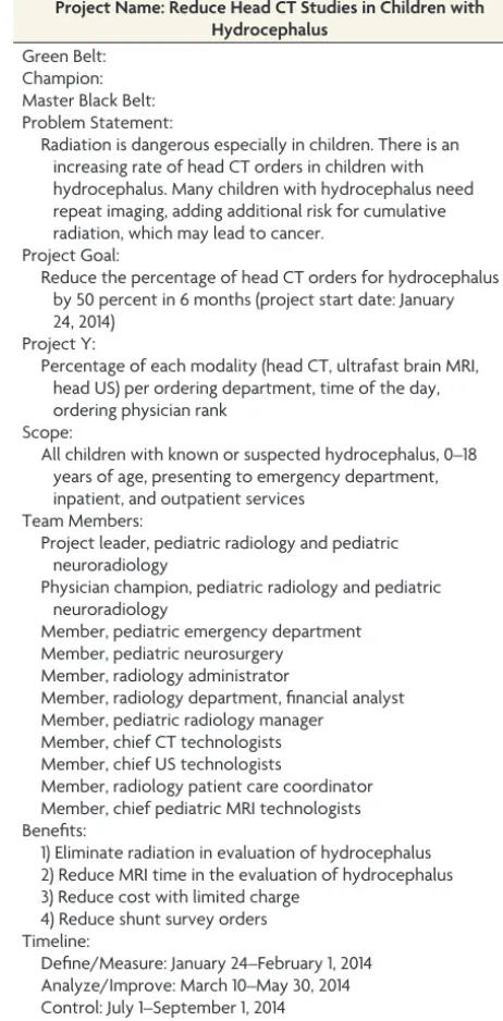
1) Define. All team members gathered around the table under the leadership of the project leader and physician champion. After hearing each group member’s input, a decision was made on the project title, problem statement, and scope of the work; the project goal was aligned with institutional and departmental priorities (Table 1).
2) Measure. Benchmark data were requested from 2 major na-tional children’s hospitals because they had comparable pa-tient profiles, inpapa-tient/outpapa-tient distributions, practitio-ners, and scanner capabilities compared with our children’s center. Both hospitals used a charge modifier to reduce the patient cost for UF-MR imaging by approximately 20%. The benchmark data were used to better understand the
[image:2.594.299.530.60.529.2]perfor-mance with regard to UF-MR imaging. Optimal and practical metrics for defining and measuring the process using institu-tional/departmental resources were discussed. The depart-mental radiology information system was searched for the fol-lowing criteria/key words: hydrocephalus; 0 –18 years of age; and type of imaging performed being head CT, head US, rou-tine brain MR imaging, and UF-MR imaging (with respective examination codes), with date and time of examination and ordering physician rank. Exclusion criteria included acuity of the medical condition requiring an immediate need for trans-fer to pediatric intensive care unit; the presence of a cochlear implant, lumbar implants/drain, or a Codman device (Cod-man/Johnson & Johnson, Raynham, Massachusetts) requiring adjustment after MR imaging; and extreme claustrophobia. Baseline data were collected retrospectively for 3 months on a monthly basis and continued to be collected prospectively Table 1: Project charter
Project Name: Reduce Head CT Studies in Children with Hydrocephalus
Green Belt: Champion: Master Black Belt: Problem Statement:
Radiation is dangerous especially in children. There is an increasing rate of head CT orders in children with hydrocephalus. Many children with hydrocephalus need repeat imaging, adding additional risk for cumulative radiation, which may lead to cancer.
Project Goal:
Reduce the percentage of head CT orders for hydrocephalus by 50 percent in 6 months (project start date: January 24, 2014)
Project Y:
Percentage of each modality (head CT, ultrafast brain MRI, head US) per ordering department, time of the day, ordering physician rank
Scope:
All children with known or suspected hydrocephalus, 0–18 years of age, presenting to emergency department, inpatient, and outpatient services
Team Members:
Project leader, pediatric radiology and pediatric neuroradiology
Physician champion, pediatric radiology and pediatric neuroradiology
Member, pediatric emergency department Member, pediatric neurosurgery
Member, radiology administrator
Member, radiology department, financial analyst Member, pediatric radiology manager
Member, chief CT technologists Member, chief US technologists
Member, radiology patient care coordinator Member, chief pediatric MRI technologists Benefits:
1) Eliminate radiation in evaluation of hydrocephalus 2) Reduce MRI time in the evaluation of hydrocephalus 3) Reduce cost with limited charge
4) Reduce shunt survey orders Timeline:
during the postintervention/improvement phase and control phase in a similar fashion.
3) Analyze. The percentages of head CT, head US, routine brain MR imaging, and UF-MR imaging examinations were calcu-lated during the baseline, improvement, and control phases. The percentage of the above-mentioned orders was analyzed per ordering unit (PED, inpatient, outpatient), per time of practice (routine day time, after hours, and weekends), and per rank of ordering physician (resident/fellow versus attend-ing), to identify the target areas for improvement.
4) Improve. A visual flow chart for improved hydrocephalus im-aging workflow was created (Fig 1) by the team and was im-plemented during the postintervention phase. Gemba was vis-ited. Four major interventions were identified. Data collection continued for 3 months.
5) Control. Existing interventions were consistently pursued, and data were recorded. The electronic ordering systems were revisited during the control phase.
Lean Interventions
Lean interventions were categorized as follows: workflow changes and communication, technology improvement, equipment/process improvement, and reducing patient charges for UF-MR imaging.
Workflow Changes and Communication. Education of all units with the new workflow was performed via an in-person visit to the PED, inpatient, and outpatient units during their internal depart-mental meetings by the project leader. The radiologic value of
UF-MR imaging in pediatric hydrocephalus was explained in de-tail. Limitations of UF-MR imaging in clinical presentations other than hydrocephalus were explained in detail, such as but not lim-ited to assessment of hemorrhage or ischemia, postoperative tu-mor assessment, and developmental milestone evaluations. In ad-dition an electronic e-mail alert was sent to all residents, fellows, and attending physicians summarizing the new workflow (Fig 1). The new workflow was printed and placed over each ordering station for easy visual display of information. The electronic or-dering system was revisited by the PED physician and pediatric neuroradiologist prioritizing head US and UF-MR imaging in the list of examination choices in the hydrocephalus imaging order-ing package with a “pop-up” information box summarizorder-ing the improved imaging workflow.
Technology Improvements. Optimization of the existing UF-MR imaging protocol was undertaken to reduce the total MR imaging time to⬍5 minutes while improving image quality using a tripla-nar T2-weighted HASTE sequence (Fig 2). Due to the short ac-quisition time, no sedation or anesthesia was used. This process was led by the project leader, physician champion, and the chief MR imaging technologist. The chief MR imaging technologist updated all MR imaging scanners with the optimized protocol and trained/informed all technologists.
[image:3.594.96.494.56.336.2]programmer. The imaging technologists were required to respond within 5–10 minutes of placement of imaging orders in our radiology information system. MR imaging technologists were required to page the pediatric neurosurgery resident before initiating the MR imaging. All UF-MR images were obtained in the dedicated pediatric MR imaging scanner except when the scanner was occupied by other medical emergencies. In such cases, MR imaging technologists were responsible for identifying alternative available scanners and direct-ing patient care. The process did not require new staff hirdirect-ing or new job creation. Diagnostic image quality was obtained in all UF-MR imaging studies. No patient required a head CT after undergoing UF-MR imaging (ie, UF-MR images were diagnostically adequate in all patients in this study).
Reduce Patient Charges for UF-MR Imaging. The cost of UF-MR imaging was initially equal to that of routine brain MR imaging. A “charge modifier” was created by the administrator of Billing, Coding, and Compliance to reduce the technical fee.
Statistical significance in the percentage change of head CTs from baseline to improvement and control phases was calcu-lated by usingZ-test. Attribute capability analysis was per-formed to predict the long-term effects of the improved results (Minitab Statistical Software; Minitab, State College, Pennsylvania).
RESULTS
We had 794 patients, of whom 25 were excluded due to following reasons: claustrophobia (n⫽6), medical emergency to evaluate the ventricular size and shunt positioning in which MR imaging
scanner availability could not meet the medical acuity (n⫽4), concern for intracranial hemorrhage (n⫽10), seizures (n⫽3), and the presence of a lumbar drain for which the provider was uncomfortable with drain settings (n⫽2). Of the 769 patients, the mean age was 5.3⫾6 years.
Benchmark data were received from 2 major national children’s hospitals, where the distribution of head CTs was less and UF-MR imaging was more than our existing baseline data (we are not allowed to share the actual numbers or percentages), indicating the timeliness of improvement in our pediatric hydrocephalus imaging pathway. One hospital reported that most of the UF-MRs were obtained on outpatients, whereas the other hospital reported that UF-MRs were predominantly done on patients in the emergency department.
In our children’s center during baseline, the PED was the unit with the highest percentage of head CT orders (86.6%); 62% of all baseline orders including all neuroimaging modalities came from residents or fellows (Table 2). Most imaging (67%) was per-formed during regular work hours (7AMto 5PM) (Table 3); how-ever, when we focused on the PED, most the studies were per-formed after hours (5:01 PM to 6:59 AM) and on weekends (Saturday and Sunday).
There was a statistically highly significant 75% relative de-crease in the total head CT examinations from baseline to the control phase, surpassing our goal of 50% reduction (Z-test,P⬍ .0001) (Table 4). The goal was achieved at 3 months from deploy-ment of the project. UF-MR imaging examinations increased from 18% during baseline to 50% during improvement and con-trol phases, showing a nearly 3-fold increase. Although there was a minimal increase in head CT and a minimal decrease in UF-MR imaging orders during the first 2 months of the control phase, similar percentages to the improvement phase were reached at the third month of the control phase. There was a decreasing trend in head US studies, with favored use of UF-MR imaging. The weekly changes in head CT orders from baseline to control phases are summarized in a p-chart inFig 3. Attri-bute capability analysis showed that the head CT examination reduction at the fi-nal control phase almost reached the 95% FIG 2.Ultrafast brain MR imaging protocol: axial (A), coronal (B), and sagittal (C) T2-weighted
[image:4.594.56.378.396.508.2]HASTE of the brain. Note the clear visualization of the ventricular system and catheter tip.
Table 2: Number and percentage of each modality per rank of ordering physician from baseline to control phasesa
Baseline I-Phase I I-Phase II I-Phase III C-Phase I C-Phase II C-Phase III
No. % No. % No. % No. % No. % No. % No. %
Attending
RB-MRI 16 20.3 4 26.7 5 18.5 3 10.0 14 20.9 6 8.8 9 12.7
UF-MRI 18 22.8 4 26.7 16 59.3 10 33.3 31 46.3 34 50.0 38 53.5
Head CT 16 20.3 2 13.3 0.0 1 3.3 4 6.0 6 8.8 3 4.2
Head US 29 36.7 5 33.3 6 22.2 16 53.3 18 26.9 22 32.4 21 29.6
Attending total 79 38.5 15 24.2 27 31.4 30 32.6 67 70.5 68 58.1 71 63.4
Resident
RB-MRI 22 17.5 5 10.6 5 8.5 5 8.1 4 14.3 5 10.2 7 17.1
UF-MRI 19 15.1 27 57.4 33 55.9 35 56.5 11 39.3 22 44.9 16 39.0
Head CT 46 36.5 3 6.4 6 10.2 6 9.7 6 21.4 8 16.3 6 14.6
Head US 39 31.0 12 25.5 15 25.4 16 25.8 7 25.0 14 28.6 12 29.3
Resident total 126 61.5 47 75.8 59 68.6 62 67.4 28 29.5 49 41.9 41 36.6
Grand total 205 100.0 62 100.0 86 100.0 92 100.0 95 100.0 117 100.0 112 100.0
Note:—I-Phase indicates improvement phase; C-phase, control phase; RB-MRI, routine brain MRI.
[image:4.594.53.533.554.715.2]confidence bounds. Z St. analysis showed that the final control phase data almost reached 2 SDs, which was the 95% confidence bound, indicating a sustainable long-term stability of the process (Fig 4).
DISCUSSION
We reduced head CT orders by 75% within 3 months from de-ploying the LSS project, surpassing our goal of reducing head CT orders by 50% in 6 months in children with hydrocephalus. More important, subsequent data collection/analysis during postinter-vention and control phases demonstrated that these changes were sustainable.
Head CT has been the mainstay of hydrocephalus imaging pathways to as-sess the ventricular size because it is eas-ily accessible in most hospitals and pro-vides quick information with a reasonably low total charge. Long-term survival has significantly improved in children with hydrocephalus with the use of modern CSF diversion techniques and advances in the care of shunts. Low-dose CT protocols have been used in some hospitals, including our own, fol-lowing the principle of as low as reason-ably achievable16; however, children with hydrocephalus will have multiple neuroimaging studies throughout their lives, increasing their total lifetime radiation exposure and conse-quently having a greater lifetime risk of radiation-induced brain cancers.5-10 Therefore, elimination of radiation-based imaging techniques whenever possible is critical to ensure the long-term health of this population. Highlighting this belief, Koral et al7 recently found that there is an increased risk of developing fatal cancer if children with hydrocephalus undergo neuroimaging surveillance with head CTs. These and similar results have repeat-edly been reported in the lay press, and as a result, both providers and patients’ families are routinely demanding that radiation-free protocols be used whenever possible.
[image:5.594.55.531.54.276.2]FIG 3. Attribute capability analysis demonstrates that our control data are almost at the 95% confidence bounds.
Table 3: Number and percentage of each modality during different hours of the day from baseline to control phasesa
Baseline I-Phase I I-Phase II I-Phase III C-Phase I C-Phase II C-Phase III
No. % No. % No. % No. % No. % No. % No. %
Work hours
RB-MRI 27 19.7 6 14.3 6 9.4 6 9.8 12 16.4 6 8.6 11 12.5
UF-MRI 34 24.8 21 50.0 40 62.5 26 42.6 34 46.6 38 54.3 44 50.0
Head CT 28 20.4 2 4.8 1 1.6 1 1.6 8 11.0 1 1.4 6 6.8
Head US 48 35.0 13 31.0 17 26.6 28 45.9 19 26.0 25 35.7 27 30.7
Work hour total 137 66.8 42 67.7 64 74.4 61 66.3 73 76.8 70 59.8 88 78.6
After hours
RB-MRI 7 20.6 2 14.3 3 25.0 2 13.3 3 23.1 3 13.6 4 25.0
UF-MRI 2 5.9 8 57.1 5 41.7 9 60.0 6 46.2 9 40.9 8 50.0
Head CT 19 55.9 2 14.3 3 25.0 3 20.0 1 7.7 6 27.3 2 12.5
Head US 6 17.6 2 14.3 1 8.3 1 6.7 3 23.1 4 18.2 2 12.5
After hours total 34 16.6 14 22.6 12 14.0 15 16.3 13 13.7 22 18.8 16 14.3
Weekend
RB-MRI 4 11.8 1 16.7 1 10.0 0.0 3 33.3 2 8.0 1 12.5
UF-MRI 1 2.9 2 33.3 4 40.0 10 62.5 2 22.2 9 36.0 2 25.0
Head CT 15 44.1 1 16.7 2 20.0 3 18.8 1 11.1 7 28.0 1 12.5
Head US 14 41.2 2 33.3 3 30.0 3 18.8 3 33.3 7 28.0 4 50.0
Weekend total 34 16.6 6 9.7 10 11.6 16 17.4 9 9.5 25 21.4 8 7.1
Grand total 205 100.0 62 100.0 86 100.0 92 100.0 95 100.0 117 100.0 112 100.0
Note:—I-Phase indicates improvement phase; C-phase, control phase; RB-MRI, routine brain MRI.
a
[image:5.594.54.532.312.381.2]Duration of baseline was 3 months, followed by 3 months of improvement (each phase for 1 month), and 3 months of control phases (each phase for 1 month).
Table 4: Number and percentage of each modality from baseline to control phasesa
Baseline I-Phase I I-Phase II I-Phase III C-Phase I C-Phase II C-Phase III
No. % No. % No. % No. % No. % No. % No. %
RB-MRI 38 18.5 9 14.5 10 11.6 8 8.7 18 18.9 11 9.4 16 14.3
UF-MRI 37 18.0 31 50.0 49 57.0 45 48.9 42 44.2 56 47.9 54 48.2
Head CT 62 30.2 5 8.1 6 7.0 7 7.6 10 10.5 14 12.0 9 8.0
Head US 68 33.2 17 27.4 21 24.4 32 34.8 25 26.3 36 30.8 33 29.5
Note:—I-Phase indicates improvement phase; C-phase, control phase; RB-MRI, routine brain MRI.
[image:5.594.53.393.409.558.2]Use of UF-MR imaging was first published in 2005 as a radia-tion-free alternative for neuroimaging in children with hydro-cephalus and has been applied by the principal author since the late 1990s.11Since then, UF-MR imaging has been validated with similar test characteristics for the evaluation of possible shunt failure (assessment of ventricular size and catheter visualization) compared with head CT.17-19Diagnostic image quality and the short image-acquisition time of UF-MR imaging have also been reported.11-15Despite the aforementioned publications, consis-tent nationwide use of this radiation-free neuroimaging tech-nique has been somewhat limited. The palpable growing demand to improve quality and safety in imaging is now requiring radiol-ogists to take leadership responsibilities, beyond simply reading a timely accurate radiology examination.20
Health care delivery systems are complex, and each institution has its unique resources, qualities, and challenges. In our study, we deployed an LSS methodology for solving our problem. In this article, we summarize and present our results in the framework of “how to do” this methodology. Lean methodology focuses on elimination of waste, which can be defective products, overpro-duction beyond demand, increased inventories, unnecessary mo-tion/transport, and wait times as typically described in the man-ufacturing industry. In this study, the “defect” we sought to eliminate was the clinically unnecessary use of head CT. We re-duced the overall radiation exposure to children significantly by using radiation-free imaging techniques. We completely elimi-nated the use of sedation/anesthesia by optimizing/shortening the UF-MR imaging protocol and educating technologists.
Unnecessary motion of the patient and transport staff was ef-fectively eliminated by use of the emergency department MR im-aging scanner and having our MR imim-aging technologists pick up and return the patients to their beds in the PED. We further re-duced the patient’s postimaging wait times by paging the
neuro-surgery resident as soon as the patients were transferred into the MR imaging scanner. The purchase of an additional shunt programmer for the PED enabled the pediatric neurosurgery staff to im-mediately check the shunt settings for patients on completion of imaging. We placed printed improved workflow charts at the ordering workstations of each unit to provide visual display.
Although there was some increase in head CTs and reduction in UF-MR im-aging in the early control phase, the per-centage of decrease in the head CTs did not fall below 50%. The early control phase coincided with the new academic year, with influx of new residents and fellows, which, in part, may explain a temporary change. The electronic or-dering system of our hospital was re-newed during the course of this project. Therefore, before implementation of the new electronic ordering system, which coincided with the beginning of the con-trol phase and the beginning of the new academic year, the revision of the hydrocephalus imaging package with the pop-up information box was not accomplished. This likely explains the brief, transient relative increase in head CTs during this period. After implementation of the changes in the new electronic ordering system, the head CT and UF-MR imaging orders returned to a level similar to that seen during the early improvement phase. This difference highlights the importance/ value of the use of electronic health care systems for a successful execution of interventions that related to imaging appropriate-ness. Attribute capability analysis demonstrated that the control phase data almost reached the 95% confidence bounds.
Our study has several limitations. We identified our patients by using radiology information systems with the search term “hy-drocephalus,” which resulted in the inclusion of all patients with hydrocephalus regardless of whether they were shunted. We of-fered a timely service 24/7 to all providing units; however, we did not measure the total cycle time or throughput of the patients. Although we did not collect such data, the fact that the services have been used well in excess of our initial goal suggests that UF-MR imaging scans are being obtained in fashion timely enough for referring providers to effectively change their ordering patterns. We did not aim for 100% elimination of head CT, keep-ing in mind the potential need for head CT for medical reasons in select cases. We did not calculate the radiation savings for our patients, though this is a goal of our team for future.
CONCLUSIONS
We surpassed our goal of a 50% reduction in head CT orders, achieving a sustained 75% reduction by effectively replacing most head CTs within a radiation- and sedation-/anesthesia-free UF-MR imaging protocol. Building a multidisciplinary team, having a tightly scoped project outline with clear goals aligned
37 33 29 25 21 17 13 9 5 1 0.8
0.7
0.6
0.5
0.4
0.3
0.2
0.1
0.0
Week
P
rop
or
ti
on
_ P=0.1568 UCL=0.4716
LB=0 Baseline Improve Control
1
P Chart of Weekly Head CTs by Stage
Tests performed with unequal sample sizes
[image:6.594.55.372.49.285.2]with our institutional and departmental priorities, and adhering closely to the DMAIC framework of LSS were all crucial in achiev-ing our goal. Although factors may vary by hospital, similar LSS methodology could yield positive results for other indications such as adult hydrocephalus or other health care delivery systems.
ACKNOWLEDGMENTS
We acknowledge the contributions and support of each team member that we could not include in the authors list: Diane No-vak (MR Imaging Manager), Bina Patel (Chief Pediatric MR Im-aging Technologist and Manager), Beatrice Mudge (CT Man-ager), Robert DeJong (US ManMan-ager), Elyce Wolfgang (Pediatric Radiology Manager), and Allison Greene (medical student) from the Johns Hopkins Hospital; and Rich Hill from the Armstrong Institute for Patient Safety and Quality.
Disclosures: Eric M. Jackson—UNRELATED:Expert Testimony: I was an expert for a case of a neurosurgical patient with a diagnosis unrelated to this project. I have not been paid as of yet;Grants/Grants Pending: I had a grant from the institution for shunt development and have currently submitted requests for additional grants. I did not receive any money directly.* Martin Bledsoe—UNRELATED:Expert Testi-mony: West Virginia University; ninety-minute case review. Daniel J. Durand— UN-RELATED:Employment: Evolent Health,Comments: For 2 years I worked as a health care consultant, helping to improve the value delivered by imaging to patient pop-ulations. None of the pediatric MRI work described here overlaps any of my work at Evolent, however;Other: National Decision Support Company,Comments: unpaid advisor to a maker of clinical decision-support software designed to help ensure that the appropriate test is ordered for the right patient at the right time. They do not make a product for pediatric patients, and this work has no overlap with their Clinical Decision Support solution. George Jallo—UNRELATED:Expert Testimony: defense work for a law firm. Thierry A.G. Huisman—UNRELATED:Board Membership: Senior Editor for theAmerican Journal of Neuroradiology. *Money paid to the institution.
REFERENCES
1. DelliFraine JL, Langabeer JR 2nd, Nembhard IM.Assessing the evi-dence of Six Sigma and Lean in the health care industry.Qual Manag Health Care2010;19:211–25CrossRef Medline
2. Vest JR, Gamm LD.A critical review of the research literature on Six Sigma, Lean and StuderGroup’s Hardwiring Excellence in the United States: the need to demonstrate and communicate the effec-tiveness of transformation strategies in healthcare.Implement Sci
2009;4:35CrossRef Medline
3. Simon TD, Riva-Cambrin J, Srivastava R, et al; Hydrocephalus Clin-ical Research Network.Hospital care for children with hydroceph-alus in the United States: utilization, charges, comorbidities, and deaths.J Neurosurg Pediatr2008;1:131–37CrossRef Medline
4. Stein SC, Guo W.The prevalence of shunt-treated hydrocephalus: a mathematical model.Surg Neurol2009;72:131–37CrossRef Medline
5. Brenner D, Elliston C, Hall E, et al.Estimated risks of radiation-induced fatal cancer from pediatric CT.AJR Am J Roentgenol2001; 176:289 –96CrossRef Medline
6. Brenner DJ, Hall EJ.Computed tomography: an increasing source
of radiation exposure.N Engl J Med2007;357:2277– 84CrossRef Medline
7. Koral K, Blackburn T, Bailey AA, et al.Strengthening the argument for rapid brain MR imaging: estimation of reduction in lifetime attributable risk of developing fatal cancer in children with shunted hydrocephalus by instituting a rapid brain MR imaging protocol in lieu of head CT.AJNR Am J Neuroradiol2012;33:1851–54CrossRef Medline
8. Durand DJ.A rational approach to the clinical use of cumulative effective dose estimates.AJR Am J Roentgenol2011;197:160 – 62
CrossRef Medline
9. Pearce MS, Salotti JA, Little MP, et al.Radiation exposure from CT scans in childhood and subsequent risk of leukaemia and brain tumours: a retrospective cohort study.Lancet2012;380:499 –505
CrossRef Medline
10. Mathews JD, Forsythe AV, Brady Z, et al.Cancer risk in 680,000 people exposed to computed tomography scans in childhood or adolescence: data linkage study of 11 million Australians. BMJ
2013;346:f2360CrossRef Medline
11. Ashley WW Jr, McKinstry RC, Leonard JR, et al.Use of rapid-se-quence magnetic resonance imaging for evaluation of hydrocepha-lus in children.J Neurosurg2005;103(2 suppl):124 –30Medline
12. Iskandar BJ, Sansone JM, Medow J, et al.The use of quick-brain magnetic resonance imaging in the evaluation of shunt-treated hy-drocephalus.J Neurosurg2004;101:147–51Medline
13. Niederhauser BD, McDonald RJ, Eckel LJ, et al.Retrospective review of rapid pediatric brain MR imaging at an academic institution in-cluding practice trends and factors affecting scan times.AJNR Am J Neuroradiol2013;34:1836 – 40CrossRef Medline
14. O’Neill BR, Pruthi S, Bains H, et al.Rapid sequence magnetic reso-nance imaging in the assessment of children with hydrocephalus.
World Neurosurg2013;80:e307–312CrossRef Medline
15. Rozovsky K, Ventureyra EC, Miller E.Fast-brain MRI in children is quick, without sedation, and radiation-free, but beware of limita-tions.J Clin Neurosci2013;20:400 – 05CrossRef Medline
16. Pindrik J, Huisman TA, Mahesh M, et al.Analysis of limited-se-quence head computed tomography for children with shunted hydrocephalus: potential to reduce diagnostic radiation exposure.
J Neurosurg Pediatr2013;12:491–500CrossRef Medline
17. Yue EL, Meckler GD, Fleischman RJ, et al.Test characteristics of quick brain MRI for shunt evaluation in children: an alternative modality to avoid radiation.J Neurosurg Pediatr2015;15:420 –26
CrossRef Medline
18. Mater A, Shroff M, Al-Farsi S, et al.Test characteristics of neuroim-aging in the emergency department evaluation of children for cere-brospinal fluid shunt malfunction.CJEM2008;10:131–35Medline
19. Lehnert BE, Rahbar H, Relyea-Chew A, et al.Detection of ventricular shunt malfunction in the ED: relative utility of radiography, CT, and nuclear imaging.Emerg Radiol2011;18:299 –305CrossRef Medline