ORIGINAL RESEARCH
SPINE
Intramedullary Spinal Cord Metastases: MRI and Relevant
Clinical Features from a 13-Year Institutional Case Series
J.B. Rykken, F.E. Diehn, C.H. Hunt, K.M. Schwartz, L.J. Eckel, C.P. Wood, T.J. Kaufmann, R.K. Lingineni, R.E. Carter, and J.T. Wald
ABSTRACT
BACKGROUND AND PURPOSE: Because intramedullary spinal cord metastasis is often a difficult diagnosis to make, our purpose was to perform a systematic review of the MR imaging and relevant baseline clinical features of intramedullary spinal cord metastases in a large series.
MATERIALS AND METHODS: Consecutive patients with intramedullary spinal cord metastasis with available pretreatment digital MR imaging examinations were identified. The MR imaging examination(s) for each patient was reviewed by 2 neuroradiologists for various imaging characteristics. Relevant clinical data were obtained.
RESULTS:Forty-nine patients had 70 intramedullary spinal cord metastases, with 10 (20%) having multiple intramedullary spinal cord metastases; 8% (4/49) were asymptomatic. Primary tumor diagnosis was preceded by intramedullary spinal cord metastasis presentation in 20% (10/49) and by intramedullary spinal cord metastasis diagnosis in 10% (5/49); 98% (63/64) of intramedullary spinal cord metastases enhanced. Cord edema was extensive: mean, 4.5 segments, 3.6-fold larger than enhancing lesion, andⱖ3 segments in 54% (37/69). Intratumoral cystic change was seen in 3% (2/70) and hemorrhage in 1% (1/70); 59% (29/49) of reference MR imaging examinations displayed other CNS or spinal (non–spinal cord) metastases, and 59% (29/49) exhibited the primary tumor/non-CNS metastases, with 88% (43/49) displayingⱖ1 finding and 31% (15/49) displaying both findings. Patients with solitary intramedullary spinal cord metastasis were less likely than those with multiple intramedullary spinal cord metastases to have other CNS or spinal (non–spinal cord) metastases on the reference MR imaging (20/39 [51%] versus 9/10 [90%], respectively;P⫽.0263).
CONCLUSIONS: Lack of known primary malignancy or spinal cord symptoms should not discourage consideration of intramedullary spinal cord metastasis. Enhancement and extensive edema for lesion size (oftenⱖ3 segments) are typical for intramedullary spinal cord metastasis. Presence of cystic change/hemorrhage makes intramedullary spinal cord metastasis unlikely. Evidence for other CNS or spinal (non–spinal cord) metastases and the primary tumor/non-CNS metastases are common. The prevalence of other CNS or spinal (non–spinal cord) metastases in those with multiple intramedullary spinal cord metastases is especially high.
ABBREVIATIONS:ISCM⫽intramedullary spinal cord metastasis
I
ntramedullary spinal cord metastasis is uncommon, with a prevalence of up to 2.1% in autopsy series of patients with can-cer.1Yet, ISCMs are being increasingly diagnosed, related toad-vances and increased use of imaging and therapies that prolong survival in patients with cancer.2-6Prompt and accurate diagnosis
of ISCM is necessary for effective treatment, and MR imaging is the preferred imaging technique.5Despite this, large series
sys-tematically evaluating multiple MR imaging features of these masses have not been published. The literature on ISCM is limited to case reports, relatively small case series, literature reviews, and autopsy series. Because ISCM is often a difficult diagnosis to make, the purpose of this retrospective study was to perform a systematic review of the MR imaging and relevant baseline clinical features of ISCMs in a large series.
MATERIALS AND METHODS
Institutional review board approval with waived consent was ob-tained for this Health Insurance Portability and Accountability Act– compliant retrospective research study.
Subjects
The radiologic, clinical, surgical, and pathologic databases at a single institution were searched to identify a group of consecutive Received December 14, 2012; accepted after revision January 12, 2013.
From the Division of Neuroradiology (J.B.R., F.E.D., C.H.H., K.M.S., L.J.E., C.P.W., T.J.K., J.T.W.), Department of Radiology, and Department of Health Sciences Research (R.K.L., R.E.C.), Mayo Clinic, Rochester, Minnesota.
Paper previously presented at: Annual Meeting of the American Society of Neuro-radiology, the ASNR 50th
Annual Meeting, April 2012; New York, New York. Please address correspondence to Felix E. Diehn, MD, 200 1st
St SW, Rochester, MN 55905; e-mail: Diehn.felix@mayo.edu
patients with ISCM (n⫽58, 1999 –2011). Patients without pre-treatment digital MR imaging examination, patients in whom al-ternative diagnoses were suspected clinically, and patients in whom ISCM was never formally diagnosed were excluded. These 58 patients with ISCM also were the basis for a recent study, which described 2 specific enhancement features on postgadolinium MR imaging in ISCMs compared with primary cord masses.7Five
(9%) of these 58 patients were excluded for the current (and the prior) study because of the lack of available pretreatment MR imaging examination. Note that included in the current study but excluded in the prior study were 4 patients who lacked available postgadolinium pretreatment MR imaging. Several additional subject details were as reported in the prior study; specifically, 4 (8%) of the remaining 53 patients “were excluded because alter-native diagnoses were being considered clinically and ISCM was never formally diagnosed.” Consequently, only patients with a clinical diagnosis of ISCM were included on the basis of review of all available clinical, pathologic, and imaging data. This yielded a final ISCM study population of 49 (92%) of the remaining 53 patients, with a total of 70 ISCMs. In 5 (10%) of these 49 patients with ISCM, the diagnosis of a solitary ISCM was confirmed by cord mass biopsy/resection. In an additional 44 (90%) of the 49 patients with ISCM, “pathologic proof of the systemic malignancy had been obtained from the primary site or a metastatic site out-side of the spinal cord.” One (2%) of the 49 patients died “before any sampling but was clinically presumed to have an ISCM from lung carcinoma, given a classic radiographic pattern of dominant primary lung mass, with multiple pulmonary metastases, hilar and mediastinal adenopathy, distant metastases, a 50 pack-year smoking history, and a family history of lung cancer.”7
MR Imaging Review
Two radiologists reviewed all MR imaging examinations in con-sensus at an electronic workstation (one [F.E.D.] a neuroradiol-ogy faculty member with American Board of Radiolneuroradiol-ogy certifica-tion, a Certificate of Added Qualification in neuroradiology, and in full-time neuroradiology practice, and the other [J.B.R.] a neu-roradiology fellow with American Board of Radiology certifica-tion). The MR imaging examinations were predominantly from our institution but did include some from outside facilities. All available pulse sequences were reviewed. The typical examination included sagittal T1- and T2-weighted, axial T2-weighted, and postgadolinium sagittal and axial T1-weighted images.
The presence or absence of the following imaging features was analyzed, by use of pregadolinium and postgadolinium T1-weighted images: 1) number of ISCMs per patient, and for each ISCM: 2) location (cervical, cervicothoracic, thoracic, thoracic-conus, conus), 3) position within the cord on axial images (cen-tral, eccentric, exophytic), 4) morphology (well-circumscribed versus ill-defined), and cord expansion (absent versus present), 5) T2 signal intensity (hyperintense, hypointense, or isointense to the spinal cord), 6) T1 signal intensity (hyperintense, hypoin-tense, or isointense to the spinal cord), 7) convincing evidence of cystic change (nonenhancing fluid signal on T1- and T2-weighted images; rated as absent, or if present, intratumoral, peritumoral, or both), 8) convincing evidence of intratumoral hemorrhage (nonenhancing T1 hyperintensity and/or marked T1
hypointen-sity and corresponding T2 hypointenhypointen-sity and/or “blooming” magnetic susceptibility artifact on gradient recalled-echo se-quences, if available; rated as absent versus present), 9) gadolin-ium enhancement (absent or, if present, homogeneous versus heterogeneous versus peripheral [ring]), 10) maximal size of en-hancing lesion in millimeters (measured anteroposterior, trans-verse [axial images required], and superior-inferior), 11) maxi-mal longitudinal extent of enhancing lesion in number of vertebral segments, and 12) maximal longitudinal extent of spinal cord T2 hyperintensity in number of vertebral segments (sagittal T2 images required). The ratio of length of spinal cord T2 hyper-intensity to length of ISCM was calculated for each lesion. For the per-patient T2 signal extent in patients with multiple ISCMs, the length of the longest lesion was considered. By including lesions that appeared eccentric or exophytic, it is possible that both pri-mary intramedullary metastases and leptomeningeal metastasis invading the spinal cord would be included among ISCMs. Thus, eccentric/exophytic lesions were further characterized as either clearly intramedullary or possibly of leptomeningeal origin with spinal cord invasion, on the basis of which was dominant, the intramedullary or the cord surface component. In addition, for the exophytic lesions: 1) original MR imaging reports were re-viewed to assess whether the interpreting radiologist described an intramedullary mass with exophytic extension or a leptomenin-geal mass with invasion, and 2) previous categorization of “rim” and “flame” signs,7two postgadolinium MR imaging findings
specific for ISCM, was noted. Note also that leptomeningeal le-sions without an apparent intramedullary component were not considered to be ISCMs.
For each patient, the reference spinal MR imaging was re-viewed for evidence of the primary tumor and/or extraspinal, non-CNS metastases. The reference spinal MR imaging as well as any other electronically available MR imaging of the neuroaxis performed within 4 weeks before or 4 weeks after the reference MR imaging were reviewed by the 2 radiologists for any evidence of other spinal column or CNS metastases not involving the spinal cord (including vertebral column, leptomeningeal, and visualized intracranial contents). Leptomeningeal metastases were local-ized anatomically as involving the spinal cord/conus, the cauda equina, the cul-de-sac, or any combination of these. Any avail-able spinal MR imaging examinations subsequent to the refer-ence MR imaging were also reviewed for development of addi-tional ISCMs.
Clinical Review
Statistical Analysis
The association between solitary versus multiple ISCMs status and the following variables was analyzed by means of2test: primary
tumor type and presence on reference MR imaging of 1) other CNS or spinal (non–spinal cord) metastases, 2) primary tumor/non-CNS metastases, or 3) specifically, leptomeningeal metastases. Descriptive statistics were obtained by use of Excel 2010 (Microsoft, Redmond, Washington). Additional analyses were conducted by using SAS ver-sion 9.3 (SAS Institute, Cary, North Carolina). Statistical significance was defined by aPvalue of⬍.05.
RESULTS
Patient and Histopathologic Characteristics of ISCM
Of the 49 patients with 70 ISCMs, 26 (53%) were female, with a median age of 57.7 years at diagnosis (range, 7– 80 years). Histo-pathologies of the primary malignancies in the 49 patients are shown in Table 1. Regarding the category of “other,” 1 of each of the follow-ing primary tumor types were represented, for a total of 6 (12%) patients with 6 (9%) ISCMs: anaplastic thyroid carcinoma, salivary ductal carcinoma, neuroendocrine carcinoma, ovarian adenocarci-noma, lymphoma, and prostate carcinoma. The nature of the CNS-origin primaries in 4 patients is as described in the prior study.7
Sol-itary versus multiple ISCMs status did not correlate with tumor type (P⫽.1607), but the 10 cases of multiple ISCMs only occurred with the 2 most common primary malignancies: lung cancer (7/10, 70%) and breast cancer (3/10, 30%).
Clinical Characteristics
Table 1 includes additional relevant clinical features of the pa-tients with ISCM. Papa-tients had a median of 2 weeks of symptoms at clinical presentation. The most common presenting symptom
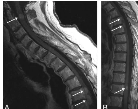
was weakness (28/49, 57% of patients). However, several patients were asymptomatic with regard to the ISCM (4/49, 8% of pa-tients); one-half (2/4) of these asymptomatic patients had multi-ple ISCMs (Fig 1). A diagnosis of primary malignancy was not always known at the time of reference MR imaging, as the clinical presentation of the ISCM preceded primary tumor diagnosis in 20% (10/49) of patients, with the actual diagnosis of ISCM pre-ceding primary tumor diagnosis in 50% (5/10) of these patients. In all (10/10) of the patients with multiple ISCMs, the diagnosis of the primary malignancy preceded the ISCM diagnosis.
ISCM Imaging Characteristics on Reference MR Imaging
Thirty-nine (80%) of 49 patients had solitary ISCM on the refer-ence MR imaging (Fig 2), whereas 10 (20%) of 49 had multiple ISCMs (31 additional ISCMs for a mean of 3 ISCMs per patient; Figs 1 and 3). The MR imaging features of the ISCMs on reference MR imaging on a per-lesion basis are detailed in Table 2. Note that most of the lesions were solitary (39/70, 56%), in the thoracic spinal cord (40/70, 57%), eccentrically located within the cord (35/62, 56%), expanding the cord (44/70, 63%), enhancing (63/ 64, 98%), T2-hyperintense relative to the cord (55/70, 79%), and T1-isointense relative to the cord (48/63, 76%). The length of cord T2 signal abnormality was often extensive (mean, 4.5 seg-ments), on average 3.6-fold greater than the length of the enhanc-ing lesion, extendenhanc-ingⱖ2 segments in 62% of lesions (43/69) and 75% of patients (36/48) andⱖ3 segments in 54% of lesions (37/ 69) and 67% of patients (32/48).
Most lesions appeared convincingly of intramedullary origin rather than leptomeningeal with spinal cord invasion. Specifi-cally, only 4 lesions were exophytic. One of these 4 cases demon-strated the “rim” sign and one both the “rim” and “flame” signs, specific signs previously described in ISCMs.7In all 4 cases, the
[image:3.594.300.532.47.229.2]interpreting radiologist described an intramedullary mass with Table 1: Clinical features of ISCMs, per-patient basis (nⴝ49
patients)
Feature Prevalence
Sex
Female 26 (53%)
Primary malignancy
Lung carcinoma 24 (49%)
Breast carcinoma 7 (14%)
Melanoma 5 (10%)
CNS origin 4 (8%)
Renal cell carcinoma 3 (6%)
Other 6 (12%)
Timing of primary tumor diagnosis ISCM presentation preceded primary
tumor diagnosis
10 (20%)
ISCM diagnosis preceded primary tumor diagnosis
5 (10%)
Dominant presenting symptoms
Weakness 28 (57%)
Sensory symptoms 8 (16%)
Bowel and/or bladder dysfunction 5 (10%)
Pain 4 (8%)
Asymptomatic 4 (8%)
Time interval, median (range) Duration of symptoms at clinical
presentation (n⫽44)
2 weeks (0.1–32)
Primary tumor diagnosis to ISCM presentation (n⫽38)
1.8 years (0–19.3)
Primary tumor diagnosis to ISCM diagnosis (n⫽44)
1.6 years (0–19.5)
[image:3.594.53.285.64.350.2]exophytic extension, not a leptomeningeal mass with parenchy-mal invasion. Of the 39 lesions that were characterized as being either eccentric in the spinal cord (n⫽35) or exophytic (n⫽4), only 3 lesions (4% of all 70 ISCMs, 6% of the 62 ISCMs for which axial images were available) appeared to potentially be of lepto-meningeal origin, given a dominant surface component, rather than originating within the spinal cord. Two of these 3 lesions occurred in the same patient.
Two findings were rare. Central cystic or necrotic change was seen in only 2 of the ISCMs (2/70, 3%) (Fig 4), with only 1 of these 2 lesions demonstrating ring enhancement peripherally about the cystic change. Only 1 lesion demonstrated convincing evidence for associated hemorrhage (1/70, 1%) (Fig 5).
Visualization of Other Metastases, Primary Tumor on Reference MR Imaging
Table 3 includes additional findings on the reference MR imaging on a per-patient basis. Note that most patients (43/49, 88%) had additional CNS or spinal column metastasis(or metastases) not involving the spinal cord and/or the primary tumor or non-CNS metastasis(or metastases) visible on the reference MR imaging (Fig 2D; Fig 3C,-D). In other words, only in a minority of patients (6/49, 12%) did the MR imaging not demonstrate at least 1 of these associated findings. When leptomeningeal metastases were present (18/49, 37% of patients) on reference MR imaging, they
most commonly manifested as diffuse involvement along the cord/conus, cauda equina and cul-de-sac (8/18, 44%); isolated involvement of the cul-de-sac did not occur. Patients with solitary compared with multiple ISCMs were less likely to have CNS/spi-nal (non–spiCNS/spi-nal cord) metastases on the reference MR imaging (20/39 [51%] versus 9/10 [90%], respectively;P⫽.0263). There was no correlation between solitary versus multiple ISCMs status and presence of either primary tumor/non-CNS metastasis or leptomeningeal metastases on reference MR imaging (P⫽.3484 andP⫽.872, respectively).
Findings on Prior and Subsequent Neurologic-MR Imaging
Forty-seven percent of patients (23/49) had evidence of other CNS/spinal metastases (non–spinal cord) on MR imaging exam-ination(s) of the neuroaxis obtained within the 4 weeks preceding the reference MR imaging. An additional 16% of patients (8/49) demonstrated such other metastases within the 4 weeks subse-quent to the reference MR imaging. The subsesubse-quent development of a new ISCM on follow-up MR imaging occurred in 22% (11/ 49) of patients.
DISCUSSION
The current study highlights several relevant clinical and MR im-aging features of ISCMs in a large series of patients. The most salient clinical features of ISCM are 1) lung cancer is the most common primary tumor, 2) the primary malignancy has not al-ways been diagnosed at the time of ISCM symptom onset or ref-erence MR imaging, and 3) patients can be asymptomatic with FIG 2. Typical solitary intramedullary spinal cord metastasis, with
vi-sualization of primary tumor. A 66-year-old man presented with 6 weeks of paresthesias, bladder dysfunction, lower extremity weak-ness, and pain. Thoracic spine sagittal T2-weighted(A),sagittal T1-weighted(B), postcontrast sagittal T1-weighted (C), and axial T1-weighted (D) images are shown. A T2 hyperintense, expansile intramedullary cord lesion(arrow)is associated with a large amount of cord T2 hyperintensity(A).The mass is isointense on T1-weighted images(arrowinB)and enhances heterogeneously(arrowinC).Also noted is a left hilar lung mass(arrowsinD),which was further evalu-ated with chest CT imaging (not shown). This hilar mass was patho-logically proved to be a grade 4 undifferentiated small-cell lung car-cinoma. Visualization on MR imaging of the primary tumor/non-CNS metastases and/or other spinal/CNS (non–spinal cord) metastases was common in this series.
[image:4.594.54.285.44.286.2] [image:4.594.301.532.46.250.2]regard to the ISCM, even in the case of multiple ISCMs. The 3 most pertinent imaging features of ISCMs are 1) almost all ISCMs enhance, 2) the associated spinal cord T2 hyperintensity is exten-sive, and 3) both intratumoral hemorrhage and intra-/peritu-moral cystic/necrotic change are rare. An additional important imaging finding is that either additional CNS/spinal (non–spinal cord) metastases or the primary tumor/non-CNS metastases are extremely common, seen in nearly 90% of patients in this series on the reference MR imaging. The prevalence of other CNS/spinal (non–spinal cord) metastases in those with multiple ISCMs is especially high.
Knowledge of these relevant clinical and imaging features of ISCM and their significance is important for radiologists and the referring clinicians. When faced with a spinal cord mass, lack of a known primary malignancy and lack of symptoms related to the mass should not dissuade one from considering an ISCM. For any
spinal cord mass, but particularly when ISCM is strongly consid-ered, the visualized lungs should be scrutinized because lung can-cer is the most common primary malignancy. Lack of enhance-ment and presence of cystic change and/or hemorrhage in an intramedullary mass should bring other etiologies of spinal cord masses higher in the differential diagnosis, such as primary cord neoplasms, in which such findings are not uncommon. The radi-ologist should scrutinize the MR imaging as well as other prior neuroaxis imaging studies for presence of other CNS/spinal (non–spinal cord) metastases and the primary tumor/non-CNS metastases. The presence of additional ISCMs should be specifi-cally sought.
The current study contributes to the literature on ISCM. Re-cently, we described 2 postgadolinium MR imaging findings spe-cific for ISCM compared with various primary cord masses, in 45 of the 49 ISCM patients from the current study.7In the present
[image:5.594.302.532.46.343.2]study, we more fully characterize the MR imaging findings for these ISCMs. Other previous series of ISCMs have generally been smaller, with the largest of these consisting of 40 patients, 27 of FIG 4. Atypical intramedullary spinal cord metastasis with central cystic change/necrosis. A 55-year-old man with recent nephrectomy of a renal cell carcinoma presented with 2 weeks of predominantly left upper extremity pain, paresthesias, and weakness, as well as global hyperreflexia. Cervical spine sagittal T2-weighted(A),T1-weighted(B), and postcontrast fat-saturated T1-weighted images(C),and postcon-trast axial T1-weighted image are shown. A mass within the cord at the level of C5 has markedly hyperintense central signal on T2-weighted imaging(A)and corresponding T1 hypointensity(B)consistent with central cystic change/necrosis. The sagittal (C) and axial (D) T1-weighted postcontrast images demonstrate the peripheral enhance-ment with lack of central enhanceenhance-ment corresponding to the region of central cystic/necrotic change. This represents 1 of only 2 cases in the current series of intramedullary spinal cord metastasis demon-strating cystic/necrotic change. The primary tumor type in the other case (not shown) was lung carcinoma.
Table 2: MRI features of ISCMs, per-lesion basis, total of 70 lesions in 49 patients
Feature Prevalence
Location (n⫽70)
Cervical 16 (23%)
Cervicothoracic 2 (3%)
Thoracic 40 (57%)
Thoracic-conus 1 (1%)
Conus 11 (16%)
Position (n⫽62)
Central 23 (37%)
Eccentric 35 (56%)
Exophytic 4 (6%)
Morphology (n⫽66)
Well-circumscribed 64 (97%)
Ill-defined 2 (3%)
Cord expansion (n⫽70)
Present 44 (63%)
Absent 26 (37%)
Enhancement (n⫽64)
Present 63 (98%)
Absent 1 (2%)
Enhancement pattern (n⫽63)
Homogeneous 31 (49%)
Heterogeneous 31 (49%)
Peripheral (ring) 1 (2%)
Size of enhancing lesion, mm, mean (range)
Anterior-posterior (n⫽63) 6.5 (1–16)
Transverse (n⫽53) 7.3 (2–23)
Superior-inferior (n⫽63) 19.9 (2–114)
Longitudinal extent, No. of vertebral segments, mean (range)
Length of enhancement (n⫽63) 1.4 (1–8) Length of cord T2 signal hyperintensity (n⫽69) 4.5 (1–15) Ratio, T2 signal/enhancement (n⫽63) 3.6 (1–14) T2 signal intensity (n⫽70)
Hyperintense 55 (79%)
Hypointense 1 (1%)
Isointense 14 (20%)
T1 signal intensity (n⫽63)
Hyperintense 10 (16%)
Hypointense 5 (8%)
Isointense 48 (76%)
Cystic change (n⫽70)
Intratumoral 2 (3%)
Peritumoral 0 (0%)
[image:5.594.56.287.67.518.2]whom were imaged with MR imaging (25 with gadolinium).8One
recent small series of 8 ISCM patients reviewed the published literature of an additional 293 cases but did not specifically assess MR imaging findings.9
The large amount of spinal cord edema compared with the length of the enhancing ISCM has been known anecdotally and
described in a smaller series with low field strength MR imaging examinations,10as well as in other small series; for instance, in 3
patients in a study by Sze et al.11However, to our knowledge, this
has not been systematically reported and quantified in a large series by use of modern MR imaging scanners.
The rarity of cystic change/necrosis demonstrated in our study conflicts with some existing literature. One review publication in the imaging literature does describe cysts to be rare in ISCM,12but
specific references are not included. Other articles state that cystic change/necrosis is common.5For instance, a study of 7 patients
reported ring enhancement suggesting central necrosis in 4; note that no images demonstrating cystic change were provided and that the authors did not include a radiologist.13A different study
reported intratumoral cysts in 10 of 19 cases, but no representa-tive image of such a case was provided.14
Other studies, including the recent comprehensive literature review by Sung et al,9have also found that lung cancer accounts
for approximately 50% of ISCM cases and that breast cancer is the second most common primary malignancy. The review by Sung et al also found a high frequency of systemic metastases, though our present study examined this specifically on the basis of what a radiologist might visualize on the reference MR imaging. Addi-tional similarities between our series and the review by Sung et al, respectively, are: propensity for ISCM to be solitary (80% versus 88% prevalence), age (wide range, with median 58 years versus 56 years), weakness as the most common symptom (57% versus 88%), symptom duration (wide range, with median of 2 weeks versus 3 weeks), frequency of asymptomatic patients (8% versus 5%), ISCM preceding primary tumor diagnosis (“synchronous presentation”) (20% versus 26%), and interval between primary tumor diagnosis and ISCM diagnosis (wide range, with median of 19 months versus 16 months). Differences between our series and the review by Sung et al, respectively, are: prevalence of sensory symptoms (16% versus 73%) and prevalence of bowel/bladder dysfunction (10% versus 43%). These discrepancies may exist be-cause we only assessed symptoms up to the time of the MR imag-ing, not for the remainder of the disease course. An additional difference between our series versus the comprehensive literature review by Sung et al, respectively, is the prevalence of thoracic ISCM (57% versus 34%). This discrepancy is probably multifac-torial. For instance, our anatomic categorization was based exclu-sively on MR imaging (rather than autopsy) findings. Moreover, we did not include patients with “intramedullary” metastases ex-clusive to the cauda equina (not involving the spinal cord and presumably leptomeningeal), which may account for the rela-FIG 5. Atypical intramedullary spinal cord metastasis with associated
[image:6.594.54.285.47.533.2]hemorrhage. A 74-year-old man with squamous cell carcinoma of the lung diagnosed 2 years prior presented with 4 weeks of paraplegia. Thoracic spine shown with sagittal T1-weighted(A)and T2-weighted (B)and axial gradient recalled-echo(C)images. Heterogeneous mildly hyperintense central signal is present within the intramedullary spinal cord metastasis on T1-weighted imaging(arrowsinA).There is corre-sponding heterogeneity on T2-weighted imaging(B).The axial gradi-ent recalled-echo image demonstrates corresponding cgradi-entral hy-pointensity (“blooming,”arrowinC),typical of hemorrhage. This is the only intramedullary spinal cord metastasis in the current series demonstrating signal changes convincing for associated hemorrhage.
Table 3: Additional MRI features of ISCMs on reference MRI, per-patient basis (nⴝ49 patients)
Feature Prevalence
Solitary ISCM 39 (80%)
Multiple ISCMs 10 (20%)
Non-cord CNS and/or spinal metastasis 29 (59%)
Leptomeningeal metastasis 18 (37%)
Primary tumor and/or non-CNS metastasis 29 (59%) Non-cord CNS/spinal metastasis OR primary
tumor/non-CNS metastasis
43 (88%)
Both non-cord CNS/spinal metastasis AND primary tumor/non-CNS metastasis
[image:6.594.300.533.63.167.2]tively higher proportion of “lumbar” spinal cord metastases (38%) seen in literature reviewed by Sung et al.
As for the development of ISCMs, several pathophysiologic mechanisms have been described, including arterial spread, ret-rograde venous spread (via Batson venous plexus), meningeal spread, perineural lymphatic spread, and direct invasion from a contiguous structure.4,8,13,15,16 Of these proposed mechanisms
for the pathogenesis of ISCMs, spread via the arterial route is generally favored as the most common, though the mechanism may differ, depending on the primary tumor cell type. The pre-ponderance of central and eccentric ISCMs in the present study and the relative lack of exophytic ISCMs also suggest that the arterial route is a more common means of spread, particularly given the robust arterial supply to the central gray matter of the cord. However, a case series encompassing all types of spinal cord metastases, ideally with microscopic pathologic correlation, would be needed to confidently achieve this conclusion. Inclusion of CSF analysis would also be potentially useful.
Limitations of this study include its retrospective nature, the use of consensus imaging review, and the lack of uniformity of MR imaging technique, because not all of the examinations were from our institution. Although our series is the largest published single institution study of patients with ISCM, the relatively small sample size remains a limitation. Only 10% of the 49 patients had pathologic confirmation. However, this is consistent with the other smaller published series of ISCM, because pathologic sam-pling from surgery/autopsy is typically not obtained. Moreover, the multiple aforementioned clinical features in the current study that were similar to published literature suggest that the patients in our series are indeed representative of patients with ISCMs. The most common reason for biopsy or resection of ISCM is for diag-nostic purposes3,7,9; knowledge of the results of the current study,
as well as of the recently described enhancement characteristics specific for ISCM,7is anticipated to decrease such diagnostic
sur-gery rates.
CONCLUSIONS
We describe pertinent clinical and MR imaging features in a large series of patients with ISCM. When considering ISCM in the dif-ferential diagnosis of a spinal cord mass on MR imaging, the re-sults of this study should be helpful to radiologists. Absence of clinical symptoms and lack of a known primary malignancy should not dissuade one from considering ISCM. Presence of cys-tic change or hemorrhage should cause one to entertain other diagnostic possibilities as more likely. Extensive edema compared with enhancing lesion size is typical. Evidence for other CNS or
spinal (non–spinal cord) metastases, the primary tumor, and non-CNS metastases should be sought because these features are common. The prevalence of other CNS or spinal (non–spinal cord) metastases in those with multiple ISCMs is especially high.
REFERENCES
1. Costigan DA, Winkelman MD. Intramedullary spinal cord metastasis: a clinicopathological study of 13 cases.J Neurosurg 1985;62:227–33
2. Mut M, Schiff D, Shaffrey ME.Metastasis to nervous system: spinal epidural and intramedullary metastases. J Neurooncol 2005; 75:43–56
3. Connolly ES Jr, Winfree CJ, McCormick PC, et al.Intramedullary spinal cord metastasis: report of three cases and review of the liter-ature.Surg Neurol1996;46:329 –37
4. Kalayci M, Cagavi F, Gul S, et al. Intramedullary spinal cord metastases: diagnosis and treatment–an illustrated review.Acta Neurochir (Wien) 2004;146:1347–54
5. Kalita O.Current insights into surgery for intramedullary spinal cord metastases: a literature review.Int J Surg Oncol2011; Epub; PMID:22312538
6. Zebrowski A, Wilson L, Lim A, et al.Intramedullary spinal cord metastases in breast cancer are associated with improved longer-term systemic control.Future Oncol2010;6:1517–19
7. Rykken JB, Diehn FE, Hunt CH, et al.Rim and flame signs: postg-adolinium MRI findings specific for non-CNS intramedullary spi-nal cord metastases.AJNR Am J Neuroradiol2013;34:908 –15 8. Schiff D, O’Neill BP.Intramedullary spinal cord metastases: clinical
features and treatment outcome.Neurology1996;47:906 –12 9. Sung WS, Sung MJ, Chan JH, et al.Intramedullary spinal cord
metastases: a 20-year institutional experience with a comprehen-sive literature review.World Neurosurg2012; Epub; PMID: 22484768 10. Crasto S, Duca S, Davini O, et al.MRI diagnosis of intramedullary
metastases from extra-CNS tumors.Eur Radiol1997;7:732–36 11. Sze G, Krol G, Zimmerman RD, et al.Intramedullary disease of the
spine: diagnosis using gadolinium-DTPA-enhanced MR imaging.
AJR Am J Roentgenol1988;151:1193–204
12. Koeller KK, Rosenblum RS, Morrison AL.Neoplasms of the spinal cord and filum terminale: radiologic-pathologic correlation. Ra-diographics2000;20:1721– 49
13. Watanabe M, Nomura T, Toh E, et al.Intramedullary spinal cord metastasis: a clinical and imaging study of seven patients.J Spinal Disord Tech2006;19:43– 47
14. Dam-Hieu P, Seizeur R, Mineo JF, et al.Retrospective study of 19 patients with intramedullary spinal cord metastasis.Clin Neurol Neurosurg2009;111:10 –17
15. Lee SS, Min KK, Sym SJ, et al. Intramedullary spinal cord metastases: a single-institution experience. J Neurooncol 2007; 84:85– 89