University of Warwick institutional repository:
http://go.warwick.ac.uk/wrap
A Thesis Submitted for the Degree of PhD at the University of Warwick
http://go.warwick.ac.uk/wrap/73917
This thesis is made available online and is protected by original copyright.
Please scroll down to view the document itself.
Studies on the enzyme glucose oxidase from
PenicilliQm am§gasakiens~
H.Geisow.
This thesis is presented in part fulfilment of
the degree of Doctor of Philosophy,University
BEST COpy
..
, .AVAILABLE
. Variable print quality
.
,.SUMMARY
A short method of purification of P.amagasakiense glucose
oxidase has been achieved. Attempts to prepare the apoenzyme were
unsuccessful. Inhibition of activity of the holoenzyme by heavy
metals was investigated. It was shown that inhibition was due to
the metal cations rather than the undissociated metal salts.
The effect of bisulphite on the enzyme was shown to be the
same as for Aspergillus niger glucose oxidase. From the kinetic
experiments with bisulphite, an ionizable group with a pK of 4.2
was found to be involved in the reaction. This pK was assigned to
of adenine
the amino grouPA of the FAD moiety of the enzyme. Experiments on the
binding of halide anions to the enzyme indicated the involvement
of an ionizable group with the sa~e pK.
P oto-c emical experiments on the enzyme revealed three
different phenomena. (1) Photo-oxidation with methylene blue or
Rose bengal as sensitiser, destroyed an ionizable group with pK
of 7.2. This was assigned to a histidine residue in the protein.
(2) At high pH in the presence of EDTA and with no oxygen present
photo-red~ction of the enzyme spectrum was obtained. The spectrum
of the oxidised enzyme was obtained when air was admitted to the
reduced species. (3) At high pH in the presence of EDTA, light
at 450 nm wavelength caused the enzyme to lose activity. This activity
loss was irreversible w~th respect to oxygen or glucose, The
presence of glucose in the reaction mixture during illumination
INDEX OF CHAPTERS
studies on the enzyme glucose oxidase.
1. Historical introduction 1
2. The purification of glucose oxidase from fungal sources 7
3. The attempted preparation of the apoenzyme of Penicillium
amagasakiense glucose oxidase, and recombination experiments 17
4.
Enzyme inhibition by heavy metals 225.
The effect of bisulphite on spectral and functional9.
Discussion of possible mechanisms for glucose oxidaseproperties of the enzyme
6.
Inhibition by halides and other anions at low pH.7. Dye-sensitised photo-oxidation
8.The photo-chemical reaction
Appendix 1. To obtain the inhibition constants of a
2-substrate reaction
Appendix 2. Titration of a protein with a monovalent ligand
77
80
Bf blio graphy.
Proteolytic enzymes from extremely halophilic bacteria
I should like to express my warmest thanks to my supervisor
Dr.B.E.P.Swoboda for his help, encouragement and forbearance during
this work. I am grateful to Professor V.lIi.Clarkfor the use of the
facilities in the School of Mo.LecuLar- Sciences, and I am pleased to
acknowledge the receipt of a S.R.C.Studentship.Finally I thank my
family and friends without whose support and encouragement this work
would never have been completed.
M1BREVIATIOlliS
The revised instructions to authors (1971) of the Biochemical
Journal, published by the Biochemical Society, London, have been
followed where ever possible. Recommended SI syrnbols for units
CHAPl'ER ONE !il§'l'ORICAL IN'fRO
ml91lQ£!
The enzymology of gluconic acid formation was investigated by
buller (1926 - 1941) using the press juices from the mycelia of the
fungi of ~. niger. and Penicilliulllg1aucuE:' Slight purification of the
enzyme was achieved by f~anke and his co-workers Deffner and Lorenz
(1937 - 1948), who also demonstrated that the activity at'the different
enzyme preparations Was proportional to the flavin content, and that
hydrogen peroxide was formed during the catalysis.
The systematic name which has been given to the enzyme is
t-D glucose: 02 oxidoreductase E.C.l.1d·4. (Report of the Commission
on enzymes, I.U.B. 1961). The trival name is glucose oxidase. Keilin
and Hartree (1946, 1948a,
b,)
have shown that the enzyme has aprosthetic group of flavin-adenine-dinucleotide (FAD) and a small
carbohydrate moiety compr ising less than 20% of the total enzyme
(Pazur, Kleppe and Cepure 1965). It catalyses an overall 2-electron
oxidation-reduction reaction between ~-D glucose and oxygen, as shown
in equation l.i. overleaf. The formation of hydrogen peroxide in
the presence of glucose and oxygen gave the enzyme a reputation for
antibiotic activity. Hydrogen peroxide has a bactericidal effect
because of its vigorous oxidising power, and. this led early workers
to call their material Penicillin A (later uotatin), penatin Or
Penicillin B.
This type of enzyme has only been isolated definitely from fungi.
Enzyme from four fungal sources has been identified and purified
:-from Penicilliwn vitale
Coulthard et al.,1942,1945
Kusai et al, 1960, Kusai 1960.
Swoboda et al., 1963, Pazur and
Kleppe 1964
Pidoplichko et a1 1965
from ~enicil~notatum
from enicillium amagasakiense
from ~spergillus niger
Several workers have carried out an extensive screening progra~me of
fungal species for glucose oxidase activity e.g. Gancedo 1967 and
Grigorov 1969. The latter has shown that glucose oxidase activity can
be obtained from all strains of !sEer~lleae, depending on the time
of incubation and carbohydrate growth source. Other glucose oxidases
have been reported from non-fungal sources, but none of them has been
purified and characterised as a flavoprotein oxidoreductase. (See the
review of Schepartz and Subers 1964).
Muller claLmed (1941) that !.niger possessed a glucose dehydrogenase
as well as an oxidase. This dehydrogenase could not use oxygen as the
electron acceptor, and the best acceptor was found to be
2,6-dichlorophenol-indophenol. Glucose dehydrogenase has been isolated from f:;.oryzaeby
Ogura (1939 - 1952). Kusai (1960) confirmed its presence in !.niger and
showed that it was not a flavoprotein.
Considerable interest has been focussed On the use of glucose
oxidase in the food industry and in clinical work. In 1949 a patent
was issued "for the removal of oxygen from sealed food containers by
glucose oxidase" (Sarett 1949). In medicine it is used to detect and
measure quantitatively glucose in blood and urine samples, after the
reported use in this field by Keston (1956).
Originally the ass~ for glucose oxidase activity utilised the
hydrogen peroxide formed by the reaction, which WaS reduced to SjW8SP
~ water by peroxidase. This Was coupled with the oxidation of a
suitable chromageri (see equations 1. i. and L
iii.).
Such chromagensincluded o-tolidine, benzidine and o-dianisidine but because of the
carcinogenic nature of these chromagens a new method has been evolved.
The hydrogen peroxide is used as an oxidising agent for 4-amino
~1'4~"
"",-.se.
(i)
glucose + oxygen glucono-d' -lactone + H202
H202 c.t..I•• c. 2'1 02 + H2O (ii)
H202 + AH2 p&ro'4lcolll.:u ..
,
A + 2 H2O (iii)~anometry used to be the prime method of followinb glucose oxidase
catalysis. Catalase was present in the assay which generated oxygen
by the breakdown of the hydrogen peroxide formed. The amount of
oxygen produced was measured and related to the peroxide formed, and
hence to the enzyme turnover. Catalase which contruninates glucose
oxidase preparations, alters the stoichrj'ilmetryof the reaction so that
only one atom of oxygen is used per molecule of glucose oxi.dis.ed,
(see 1.i. and 1.H. ). It is possible to use millil'ftOlarconcentrat ions
of cyanide in the assay which inhibit any traces of catalase present
in the enzyme preparation., This restores the stoichiometry of the reaction
to one molecule oxygen used per molecule glucose oxidised.
0JCYgen uptake is now measured polarographically using a Clark-type
oxygen electrode suitably connected to a pen recorder through a
resistance box. It is 'also ~ossible to follow hydrogen peroxide
production spectrophotometrically at 235 nm in the presence of cyanide
(Bright and Gibson 1967), or by any of the colo¢rimetric methods
mentioned previously using 1 t.I-H2S04to inactivate the enzyme after
the reaction has procee ed for a period of'time (Savage 1951, White and
Secor,1957, Pazur and Kleppe 1964).
The overall mechanism of glucose oxidase from P.notatum has been
investigated by Bentley and euberger (1949) using isotopes of oxyge
Their findings are summarised in equations l.iv. and l.v.
+
H
o't
2.•
Ol~
1 •
Thus water is not involved in the reaction, the hydrogen peroxide being
derived from the oxygen and the glucose. !.:..oreoverthey found that the
rate of gluconic acid formation was too slow t0 be part of the primary
process which was the upt ake of oxygen. The 1"irst product, the lact one,
was detected polarographically and since it was found to be in the
pyranose ring form, Bentley and leuberger deduced that the glucose must
be in the ring f'orm too. The hydrolysis of'the lactone to gluconic
acid occurred spontaneously.
Keilin and Hartree (1952) showed that glucose oxidase was highly
specific for the ~-anomer of glucose, using _!.E~~ enzyme. This
f'inding was donfirmed for the enzyme from
!:.
vitale (De[tyar, Gulii andMaizel 1965), A_. n;L.g~r(Pazur and Kleppe 1964) and
!:.
amagasakiense(Kusai 1960). The substrate specificity of the latter enzyme species
~1.1e
is shown in ~l.2. The ef'fect of'various substituents in different
positions on the glucose ring has been studied by Pazur and Kleppe
(1964). From this they concluded that either of the hydroxyl groups at
positions 1 or
3,
equatorial to the ring, might be the site at whichthe en~me is attached to the glucose.
of
Earlier glucose oxidase was described as catalyst A an overall
2-electron oxidation-reduction reaction. Bright and Gibson (1967)
used I-deutero-glucose to investigate the hydrogen transfer from
glucose to flavin. They found a kinetic isotope effect of between
10- and 15- fold associated with flavin reduction. This would be
consistent with nucleophilic attack by the glucose 0-1 hydroxyl group
on the fl-avin nucleus, followed by proton abstraction from the carbon
and electronic rearrangements (Veibel and Bright 1971). The kinetic
isotope effect would come from the proton removal from glucose,
controlled by k2 in the mechanism O'IU' I•• ~
E-~
OKE.
...
f
r4d
The substrate attack is shown at position C-4a rather than the carbo~l
group at
C-S
suggested by some workers following the model studies ofBrown and Hamilt on (1970). Massey et al (1968) and Muller, Massey et
a1 (1969) have shown that substrate attack on the oxidised flavin may
occur at position C-4a or 1-5 on the isoalloxazine ring. Weibel and
Bright (1971) have identified an acidic amino-acid residue (pKa- 10)
close to the -1 position represented by H-A in the above mechand.sr
A second mechanism, which is consistent with the isotope effect
involves hydride transfer from the carbon to the flavin (Weibel and
Bright 1971).
tc~O ...
I1,C"'O- _.
-C~
0
H,C/
o- ~-c
1'0" 'C/
0-'0-
./ '-
~
"
-
H6'---
,
'-
-
/'--HO 0 0 .... "0
"+Jf
~+
Jf
K+H
fc"'O
+
" 0-
..c:~o
H
,A-
/1'0H~
0-"c'"
~
'C
-C
C/"
'OH l+O/
'-
~'OH
UC:'-
'OH
-£/ \..._
Hydride transfer to the flavin Occurs because of the electron sink effect.
Hemmerich, Nagelschneider and Veeger (1970) have reviewed the
chemistry and molecular biology of'f'Lavi.nsand f'Lavoprotei.na, They
discussed the implications of the two possible sites for substrate
attack on the oxidised flavin. They discussed but did not distinguish
between hydride transfer and group transfer to the flavin.
Bright and Gibson (1967) did not detect any isotope effect in the
oxidative half reaction of glucose oxidase. This has led Weibel and
Bright to suggest that the oxidation of flavin proceeds via electron
transfer to oxygen followed by rapid proton dissociation from N-l and
N-5 of the isoalloxazine ring.
Most of the work on the glucose oxidase mechanism has been carried
out on A.niger enzyme. It is hoped to show, in the following chapters,
THEPURIFICATIOi_2!...QgrCOSEOgDASE FROMFUI'iGAL SOURCE§.
In tr oduct ion
The first attempt at purification of glucose oxidase by Franke and
Lorenz (1937) involved precipitating the e'nzyme from the press juices
of niger mycelia.. Toe mycelia were disrupted in a Buchner press
at 300 atmospheres pressure • The enzyme was precipitated by the
addition of a twelve-fold excess of a 2:1 ixture of alcohol and
ether to the press juices. The specific activity of the preparation
of Franke and Deffner (1939) at 30°C was 37 }.Lmoles/min/mg dry weight.
Coulthard and co-workers (1945) using a culture filtrate of
EenicilliQm not~, concentrated it by acetone precipitation. Then
they either precipitated the resuspended extract by add it ion of
tannic acid, or Reinecke's salt to obtain a fairly pure product.
Further purification was by acetone precipitation and a~monium
sulphate i'ractionat ion at approximately 80% saturat ion. The specific
activity of their preparation at 300C was 40 01 oxygen/min/mg dry (194~)
weight. Using the same tec~nique, Keilin and HartreeAobtained a
preparation with a specific activity of 73 (39°C).
Both Pazur and Kleppe (1964) a d Swoboda and .assey (1965) have
used a eonmer-cLal- A. niger mycelial preparat ion as their source of
the enzyme. Pazur and Kleppe (1964) dialysed a solution of mycelial
powder against water and using anllnoniumsulphate, obtained a fraction
(between 60% and 877~al11.l1l0niurl1sulphate saturation) which contained a
high percentage of the original oxidase activity. This dialysed
fraction was applied to a DEAE-cellulose colu.m and eluted with 0.1
1.1-acetate buffer pH3.7, carbohydrate and catalase cont~ninants having
been eluted earlier with 0.1 11 -acetate buffer pH4·
5·
The yellow _,:'ractionwas r echr o at ognaphed on a smaller column of' DEAE-cellulose.
In contrast, Swoboda and :assey dialysed their ext.r act against
Q) [J) o o ~
.
r-i
.
f'C'\
r-i o o
...-o
.
CO
o
.
~
o
.
No
"')
.
Nc o
.
~
o
~
0.1 M-acetate buffer pli :4.5 and removed the precipitate formed
during the dialysis by centr ifugat ion. The supernatant was applied
directly to a column of Arnberlite-CG 50 (suitably treated). 'I'he
column was washed with O. 1 M-buffer pR 4.5, and the enzj rne was eluted
with O. 1 M-acetate buffer pH
5.
O. Protein was precipitated from theeluate by ammoniumsulphate t'ract;ionat ion (80-9CJ./& saturat ion) ,
redissolved, dialysed against 0.1 l1-l,hosphate buffer pH 6.0 and
applied to a DEAE-cellulose column. The enzyme was finally eluted
by 0.2 M-phosphate buffer pH 6.0, to be followed by concentration
with ~moniQm sulphate and dialysis. A table comparing the
recovery of specific activity from the starting material and yield
of total units in each of these two met uods has been drawn up
(table
2.1.).
A simple industrial purification of commercially available
glucose oxidase giving a yield of
90%
of initial activity relies onthe precipitat ion of the enzyme by addition of' methanol to an
aqueous solution of the enzyme extract. A pH of 7.0 ~'" continuously
maintained. Hyflo-supercel is added and the precipitate is separated.
The filter cake is homogenised in cold water pH 6.0, filtered and
lyophilised. An enrichment of enzyme material of 300% is claimed
possible (Bergmeyer et al
1%9).
Purification of enzyme from ~ale has been achieved by either
putting the culture filtrate down a column of SG-I (equivalent to
Amberlite-CG 50) and eluting with 0.6 M-acetate buffer pH5.0
(Pokrovskaya and Chistyakova 1965) or by absorption at' the enzyme
from the culture fluid by anhydrous aIumi.na pR 6.5-7.0 and elution
by shaking with a
9%
solut ion of ammoniumchlorideID
0.3 M-potassiumdihydrogen phosphate (Gulii and Degtyar
1962).
In each case theenzyme has been precipitated from the concentrated eluate by ethanol
r("S~)I.
koldt.t"---+-i~bu 0 - ,.;",
---+--1\
T4\ol'l.
~t.Mb"4A~ --- __c..olllr'~
hp
4
de.c..kbde.~F'g.2.1.
~=~=======g;;;?)----1I-
M0'5"t...·1(. st-irl'tJ'~Uo ..."
i\YU c'\(c.trocit.
lectrodc used for
Only Kusai has published a purification procedure for Pencil~
amagas~~ (Kusai 1960, Kusai et al 1960). He originally grew
a culture of this fungus which he found to exude glucose oxidase
in to the culture medi ' A crude extract of this culture medium
is now available conmerc Lal Iy under the name "Deoxi.ne'' (Nagase and
Co., Ltd.) Kusai dialysed the crude extract over-night against 0.1
-acetate buffer pH 4.5, centrifuged to clear the solution and
passed all the supernatant down an A.mberlite-CG 50 column (5 x 25 em)
buffered with the same buffer. After washing with 2-3 1 of buffer
until the elute was clear, during which process half the glucose
oxidase activity was eluted in the first washing, the rest of the
enzyme was eluted with 0.1 M-acetate buffer pH 5.0. After
fractionation and dialysis against 0.05 M-acetate buffer pH 5.0,
the solution was passed through a second column 01' A.rnberlite-CG 50
(2 x 70 em) equilibrated with the same buffer. The enzyme was
eluted with the same buffer, was fractionated with rurunoniQrnsulphate,
dialysed against water and crystallised with amrnoniurnsulphate.
The other glucose oxidase fraction from the first column would not
crystallise on further pJTification and was thought by Kusai to
contain partially modified enzyme.
Jaterials and ethods.
a). r.iethod of' measuring glucose oxidase activity.
l'easure ents of activity were made using a Clark type oxygen
electrode as shown in fig.2,1. It consisted of a platirnlrn electrode
with a silver electrode on either s ide set in an inert resinous
material. The tip of this assembly was covered by a thin Teflon
membrane held in place by a rubber O-ring. Between the electrOdes and
to an adjustable potentiometer box which was connected in turn to
a pen recorder.
The electrode assembly was screwed into a solid perspex rod
which just fitted a cylindrical glass electrode cell. The
di~ensions were sO arranged that when the perspex electrode
holder was fitted into the top of the cell, there was a space
beneath the electrodes to contain exactly
5
ml. reaction mixture.Down the side of the perspex holder was a groove just large enough
to allow a microsyringe needle to be introduced into the electrode
cell.
The electrode cell was held Ln a glass water jacket connected
to a thermostatted water bath and purnp , and a te!ilperatureof 25°0
was maintained. Additions of enzyme, substrate or inhibitor were
made to the reaction mixture in the cell by means 01' a microsyringe
used as a microburette (Scientific Glass Engineering,Melbourne,
Australia) whose needle was passed down the groove in the electrode
casing as described above.
For normal assays, unless otherwise stated, the assay mixture
contained 0.01 -glucose and 10-3 M-EDI'A in 0.1 M-acetate buffer
pH
5.6.
During enzyme purification procedures, excess catalase(8 ug/ml., Sigma type C-1OO) was added because 01' the presence of
contaminating catalase in small quantities. Using excess catalase,
activity values for glucose oxidase were a half those obtained with
no catalase present, because of the changed stoichiometry of the
reaction (see equations 1.i.and l.ii.) .
The initial rate of oxygen uptake was obtained by measuring
the initial gradient of the recorded curve of oxygen uptake against
time, which was drawn out by the pen recorder. The units of
specific activity used are fA-mol of oxygen/mi.n/rngof protein.
(Oxygen saturation of aqueous solutions in equilibrium with air at
250 and atmospheric pressure is 2.6 x 10-
4 ).
b) Method of estimating protein and enzyme concentration.
Protein absorbance was measured on a Unicam sp600 spectrophotometer
(Unicam-Pye, Ca.rnbridge). 1 mg/ml (dry weight) protein has an optical
density at 280 ~rn of 1.85 (Kusai 1960). During the preparation of the
enzyme, protein was estimated by the biuret method (Gornall, Bardawill
and David, 1949).
Enzyme concentration was calculated from the absorbance of the
enzyme at 450 nrn,using the millimolar extinction coefficient of
11.3 for active site concentration, and 22.6 for the millLrnolar
concentr at ion.
c) Treatment of DEAE-cellulose for making chromatography columns.
D AE-cellulose was treated with 2 } -HC1, distilled water, 2 'I-NaOH
and finally distilled water to charge the cellulose. During these
procedures, fines were removed at each stage. 'I'hecharged material was
suspended in 0.1 -acetate buffer pH 5.6 (1.5 -sodium acetate adjusted
with 1.5 l-acetic acid to the required pH and diluted with distilled
water). The material was packed into a column and equilibrated with
.01 -acetate buffer pH 5.6.
d) Treatment of dialysis tubing.
Before use all dialysis tubing was left in 5 x 10⁻⁴ neutralised
EDTA for 30 min. and then washed thoroughly with distilled water.
e) Purification of glucose oxidase.
All steps were carried out in a cold room at 4°C. Deoxine
( agase and Co. Ltd. ,Japan) was treated according to the method of
Kusai et al (1960) to the end of the first chromatography step on
Amberlite CG-50. Since the Deoxine powder as supplied was found to
set like cement on addition of buffer, it was necessary to
leach out the yellow coloured material. This was done by making
several additions of buffer, of about
5
ml, to the Deoxine powderwhich was ground up in a mortar and pestle. The voLumes of yellow
liquid were pooled and dialysed against the same buffer (0.1 M-acetate,
pH 4.5) overnight, and cnromatographed on Amberlite as described by
Kusai., From this 'column all yellow fract ions were collected and
assayed for glucose oxidase act ivity and protein content, as
described in a) and b) above. ctive fractions were pooled and
divided, to be treated in one of two
ways:-1. Continuing with the Kusai metood of purification.
2. Adding a:,LTJloniumsulphat e t0
8OJ~
saturat ion (at 4°C). Theprecipitate was taken up in 0.02 M-acetate buffer pH 5· 6 and
dialysed against dist.i.lled water overnight. The non-diffusi.ble
material was applied to a colQmDof DEAE-cellulose~Vnatman type
22) 30 x 4 cm which had been pre-equilibrated with 0.02 M-acetate
buffer pH
5.6.
The adsorbed enzyme material was washed with onelitre of the same buffer and very slowly eluted wit ~ 0.05 M-acetate
buffer pH
5.6.
Subsequent purifications of the enzyme were carried out using
a sLmpler technique involving only one chromatographic step. The
solution of yellow material from Deoxine powder was dialysed
exhaustively against 0.01 Ii-acetate buffer pll
5.6
for3
days. Aftercentrifugat ion to clear the non-diffusi ble material, the supernatant
was tJ\At. onto a coIumn of DEAE-cellulose (Whatman type 52) 20 x 5 cm,
pre-equilibrated with the same buffer. After washing with 2-3 litres
of buffer, the yellow band was eluted with 0.05 I-acetate buffer
pH
5.6.
50-100 ml fractions were collected and assayed for enzymeactivity and protein content. Active fractions were combined, and
the total volume was reduced five-fold by pervaporation. That is,
a length of Visking tubing containingfue solution was suspended in a
forced draught of cold air (Sober
1965).
The enzyme was precipitated;...
~ ~ .-I
> co II)
.
0 +> +> N 0
0 0
....
r:ocv ..
"
H :::s....
'SS1.o ;... N +>....
,l;l> ... .-I
...
.
"0....
Cl 'CCV 0 +>
~
..
(I) ,.c; C)
....
t: +> CO
"
!II (I) ..-I 'rl e C) e
-a
..-I <,.8
..-I ... .-I
(I) CO
....
0 0 c:tU II) C) a \l'I
....
0:::s cv cv I
...
~ 0. '-J
"
\l'I 0 >. >....
tfl
....
U .-I s:...
cv >< .;;:f
.,
0. .c '0CJ ,.c;
2
III ..-I ID +> "0...
....+> :x:: +> (I) "0 'H
....
~ )I
...
Q Q.
....
+> Cl lI'..,
uIE CH ..-I to 0
0 0 t.. M
...
C...
c: g. c:H
....
(I) ~ 4J 0 Q 0 0...
Cl....
"
0....
.0 C)....
El....
...
....
•
0
....
Q +> 0...
(I)...
....
!WQ)
....
tn :::st
.,
H ::J....
..
.,
....
(I) +>
....
.0 :t:
"
..c ..-I....
C 11 (Ja) IU 11) r:: 0 u :::s 0
"0 C) ;... ¢I 0
....
....
D....
+>
'M
....
.-I +> +>,...
:
..
><
....
a) IU ;... 0 Cl u0
....
....
+> ,.c; 0•
k•
...
....
"0 "0 (I)
~ .c: "- Cl 0
...
..,
;J:.Q) 0 0
...
11\ ...+>
...
...
fI) El Q) co H
....
Cl) 0
0 > I Cl ,~ er: :. M 'U
C) tU '0'1 ;:0: 0 0. 0
...
•
5
:::s +> +>
"
..
W I....
.-I Cl (I) .-I II) 0
"
N..
(I :II.:c :::s El
....
cv-
C~
-
>. "0•.-4 co 0 0 +>
....
4....
....
(fl
~
"
:::s
....
£
III 0~ '
..
::>
"
.c...
:::s G ...' \11'0'1 U .0
....,
.,
Cl 0-....
....
8
>."
Cl (!J 41..-1""
> co 11\ ...0
(fl o+> (fl
"0 C) 0 +> N
....
N0 Q)+>
....
co .erN
..t:: k
"
C;+> ... :::s
0 Q) blto
....
El...
UC s:: III 0 »"
•.-4 +>
....
+> •.-1 tU ... > ...
~
C) 0 •.-4 Cl C"\ ... ..:t 0 0
•.-4
'"
...
a \l'I'"
,...
QC)....
'.-I
'"
C) "-...
·rl .-I IU
"
k
....
1>\
:::s .-I C) SI
IU -.-I
"-tW
....
...0
III +> •.-4 0
.c:: III C)
e
k
+> cv 0
·rl 0.
.0
....
IU III 0 11'1'"
"
00 III \l'I
....
0
....
~ Q)
6
10~
....
11\..
Cl '0"
t: Il\ ~...
:..0 .._,
1<
u
....
::s :x;...
.,
(fl .::t
•
"
....
Cl. III....
·rl "0 0 (I)
....
(I)....
•
.,
k 0 (I) IX: +> Q) ."
....
...
"0...
tU ,.c; 0.
....
...
'H Cl
..
.Q...
.-I tU :::s ;... ~ +i0
e
(j) s.....
+> C .0 .Q
...
t::•
0 El
....
Q) cv (j) 0 Q.::J 0
•
() 0
....
.0 ()
....
Q) D....
>. c
·rl
....
~ CIS
...
+>"
...
.c: 0«
co(I)....
I/) :::s I «I III Cl•
CL....
.0 ).!i;
"
...
0...
"
...
:::s III tl 0 Q)
...
.,
0
...
, u.
~ >. (j) 0 .-I....
u III.,
....
et
N (j) .-Ias +>as
...
III...
0...
>. 0 0 1
e
u u +II....
N ,.c; 'M +> ..c::
0 as
....
Cl 10...
.,
"'"
+> ." (j) 0. ,.c;
,..
..Cl•
'"
cv 0 a)...
1.(\....
....
u ~,..
...
0 ,,::
....
Cl cv tU...
....
.
•
Ii;...
.0 Q > I 01
..
IX: ..t::t 0
.,
tU •.-4
....
;;;:0 c. 0
...
0g
u
E-< Cl)
...
+>"
Vol...
....
.
...
0
;:> (I) ... as 0
...
N Ii)....
0 N
a
:::s El
....
Cl)-"
...
-
.Qas 0 0 +>
....
4....
II .£'I .At -ri...
l:I~
...
:::s....
fi]
10 .D...
i
004"
,.c;....
:::s Ot....
CII...
..
....
u Q) .0....,
Cl )Ifrom solution by addition of solid ammonium sulphate to 90ft
saturation and was dialysed against distilled water overnight.
Other methods of concentrating the column eluate were tr ied
including absorption of water by Sephadex G-50 (Pharmacia, Sweden)
and a simo Le form of ultrafiltration where a negative pressure was
applied outside a visking tube containing the solution and supported
by a nylon bag (Sober et al 1956, Sober et al 1965).
f) Criteria of purity of the sample.
The purity of the enzyme was investigated with respect to
cat al.ase, Assays of glucose oxidase act ivity were performed as
described in a) above, with and without excess catalase being
added. In addition assays were carried out with no added catalase,
but 10-3 M-potassiQillcyanide present to inhibit any traces of
contaminating catalase.
The enzyme was also examined spectrophotometrically between
300 and 550 nm using a Cary mOde1-14 spectrophotometer. The record
obtained was compared with that of'Kusai (196:)), when any
chromophoric impurities would have been noticed. The specific
activity of the final preparation was compared with that of Kusai
(1960) who investigated and COnfirmed the homogeneity of his
preparation by ultracentrifugal and electrophoretic methods.
B~~
Neither the Kusai method nor the modification of it gave a
satisfactory purification of the enzyme. A table comparing the
effectiveness of the two methods is given in table 2.2. For all
subsequent purifications only the one using one chromatographic step
on DEAE-cellulose was used. Adsorption onto and elution from an
ion-exchange column (DEAE-cellulose) achieved considerable purification.
When the dialysed fluid was passed down the column, the yellow
-..
>.
...,
...
-
Q<.> Q ~
•.-1 •.-1 00 .::t 0.. ...,
+> S l"- <Xi P'\ N
C) <, l"- 1."'\
....
to r-f N N
...
III0
.,
r-f a ~
to
3-...,
g
>. D >....,
... (0'\ > -•.-1 Q...,
•.-1 0C) El 0 0 0 0
to <,
.
.
.
Cl ~ Q
-2
'"
....
C) El .::t -0 et
•.-1 <,
....
'H r-f
....
•.-1 0
C) El
Q) X.
0, ..., ..c
Ul ~
....
0 ]r...,
to "0 El0 ~ J..
H 5:
-
'0 II.r::: •.-1 r-f .::t r-f GO 0 Q.
(J Q) a
.
...,
<, rl 0 0 l"- 0Q) 0 Cl u
r::: M
s
0 P. ..."
0 N r::: ..-1
•
>•
r-f II 0 >.,
~ Q)""'
0 0 0\....
•.-1
3
r-f r-f~
.
...
IE r-f N k
(j) r-f '-" :$
UI 0
to :.>- rl
'0 II
'M ~
~ H 0
0 Q)
...
Cl 'H Q)
(l) 'H
...
t,04UI 0 ;:1 to 0
0 r-f .0
...
+>(J I Q) .c >.
;j El Q) Cd (J at r..
..
r-f 0
...,
to 00....
.,
Cl'"
to I 0 s:....
:-....
+> Q ::t:.
s.- III 0'H Cl 0 )I
~
0 r-f (J c: rot >
to lO 0 0 .c 0 '0 N
5: ...
•
.
...,
.,
0 M ~ '0 0
.,..
.
r-f'M Q) Q) ]r s: rl s::
...
....
r-f .c ..c.
0 ......
Cl! to 0 0- '0 \l\ ... +>
(J ~
.
III •.-1 III...
III C•.-1 0 M ]r
...,
<0....
0'H Q) en ::7 0. +> '0
...
•,-4
~
+> UI 0 "0 r-f
....
+>M UI >.
...,
Q) III M Q,8.
c: III III .: (II....
41 UQ •.-1 '0 El Ul ~
....
() g ,...ID III 0 Cl) 'C 'H c: Cl
....
en CI'\ M ]r ::J 0
...
"
....
'H <Il .c \l\ .0....
Q, Q...
C'\ 0
...
(J..
...
::s.
0 III II' III At'"
N Cl) (II 'H r-f UI 0,
....
k 0 c.,-4 r::: III 0 10
8.
'l.I<II Ul •.-1 'C ... r-f M
...,
N III....
rl >. M r.. ::7 (-,) (II 10
...
.c.0 r-f
g
1(\ III r-f 'H () >...
...
«I «I +> rl
....
<Il k~
....
Eo< ... 0 ~ to (II ::J I Cl) c:
component formed a discrete band at the top of the column. No
yellow fraction was lost during the washing process; indeed until
the strength of the buffer was increased from 0.01 M to 0.03 M the
yellow band did not move. Addition of the 0.03 M-buffer caused the
yellow band to spread, but 0.05 M kept the band more Or less intact
as it eluted off the column. The use of 0.08 M-buffer ensured
that the yellow band eluted in a minimum volQme. If much stronger
buffer solutions were used some of the yellow band streaked down the
column ahead of the main band (possibly analogous to the partially
modified enzyme of Kusai.)
As the yellow band eluted a wide dark brown band was left at
the top of the column. It was seen to move only very slowly under
the eluting conditions described, and was thought to be the major
concentration of catalase, the major contaminant of Deoxine and
other sources of glucose oxidase. Swoboda (1963) noted that two
peaks having catalase activity were obtained in his final elution
off the DEAE column. The first eluted with 0.1 M-phosphate buffer
pH 6.0, the second peak eluted with 0.2 M-phosphate buffer pH 6.0 at
the front of the activity peak of glucose oxidase.
The recovery of total activity and enhancement of specific activity
at each step are shown in table 2.3. for an average preparation.
The protein content of the yellow fractions was rather low
(0.1-1.0 mg/ml protein) and contained in large volumes of eluting
buffer. AmmoniQm sulphate fractionation is inefficient when very
dilute protein solutions are used. Therefore attempts were made to
reduce the volQme. Surrounding the solution in visking tubing with
Sephadex was effective but expensive and slow (Sephadex takes up
2-5
gwater/g dry weight). There was also the possibility of ultra-violet
absorbing impurities diffusing from the Sephadex into the solut ion (R.Brown
SpectlUi'I iro n 0)
1.0
w
U
Z <C(
co
~
o
Vl co -et
300 350 400 450 SSG om
0.0
Fig.2.2. ne spectra oL glucos
technique led to the pervaporation method being used. A five-fold
reduction in volume was obtained.
The enzyme solution was concentrated further by ammonium sulphate
precipitation. All the protein and enzyme activity were found in the
precipitate at
90%
saturation. 0 precipitate was obtained at lowersaturation. Hence this step was one of' concentration rather than
pur if'ication.
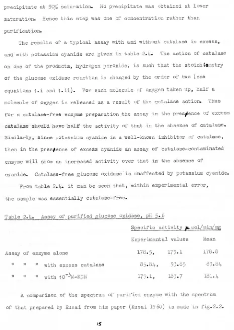
The results of'a typical assay with and without catalase in excess,
and with potassium cyanide are given in table 2.4. The action of catalase
on one of the products, hydrogen peroxide, is such that the stoich:~.metry
of the glucose oxidase reaction is changed by the order of two (see
equations 1.i and 1.ii). For each molecule of oxygen taken up, half a
molecule of oxygen is released as a result of the catalase action. Thus
for a catalase-free enzyme preparation the assay in the pres;ence of excess
catalase should have half the activity of' that in the absence of catalase.
Similarly, since potassium cyanide is a well-known inhibitor of'catalase,
then in the prestence of excess cyanide an assay of catalase-contaminated
enzyme will show an increased activity over that in the absence of
cyanide. Catalase-free glucose oxidase" is unaffected by potassiQm cyanide.
From table 2.4. it can be seen that, within experimental error,
the sample was essentially catalase-free.
~able ~~~~~rified glucose oxidas~, pH
5.6
§Eecific a~vit,~~~~~~~
ExperLmental values Mean
"
"
II with excess catalase" with 10-
3
!i-KCN178.5,
85.84,
179.1,
178.8
89.84
181.4
Assay of enzyme alone
"
"
A comparison of the spectrum of'purified enzyme with the spectrum
[image:27.497.27.492.135.791.2]Discussion.
The purity of an enzyme is established by showing that the final
product of purification consists of only one protein. If the product appears
homogenous by several different tests then there is a high probability that
it is the pure enzyme. i'iostof the tests are based of the physical properties
of proteins, but they ~ay include tests on catalytic properties also. Kusai
et al (1960) established the purity of their glucose oxidase preparation in
the following
waySj-1. By measuring spectrophotometrically the absorbance at 280nm per absorbance
at 460 nm. This ratio measured the protein content of the preparation per
flavin mOiety, and was found to be constamt for the final fractionation.
crystallisation and recrystallisation. Similarly lack of contamination by
coloured impurities was measured by the ratio of absorbance at 380 to 460 mn.
2. Ultracentrifugation was carried out in 0.1 M-acetate buffer pH
5.6,
andfrom the pattern obtaned the enzyme was said to be homogenous.
3. Electrophoresis in 0.1 M-acetate buffer pH 4.85 again showed the enzyme
to be homogenous.
None of these tests was carried out for the present enzyme preparations.
During chromatography it was noted that as the discrete yellow band with
glucose oxidase activity was eluted from the column, a brown coloured material
possibly catalase, remained at the top. Assays of the final product with
excess catalase or with 10-3 M-KCN (table 2.4.) showed that the enzyme was not
seriously contaminated with catalase. The spectrum of this enzyme was very
similar to that of Kusai's preparation. The latter had a specific activity of
of 100 mol oxygen uptake/mg protein/min compared with an average value of 65
for this preparation.
Unfortunately enzyme purity was not fully established and final specific
activity was low. Considerable loss of activity occurred at the chromatography
stage and further work should have been done in this area if this shorter
purification method is to supersede that of Kusai.
0.1
,
;'
0.5
z
o
I-U
Z
....
x w
I
I
I
/
,
D,
"
"
/
\ ;'
,
~,"
0.3
350 450
sso
om
Fig.3.1. ru~ of rcconstitu
and F'.J" or F.'1!J.C'us at 19f')
Curve A F:.1'-enzy:ne, oxidise fOrti.
Curve C.
"
" r uced ~ith glucose.Curve B FAl:i-erzyme, oxidised for.n.
Curve D It
QRAPrER THREE THE ATTEMPTED PREPARATION OF THE J\POEN2YLiE OF P. Al. GASAKIENSE GLUCOSE OXIDASE .AND REC01.lJ3INATION EXPERD,IENlS WITHT'I-IE .APOENZYME Olt' A. lUGER GLUCOSE
OXIDAS~.
-In~ction.
Kusai (1960) described the preparation of the apoprotein of
~.amagasakiense glucose oxidase, by an acid-a~monium sulphate method
at 0°. He was able to recombine the apoprotein with both l!'ADand FMN
and obtained re~ation of 93% initial activity in the former case.
~ did not reactivate the enzyme, but it was found to combine with the
apoprct ei.n, Addit ion of glucose to both the FAD-enzyme and the
FlvIT'I-enzyme brought about reduction of the prosthetic group and a change in
their absorption spectra (see fig.3.1.). However the results showed
that the newly synthesised enzyme contained
6
moles flavin per moleenzyme, of which 2 moles flavin were reduced by glucose addition.
He did not obtain enhanced flavin content when native enzyme was
incubated with excess FAD. To account for these facts, it was
suggested that the protein Was probably modified by the acid-ammonium
sulphate treatment, enabling a maximum combination of 6 moles flavin
per mole enzyme. But further experiments failed to confirm that the
protein moiety had been modified sufficiently to be more susceptible
to proteinase digestion.
Although acid-arll.moniu.msulphate treatment is most comrnonly used to
split t is flavoprotein enzyme into its componerrt parts (see Pazur and
Kleppe 1964, Swoboda 19696.)it has been reported that ultra-violet light
will eLi.cit dissociation of the FAD- protein complex 0 the P. vitale
enzyme (Levina et al 1965). No attempt was made by these workers to
reactivate the enzyme. Klarner and co-workers (1969) have demonstrated
the dissociation of part of their s~~ple of glucose oxidase from ~ger,
into flavin and ap opr ot e i,n after d.ia Iy s i.s against 0.02 M-phosphate
buffer pH 11.4. They claim that full activity was regained on
neutralisation of the solution containing both parts of the enzyme.
Dialysis against 1 M-potassium bromide has effected the
dissociat ion of the flavoprotein enzyme D-amino acid oxidase (Massey
and Curti 1966), while Komai, Massey and Palmer (1969) have obtained
deflavo xanthine oxidase by treatment of the holoenzyme with calcium
chloride at high concentrat ion, when the flavh\ was released as FiVm.
(High concentrations of calcium or magnesiwn ions cause hydrolysis
of FAD.)
Muller (1941) reported that his enzyme preparation oxidised
glucose both aerobically and anaerobically using 2,6-dichlorophenol
indophenol (DCPIP) as electron acceptor. Keilin and Hartree (1946)
have confirmed this as has Kusai, who found that anaerobic oxidation
of glucose occurred at a rate only 3.3JQ of that in the presence of
c!!d.ygen. He also showed not only that the ]:MNcontent of his Ii'11l'J-enzyme
could be reduced by a third on addition of glucose, but that anaerobic
reduction of added dye was faster with the :B1JvIN-enzymethan wit: the
FAD-enz me. Swoboda (196~ showed that for glucose oxidase from A.niger
}1iN does not bind to the apoprotein. In experiments where apoenzyme
was added to a mixture of F11Nand FAD, and the mixture assayed
Ll11.mediately for regenerated glucose oxidase activity, Film was found
not to inhibit the -bi.ndi.ng of apoenzyme and FAD. Moreover , in
sedimentat ion experiment s the sedimentat ion pattern was the same for
apoenzyme and FMNtogether as it was for apoenzyme alone. The pattern
of sedimentation for apoenzyme and FAD together was quite different.
Pazur & Kleppe (1964) noted on reactivating apoenzyme with FAD and FMN,
that only the incubated FAD plus apoenzyme had glucose oxidase
activity-~ethods and
materials-a) The purification of FAD.
The purification of commercial FAD (Sigma) was carried out by the
chromatography on a DEAE-cellulose column as described by Massey and
Swoboda
(1963).
However, gradient elut ion was used with phosphatebuffer between 0.1 M and 0.6 For all yellow fractions
froM
theFLUORESCENCE (orbilfO'Y unils)
(,
r0 ..
an ~ M «"! "":
0 0 c:i 0 Cl
0 .n _
....
...,
1
0 w 'r
....
V s wCl ....
...
\n 0 <t-v • Cl III
~
<
::>E
_, M W c,'"
'"
.I> 11 0 ell N ~ ~....
til 0 ...,..
o N'"'
OJ E:: E 0 o Cl "0 ." Q) N .c ro s.. Q), 0 0 () ...., c til Q) E o 0 (I) :.. Q) .c s.. () 0 ::l....
....
0 CHs:: Cl
...
:.. N QI ...., ...., CO c:: 0 Cl
....
'"
....,...
:l .-i QI Q)eS
0....
...
.
N.
(\"\ 01....
~ ('II "": 0 0I
I
100
• I
I
E :;)
50
E
)( 0
E
....
0
Cf<
~ >- to->
t-U
<Cl:
FAD CONCENTRAT ION. (M)
column the absorbance at 450 nm was measured. The fluorescence of
these same fractions waS measured in a Farrand spectrofluorimeter mark
II (Farrand Optical Company Inc.) using an excitation wavelength of
460 nm and measuring the emission at 520 rm, The elution pattern is
shown in fig.3.2.
b) Preparation of apoenzyme from ~.amagasakiense glucose oxidase.
The methods of Kusai (1960) and Swoboda (1969a) were used. Both
methods utilise acid-w.LmoniQm sulphate treatment under slightly different
conditions of temperature and pH. In each case the final preoipitate
was assayed for residual glucose oxidase activity and for regenerated
activity after incubation of the precipitate with 10-3 M-FAD for 30
min at 250• Details of the assay are given in "Methods" chapter 2.
The method of Komai, Aassey and Palmer (1969) was tried, but
without the initial dialysis, which was designed to remove the
contaminating phosphate from their enzyme preparation.
c) Preparation of apoenzyme from A.niger glucose oxidase.
The method of Swoboda (1969a) was used. The final precipitate was
assayed as described in (b) above and the results snown in table 3.1.
Titration of the apoenzyme with FAD was carried out by incubating
equal volumes of 2.5 x 10⁻⁵ M-apoenzyme and FAD (var ious concentrat ions)
for 30 min at 25°C. The mixtures were then assayed for glucose oxidase
activity, and a graph was plotted of activity against concentration
of FAD (fig.3.~.)
Results.
The results of the chromatography of B'AD are shown in fig-3.2. Two
peaks of yellow, fluorescent material were eluted from the column. The
ratio of fluorescence to absorbance at 450 nm of the first peak was ten
times greater than that of the second peak. Hence the first peak was
",U"l" and the second w.as FAD.
Attempts to prepare the apoenzyme of glucose oxidase from
~.amagasakiense proved unsuccessful using both the Kusai and Swoboda
Table
3.1.
Results of an a120enzyme preparation using •• niger
E~ucose oxidase.
Protein Volume Specific activity
(biuret) ml
14
mol/miry'DJgIU,l!jml
Holoenzyme 14.0
4
36.0Apoenzyme
4.0
3
Apoenzyme after in~bation with
2.0 6 12.7
10 Jot-FAD
Recovery of specific activity =
30%
methods. In each case when the final precipitate was tested for
activity in the absence of added FAD, activity was found. This
activity was not significantly enhanced by incubating the precipitate
with excess FAD before assa,ying for activity. It was noticed that
most of the precipitate was still yellow even after five cycles of the
Kusai or Swoboda procedures.
An apoenzyme preparation by Dr-sSwob oda using the method of Komai
and coworkers, yielded about 1
07~
apopr ot ei n, However, the sma.lLquantity of holoenzyme avail&)le prevented further attempts at
apoenzyme preparation. The results of' a typical apoenzy: e preparation
of
1::.
niger enzyme using the Swoboda method are given in table3.
1•It was observed that most of the yellow flavin was removed from the
enzyme during the first treatment by acid-runmonium sulphate at
-50.
Not more than three cycles of the procedure were required in total to
remove the FAD content of the holoenzyme. 'I'he results of the t itrat ion
of the apoenzyme with FAD are shown in fig.3.~. From this it can be
seen that the end point of the titz-at Lon was 5 x 10⁻⁵ M-PAD for the
2.5x10⁻⁵ M-apoenzyme present. 'I'hi.s gives a maximam binding of 2 mol
FAD/mol apoenzyme.
Discussion
No preparation of ~.amagasakiens~ apoenzyme produced sufficient
yield for further experiments. Using
1::.
niger apcensyme it was confirmedthat there are
2
molesFAD
per mole protein in the enzyme.Swoboda
(1969a)
discussed the possibility of the apoenzyme existingin two forms Pl and P2, both of which were able to combine with FAD
as
follows:-PI ~ P2 ...• (i)
P I-FAD - P 2-FAD •••••
(ii)
P2-FAD ••.•.•••.••••.•••
(1.1.1.)
Kusai
(1960)
showed that whereas the P. amagasakiense apoenzymecombined with both
m
and FAD, only the latter reactivated the enzymetowards glucose and oxygen. However, the newly-synthesised FMN-enzyme
was more effective in the DCPIP assay than was the FAD-enzyme.
It had been hoped to distinguish between mechanisms
(ii),
and.(i) and (iii) above, by investigating DCPIP and oxidase activity as
the apoenzyme combined with tAD and li'lrn at low temperatures. It has
been shown by Kalse and Veeger (1968) and Visser and Veeger (1968) that
in the case of 1ipoarnide dehydrogenase there are two stages of
regeneration of activity. When the apoenzyme monomer and FAD are
incubated together the reconstituted enzyme has high DCPIP activity
and low activity towards oxidised lipoate. Prolonged incubation gives
holoenzyme (possibly a d.imer ) with low DCPIP activity but high activity
towards oxidised lipoate.
CHAPrER FOUR ENZYME INHIBITION BY HEAVY II1!£.&§.
.!,illroduction
The sensitivity of proteins to metals at very low concentrations
often results in denaturation and acc onp anyi.ng structural changes of the
protein molecule. In some cases the effect of these metals in bringing
about changes in the secondary and tertiary structure of the proteins has
been correlated with the catalytic oxidation of sulphydryl groups in the
protein. For example, Casola, Brumby and Massey (1966) and Casola and
Massey (1966) have shown that catalytic oxidation by metal ions of
sulphydryl groups in the enzyme lipoyl dehydrogenase results in a change
in the activity of the enzyme. Inhibition of enzymes by metals may be due
to structural changes in the protein chain, either around the act ive site
or at a site distant from the active site but involved in the catalytic
activity of the protein, the catalytic site.
In addition to the effects of metals on the protein constituents of
an enzyme, there are some reports of the complexing of metals to co-enzymes.
Albert
(1953)
reported a series of studies on the avidity of some naturalsubstances for trace metals. These natural substances included riboflavin,
a precursor of FAD, which showed a greater avidity for the ferrous ion than
for the cupric ion. In each case the avidity is thought to have been due
to the presence .of an ionisable hydroxyl group at the C-4 position, peri
to the tertiary heterocyclic nitrogen atom at position
5
in the isoalloxazineBamberg and Hemmerich (1961) have invest igated complexes of' silver
and isoalloxazine. Their results have shown the nature of the interact ion
between the ions of silver (1), copper (11) and mercury (11) but with no
other metal ions. Degtyar and Gulii (1967) have reported that while glucose
oxidase from ~~~ is inhibited by mercuric inhibitors, it is activated
by calcium and manganese salts. This latter finding is rather surprising
since Komai, Massey and Palmer (1969) have used high concent r at ions of
calciQm and of manganese to prepare deflavo xanthine oxidase. They found
that these salts caused hydrolysis of FAD.
Kusai (1960) has shown that for P.amagasakiense enzyme the catalytic
activity is strongly inhibited by sulphydryl reagents such as pC:MB,
HgC12' AgCl, CuS04. For the !.niger enzyme species Swoboda and Massey
(1965) could not detect any sulphydryl groups in either the oxidised or
reduced native enzyme when they reacted the enzyme with p-mercuri-acetate
and measured the excess mercurial compound polarographically. After
denaturation by heat or urea they obtained evidence of One cysteine (SH)
and two cystine (S-S) residues per molecule of' enzyme. Kleppe (1966)
confirmed that this glucose oxidase species was not inhibited in either
the oxidised or reduced form by mercurial compounds. Nakamura and Fujiki
(1968)
have carried out a complete amino-acid analysis of the glucoseoxi.dases from
!.
niger and P.amagasakiense and have found that there are7 sulphydryl residues per molecule of enzyme frtM_either source.
Nakamura and Ogura
(1968)
have obtained inhibition of' activity of theA niger species by mercury
(II),
silver (1) and copper(II)
salts, and bypCMB and p-mercuriacetate. They have shown that the effect of these two
latter inhibitors is reversible by dilution.
Iat er ials and meth ads
Inlilbition studies were carried out in two ways, using the oxygen
electrode to assay for residual act ivity as described in "Materials and
(i)
Preincubat ion method.The enzyme and inhibitor were incubated together in the electrode
cell for various lengths of time before the addition of glucose to init iate
the react ion.
(ii) on-incubation method.
With the glucose and inhibitor in the cell, the enzyme solution was
added last of all to start the reaction.
The first method was used to obviate the lag period in inhibition
experiments. The latter method should give identical results if the
inhibition is reversible. If part of the inhibition was a slow reaction,
then the preincubated assay would show a different rate of initial
activity to that of the non-incubated assas·
In some experiments potassium cyanide (10-4 M-final concentration)
was added to inhibit any traces of catalase with which the enzyme might
be contaminated. Such a contarnination would upse t the stoichiometry of the
reaction by regenerating oxygen in the electrode cell.
Most experiments were carried out in 0.1 M-phosphate buffer pH 6.0 at
Z50C. In experiments with pC$lB 0.1 M-pyrophosphate buffer pH 7.0 was used.
With copper sulphate as inhibitor 0.01 M-acetate buffer pH 6.0 was used.
The final enzyme concentration was 3.0 x 10-
9
M (active site concentration).Results
---a) Effect of time of incubation on inhibition by HgC12
The effect of incubating enzyme and inhibitor for various tLmes before
assaying for activity is compared with no preincubation in the following
table.
The effect of_Ere incubat ion on inhibition
~reincubation (miEl , ~O__ 1
5
10 _ _1.217
19
2117
17
Similar results were obtained at higher inhibitor concentrations. It can
be seen that no difference was observed in the percentage inhibition
obtained using either the preincubation method or the non-incubation
method.
b) Effect of concentration of mercuric salts on the extent of inhibition ..
The results are summarised in the following table. Five minutes
preincubation was used throughout these assays ..
l~~n by various mercuric salts
Inhibitor final
_ cone. ( 11) ________ ~10~-_4 10-
5
___ _12-4+ _
Inhibitor
--____ -__1~bition ('(0) _
HgC12
HgBr2
Hg( ~D3)2 basic
pCMB
81
19
88
7623
72
88
45
75
96
+ values from Kusai 1960
Various concentrations of Hg(C )2 had no inhibitory effect
c) Effect of KC on the inhibition of activity
As can be seen from the following table, the presence of a ten-fold
excess of cyanide in inhibition assays caused reversal of inhibition
The effect of KCN on inl1.ibition
%
inhibition-HgBr2 + enzyme
23
"
+ 10-3 -KC + enzymeo
93
-2
+ 10 M-KCN + enzyme
o
This reversal of inhibition was also demonstrated in the following way.
An
inhibition assay was set up as described in "Materials and Met hods" and the
progress of the assay was followed on a pen recorder. After 100 sec the
addition of a ten-fold excess of cyanide over inhibitor was made to the
electrode cell. A change in the rate of oxygen uptake by the enzyme was
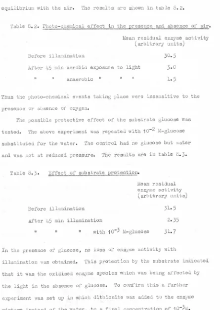
seen immediately. This is shown in fig.4.l.
100
o 100 200
Fig.4.l. ddition of 10-
3
M-KC to an inhibition assas containigg10-4 -HgC12.
In this experiment the inhibition assay showed that the enzyme had lost
88%
of its activity. On addition of the ten-fold excess of KCl glucoseoxidase activity was restored to 100% as if no inhibitor was present.
d) Effect of inhibition by silver salts
In all cases when the silver salt was dissolved in 0.1 M-phosphate
buffer pH 6.0, a fine yellow precipitate was formed. This may have been
silver triphosphate, which is a yellow salt, and it rendered the inhibitor
solution unusable. It is likely that silver salts are not precipitated by
other buffers but none were tried. Kusai (1960) reported that silver
chloride brought about 88% inhibition at 10-4,.
e) To investigate whether cation inhibition is due to a dipole or metal
ion effect.
For a metal salt consist ing of metal Ii and ligand L, the following
equation can be applied to its
dissociation:-ML
where Keq is the equilibrium constant
When the concentration of undissociated salt (ML) is held constant in
solution there will be a fixed proportion of dissociated salt in the form
of metal ion and ligand, the proportion being determined by the equilibrium
constant.
Copper sulphate was chosen as an easy inhibitor to use. Kusai (1960)
obtained
30%
inhibition at 10-4 M and 0 at 10-5
'1. For copper sulphateK
=
223.9. Thus using a constant concentration of CuS04(5
x 10-5M) ineq
I Q) ..::t a 0 ~ Cl) ::s ~ o
,
l..C\ENZYME ACTIVITY (%) ~
I
0
g
0">0 po I r-l 0 ><
t
.-i>:r;-t C"\ ~
.~
0 -'M 0 rJl ~ ::s \0 ~e
tIl 0 Z Co 'M <I) +> -c J.t 'n w Q) .0 0.:....
'M U....
.c Z ::s s:: .0 04
'Ms:: Q) \t'\
~ N 0 co 'M +>
....
+' Q) III co Uo 10 ..:::t
I 0
....
Ul 0 ::s .-i U Q) 0 I 1-0.
~ ::s 0 +' l..C\ co I~ It! 0
s::
....
Q) 0 .c 'n I<....
....
..,.. \t'\ ~ '"0 0 ~-.c 0 s::
If.
U 0
..-1
0 »
+'
+> co III
It! J.t
(j)
{I) +'
:> co ~
M Q)
::s CII o
o '"0 ~
0 0 r-l J.t () (c +> +J U Q) s:: CII +' Q) r-l
....
S Q) <II
...
M s:: Q)
III
Q) :>
c~ 0)
....
» +'
~ll >: o
0 0 co
of undissociated salt by adding various calculated amounts of sulphate in
the form of sodiQm sulphate to the ass~ mixture. When the percentage
undissociated Ou804 is plotted against sulphate ion concentration, the
following curves can be predicted for the concentrations of free metal ion
and salt in the assay mixture.
100 100
1
o1
Fig.4.2. Predicted curves for dissociation of metal salt.
As the proportion of ligand is increased in the assay mixture, the amount
of free metal ion decreases while the amount of undissociated salt increases.
Fig.4.3. shows the experimental curve obtained, and as can be seen, enzyme
activity increased as the concentration of free metal ion decreased. That
is, the enzyme was inhibited by free metal ions in solution and as the
metal ion concentration cic.c~ •• ci Ithe enzyme recovered its catalytic
activity. Therefore inhibition by Ou804 is due to the dissociated
copper cation and not to the salt. The presence of weak acetate buffer was
thought to have no effect on the copper ion concentration since copper
acetate is soluble in aqueous solution up to 0.35 molar.
Qiscussion
The inhibition of P.amagasaki~ glucose oxidase by metal salts
has been described by Kusai
(1960).
In this paper he showed that at thesame molarity, GuS0
4,
AgCl and HgC12 brought about the same degree ofinhibition of the enzyme. For copper sulphate it has been shown that
inhibition is due to the copper cation. It seems likely therefore that the
free cations of silver and mercury, and not the undissociated salts, are
responsible for inhibition shown by silver and mercury compounds respectively.
Inhibition by mercurial compounds was confirmed experimentally at
concentrations similar to those used by Kusai. Both the preincubation and
the non-incubation experLments gave identical results indicating that there
was no slow reaction between the enzyme and inhibitor.
From kinetic studies, Nakamura and Ogura (1968) have shown that
mercury (II) competes with oxygen for the reduced form of the enzyme,
and not with glucose for the oxidised form. They analysed the inhibit ion
by mercury (II) and silver (I) compounds using the double reciprocal plot
method (see appendix 1). From the graphs obtained the inhibition by silver
was shown also to be competitive with oxygen for the reduced enzyme species.
This was confirmed by stopped-flow experiments. Using difference absorption
spectra they also showed that the change in spectrum of free FAD when
mercury (II) was added to it was sLmilar to the difference between the
spectrQm of the enzyme and that of enzyme + mercury (II) or enzyme +
silver (I). However using kinetics to investigate the binding sites of
these two metals, they found that the binding sites are different for the
.metals.
It has been shown that free cupric ions are responsible for the
inhibition when CuS04 is used, and has been suggested that mercuric ions