ISSN Online: 2164-2656 ISSN Print: 2164-2648
DOI: 10.4236/aid.2018.81002 Jan. 26, 2018 10 Advances in Infectious Diseases
Progressive Multifocal Leukoencephalopathy
—A Case Report in an Immunocompetent
Patient
Ana Faceira
1,2*, Daniela Magalhães
2,3, Filipa Ceia
2,4, João Nuak
2,5, Carina Reis
6, Susana Ferreira
11Internal Medicine Department, Hospital de São João, Porto, Portugal 2Faculdade de Medicina da Universidade do Porto, Porto, Portugal
3Endocrinology, Diabetes and Metabolism Department, Hospital de São João, Porto, Portugal 4Instituto de Investigação e Inovação em Saúde, Universidade do Porto, Porto, Portugal 5Infectious Diseases Department, Hospital de São João, Porto, Portugal
6Neuroradiology Department, Hospital de São João, Porto, Portugal
Abstract
Progressive multifocal leukoencephalopathy (PML) is a demyelinating disease of the central nervous system due to the reactivation of the JC virus, which usually occurs in immunocompromised patients and is a major opportunistic infection associated with HIV infection. We report a case of a previously healthy patient who was diagnosed with PML.
Keywords
Immunosuppression, Immunocompetent, JC Virus, Progressive Multifocal Leukoencephalopathy
1. Introduction
JC virus (JVC) is a double-stranded circular DNA virus, which belongs to the Polyomaviridae family and to the Polyomavirus genus. The polyomaviruses are ubiquitous in nature and are species-specific, similar to the JCV in humans.It is assumed that the major routes of the virus’s transmission are inhalation and in-gestion of contaminated water [1][2].
JCV is the etiologic agent of a demyelinating disease of the central nervous system (CNS)—progressive multifocal leukoencephalopathy (PML). The patho-genesis of PML is divided into 3 phases. The primary infection is usually as-ymptomatic, with approximately 85% of the adult population showing antibod-How to cite this paper: Faceira, A.,
Ma-galhães, D., Ceia, F., Nuak, J., Reis, C. and Ferreira, S. (2018) Progressive Multifocal Leukoencephalopathy—A Case Report in an Immunocompetent Patient. Advances in Infectious Diseases, 8, 10-16.
https://doi.org/10.4236/aid.2018.81002
Received: December 17, 2017 Accepted: January 23, 2018 Published: January 26, 2018
Copyright © 2018 by authors and Scientific Research Publishing Inc. This work is licensed under the Creative Commons Attribution International License (CC BY 4.0).
http://creativecommons.org/licenses/by/4.0/
DOI: 10.4236/aid.2018.81002 11 Advances in Infectious Diseases ies against the JCV, and thereafter the virus remains as a persistent latent infec-tion in the urinary tract, bone marrow and spleen. The final phase is related to the reactivation and dissemination of the virus with an almost certain hemato-genous spread to the CNS [1][2][3].
2. Case Report
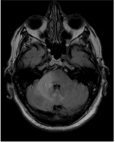
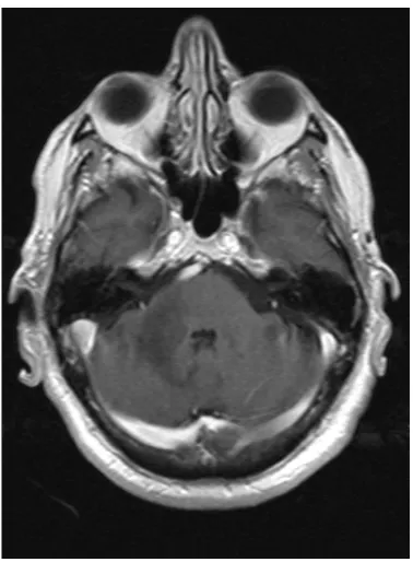
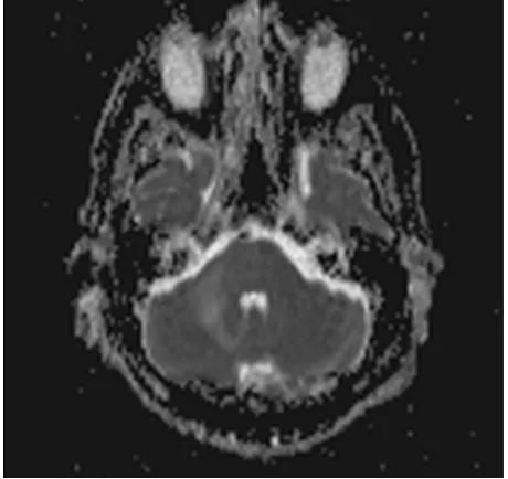
[image:2.595.279.469.473.707.2]A 68-year-old Caucasian male was evaluated in a medical consultation with a history of dysmetria of the right limbs, dysdiadochokinesia, limb ataxia and dy-sarthria, which lasted for approximately one month. The brain magnetic reso-nance imaging (MRI) showed a T2 hyperintense (Figure 1, Figure 2) and T1 hypointense lesion in the right cerebellar hemisphere and middle cerebellar pe-duncle, with no mass effect or gadolinium enhancement (Figure 3), and diffu-sion-weighted imaging (DWI) showed predominantly facilitated diffusion of water molecules, with patchy peripheral high signal but with no apparent diffu-sion coefficient (ADC) lowering—no restriction (Figure 4, Figure 5). An exten-sive complementary study was carried out, including: cerebrospinal fluid (CSF) analysis (microbiological, immunophenotypic and cytological studies with ma-lignant cell research), immunological study (antineutrophil cytoplasmic anti-bodies, antinuclear antianti-bodies, anti-double stranded DNA antibodies) and in-vestigation to exclude a solid neoplasia/lymphoma (upper gastrointestinal endo-scopy, colonoendo-scopy, thoraco-abdominopelvic computed tomography scan, se-rum protein electrophoresis) or an infectious disease (particularly HIV infec-tion). This study was inconclusive and did not allow an accurate diagnosis. Thus, because it was not possible to exclude that it was a subacute cerebral infarction, antiplatelet therapy with clopidogrel and motor rehabilitation treatment were set.
DOI: 10.4236/aid.2018.81002 12 Advances in Infectious Diseases
Figure 2. Coronal T2 TSE.
Figure 3. Axial post gadolinium T1 SE.
[image:3.595.279.468.326.583.2]DOI: 10.4236/aid.2018.81002 13 Advances in Infectious Diseases
Figure 4. Axial DWI.
Figure 5. Axial MAPA ADC.
To what concerns the previous medical history, the patient had hypertension, dyslipidemia, degenerative osteoarticular disease and benign prostatic hyperpla-sia. The patient had no history of immunosuppressive therapies. The epidemi-ological context and family history were irrelevant. There was no consumption of tobacco, alcohol or illicit drugs and no sexual risk behaviour.
[image:4.595.259.489.323.540.2]DOI: 10.4236/aid.2018.81002 14 Advances in Infectious Diseases showed the presence of dysarthria (without aphasia), right dysmetria in the fin-ger-to-nose and heel-to-shin tests (without worsening when closing the eyes), dysdiadochokinesia in the rapid alternating movement test and ataxic gait in the heel toe walk test.
Laboratory exams revealed haemoglobin of 13.0 g/dL, leucocytes of 6.19 × 10⁹/L (neutrophils of 4.18 × 10⁹/L and lymphocytes of 1.28 × 10⁹/L with 953
CD4+), C-reactive protein of 12.5 mg/L, a normal LDH, and no hepatic or renal
dysfunction. Serum levels of immunoglobulins and complement were normal. The peripheral blood immunophenotyping analysis revealed a polyclonality of B cells, with no changes in the T cells. During the course of hospitalization, tran-sient lymphopenia was observed with a minimum lymphocyte count of 0.82 × 10⁹/L.
Taking into account the clinical course and particularly the imaging features, it was hypothesized that it could be a JC virus infection. HIV serology (HIV 1/2 screening with an antigen p24 plus antibodies test) was negative and HIV RNA was not detected. A lumbar puncture was performed which revealed total cells 2/μL, proteins 0.85 g/L and a normal CSF/serum glucose ratio. The JC virus DNA was positive in the CSF (1.47 × 10⁴ copies/mL) and in the blood (<150
copies/mL), determined by polymerase chain reaction (PCR). Accordingly, the definitive diagnosis of PML was assumed and treatment with mirtazapine was initiated. An obvious and progressive clinical and neurological worsening was observed and one month after the diagnosis the patient was aphasic and without the ability to have an autonomous gait. No further treatment was attempted, nor any further diagnostic or invasive intervention were performed. The death was occurred 1 year and 5 months after the diagnosis.
3. Discussion
PML occurs more frequently in immunocompromised patients and is a major opportunistic infection associated with HIV infection, making up approximately 80% of patients with PML. Other less common causes associated with PML are haematological malignancies (13%), organ transplant recipients (5%) and auto-immune diseases treated with immunomodulators (3%) [3]. However, PML is no longer limited to the aforementioned groups, being described in patients with conditions associated with minimal immunosuppression, such as renal failure or liver cirrhosis. In addition, there are some reported cases of PML in patients without any apparent cause of immunosuppression [3]. After a review of the lit-erature, we found about twenty-four cases described in individuals with no co-morbidities and without known cause of immunosuppression, but we emphasize that in half of these cases the HIV serology was not reported.
ab-DOI: 10.4236/aid.2018.81002 15 Advances in Infectious Diseases normalities, and language and speech disturbances [4]. PML is typically a bilat-eral, asymmetric and supratentorial white matter disease (typically parietal and frontal lobes), although it may be unilateral and with just a single lesion. The white matter of the posterior fossa is the next most commonly involved area, classically the middle cerebellar peduncles and adjacent pons and cerebellum, often synchronous with supratentorial lesions, being limited do infratentorial structures in about 10% of cases [2].The MRI is the technique of choice, which reveals hyperintense lesions on T2-weighted and T2 FLAIR images and hypoin-tense lesions on T1-weighted images in the involved brain regions, and periph-eral patchy diffusion restriction may be present. Mass effect is infrequent and mostly minimum, and lesions may enhance in about 10% of cases, typically at the periphery.Although the brain biopsy is the gold standard for the diagnosis of PML, if the neurological and neuroradiological findings are suggestive, the di-agnosis can be established by demonstrating the presence of JC virus DNA by PCR in the CSF [2][4]. Thus, in this case the brain biopsy was not performed.
Although obvious immunosuppression exceedingly increases the risk of PML, many chronic diseases might be associated with a transient immune dysfunction which may promote the reactivation of the JC virus by impairing the ability to mount a T cell response. Another cause that has been associated with PML is idiopathic CD4+ T-lymphocytopenia [5]. In this case, the initial neurological
symptoms and the first brain MRI led to a misdiagnosis of a stroke. Subsequent clinical evolution and the imaging progression of the lesion raised other diag-nostic hypotheses. Taking into account the extensive complementary study car-ried out, it was possible to exclude with some certainty that it was a neoplastic, lymphoproliferative, autoimmune disease. We emphasize that transient lym-phopenia was observed during hospitalization (an immunophenotypic study was not performed at this time because this analytical change was only observed in a single study), and was not observed during subsequent analytical monitoring.
4. Conclusions
Associating the positive CSF JC virus PCR result to the highly suggestive clinical and imaging findings, it was possible to establish the diagnosis of PML. The cause of viral reactivation in our case remains unknown.
Currently, there is no specific antiviral agent for the JC virus infection and the existing treatment goal in PML is to restore host adaptive immune response (for example, antiretroviral therapy if HIV infection). Although the results were un-satisfactory, a number of specific drugs against the JC virus (as well as cidofovir, topotecan, cytarabine, mefloquine, mirtazapine) have been investigated. Mirta-zapine has been implicated in preventing the progression of PML in some case reports [2][5], which was not observed in our case.
Conflicts of Interest
DOI: 10.4236/aid.2018.81002 16 Advances in Infectious Diseases
References
[1] Tan, S.C. and Koralnik, I.J. (2015) JC, BK, and Other Polyomaviruses: Progressive Multifocal Leukoencephalopathy (PML). In: Bennett, J.E., Dolin, R. and Blaser, M.J., Eds., Mandell, Douglas, and Bennett’s Principles and Practice of Infectious Diseases, 8th Edition, Elsevier Saunders, Philadelphia.
[2] Bag, A.K., Curé, J.K., Chapman, P.R., Roberson, G.H. and Shah, R. (2010) JC Virus Infection of the Brain. American Journal of Neuroradiology, 31, 1564-1576. https://doi.org/10.3174/ajnr.A2035
[3] Gheuens, S., Pierone, G., Peeters, P. and Koralnik, I.J. (2010) Progressive Multifocal Leukoencephalopathy in Individuals with Minimal or Occult Immunosuppression. Journal of Neurology, Neurosurgery & Psychiatry, 81, 247-254.
https://doi.org/10.1136/jnnp.2009.187666
[4] Berger, J.R., Aksamit, A.J., Clifford, D.B., Davis, L., Koralnik, I.J., Sejvar, J.J., et al. (2013) PML DIAGNOSTIC CRIteria: Consensus Statement from the AAN Neuro-infectious Disease Section. American Academy of Neurology, 80, 1430-1438. https://doi.org/10.1212/WNL.0b013e31828c2fa1