ORIGINAL RESEARCH
Contribution of Diffusion-Weighted Imaging in the
Evaluation of Diffuse White Matter Ischemic
Lesions in Fetuses: Correlations with
Fetopathologic Findings
F. Guimiot C. Garel C. Fallet-Bianco F. Menez S. Khung-Savatovsky J.-F. Oury G. Sebag A.-L. Delezoide
BACKGROUND AND PURPOSE:The sensitivity of fetal MR imaging is poor with regard to the evaluation of diffuse ischemic white matter (WM) abnormalities. Our purpose was to evaluate the contribution of diffusion-weighted imaging (DWI) in the analysis of microstructural changes in WM and to correlate neuroimaging with neurofetopathologic findings.
MATERIALS AND METHODS: We included fetuses with MR imaging, DWI, and a fetopathologic exam-ination. In a region of interest defined by MR imaging, where T1 and T2 intensities were abnormal, the apparent diffusion coefficient (ADC) was measured and immunohistochemical analysis was per-formed. In fetuses with no WM abnormality in signal intensity, region of interest was defined at random. Histologic reading was performed with a complete blinding of the MR imaging results and ADC values. Three degrees of histologic appearance were defined with regard to vasogenic edema, astrogliosis, microgliosis, neuronal and oligodendrocytic abnormalities, and proliferation or congestion of vessels and were compared with a2test in groups A (normal ADC) and B (increased ADC) fetuses.
RESULTS: We included 12 fetuses in group A and 9 in group B, ranging from 29 to 38 weeks of gestation. All group B fetuses and 1 group A fetus demonstrated WM abnormalities in signal intensity. WM edema and astrogliosis were more common in group B than in group A (7/9 vs 2/12 and 8/9 vs 4/12, respectively). No significant difference was observed between both groups with regard to the other parameters.
CONCLUSION:This study showed a strong correlation between increased ADCs and 1) WM abnor-malities in signal intensity on MR imaging, and 2) vasogenic edema with astrogliosis of the cerebral parenchyma.
T
he contribution of fetal MR imaging in the evaluation of cerebral ischemic lesions has already been evaluated.1MRimaging with the use of T2, T1, and T2*-weighted sequences has proved a valuable tool during the third trimester in the detection of small focal parenchymal lesions (such as cavita-tions and calcified leukomalacia) and ischemic lesions involv-ing the cortex (4-layered polymicrogyria and laminar necro-sis).1,2Conversely, the sensitivity of MR imaging was poor in the evaluation of diffuse ischemic white matter (WM) abnor-malities.1However, the detection of such lesions, before the
establishment of irreversible damage, is challenging in at-risk situations.
Injury to the WM gives rise to neuronal hypoxia and, sub-sequently, to neuronal death, which results in disruption of the cell membrane and the release of some neurotransmitters such as glutamate. The latter has been demonstrated to have a cytotoxic effect on neurons.3From this step begins microglial
and astrogliosis responses. Microglial cells produce proin-flammatory cytokines and reactive oxygen and nitrogen spe-cies that contribute to the strength of this deleterious effect.4
Astrocytes are recruited to repair the injured tissue and to reduce the effect of glutamate, resulting in a cellular swelling that corresponds to cytotoxic edema. A secondary vasogenic edema appears, caused principally by lysis of the astrocytic cell membrane and an opening in the blood-brain barrier, which induces an increase of the amount of water in the extracellular space.5 This vasogenic edema is present 1 week after the
insult.6
The purpose of this study was to evaluate the contribution of diffusion-weighted imaging (DWI) in the evaluation of dif-fuse ischemic WM changes in fetuses, by correlating DWI and MR imaging with neurofetopathologic findings obtained by standard histologic and immunohistochemical analyses of the main cellular populations of the brain.
Materials and Methods
Experimental Procedures
All patients included in this retrospective study underwent fetal cere-bral MR imaging with DWI. Termination of pregnancy (TOP) was performed in accordance with the French legislation and was fol-lowed by a fetopathologic examination. For each fetus, we recorded the main ultrasonographic and MR imaging findings, including the region of interest, where we measured the apparent diffusion coeffi-cient (ADC). The gestational age at which TOP was performed, and the delay between MR imaging and fetopathologic examinations was also recorded. We performed a complete neuropathologic examina-tion and undertook immunohistochemical studies at the level of the region of interest, to achieve histologic and MR image matching. We obtained histologic results with a complete blinding of the MR
imag-Received May 12, 2007; accepted May 15.
From the Service de Biologie du De´veloppement (F.G., F.M. S.K.-S., A.-L.D.), Service de Radiologie (G.S., C.G.), and Service de Gyne´cologie-Obste´trique (J.-F.O.), Hoˆpital Robert Debre´, AP-HP, Paris, France; and the Service d’Anatomie Pathologique (C.F.-B.), Hoˆpital Saint-Anne, Paris, France.
Please address correspondence to Anne-Lise Delezoide, Service de Biologie du De´veloppe-ment, Hoˆpital Robert Debre´, 48 Boulevard Se´rurier, 75019 Paris, France; e-mail: [email protected]
ing results and ADC values. We subsequently performed confronta-tion of MR imaging and histologic results.
MR Imaging and DWI
We performed MR imaging on a 1.5T unit (Intera; Philips Medical Systems, Best, the Netherlands) 30 to 40 minutes after fetal sedation was achieved with maternal oral administration of flunitrazepam (1 mg). We performed a brain examination with a phased-array abdom-inal coil, T1-weighted spin-echo, spectral presaturation inversion re-covery, fat-saturated sequences (697/14/2; flip angle, 90°; matrix: 256⫻256; FOV, 320 mm; rectangular FOV, 75%; section thickness, 4 mm; acquisition time, 2 min 56 s) and T2-weighted single-shot turbo spin-echo imaging (24,617/100/1; flip angle, 90°; turbo spin-echo fac-tor, 84; matrix: 256⫻256; FOV, 280 cm; section thickness, 3 mm; acquisition time, 24 s). DWI was acquired without maternal breath-hold on transverse sections with a multishot echo-planar imaging sequence with diffusion gradient (TR, 3341 ms; TE, 88 ms; flip angle, 90°; matrix, 95⫻256 mm; FOV, 280 mm; section thickness, 5 mm; b-values of 0 and 700 s/mm2applied in 6 noncolinear directions;
amplitude, 30mT/m; acquisition time, 53 s) (Fig 1). The same radiol-ogist (C.G.) manually traced a rectangular region of interest (region of interest size varied from 100 to 150 mm2) in the region where the
WM signal intensity was abnormal, on the image obtained with b⫽0 s/mm2. If the signal intensity was normal, the site where ADC was
traced was sampled at random. The ADC was calculated according to the equation: S(b)⫽S(0). exp(-b x ADC) where S(b) is signal inten-sity at a b value and S(0) is signal inteninten-sity without diffusion gradients. In reference with normal published data in fetuses,7,8ADC was
con-sidered normal when it was strictly inferior to 2.10m2/ms and
in-creased when it was superior or equal to this value. Two groups were defined: fetuses with normal ADC (group A) and fetuses with in-creased ADC (group B).
Termination of Pregnancy
We obtained TOP by performing feticide (injection of 5 U/mL of pentothal or 5 U/mL of KCl into the vein of the fetal umbilical cord) followed by induction of labor. Delivery usually occurred within 24 hours after feticide, and a fetopathologic examination was performed within 24 hours after delivery, which allowed good brain preservation.
Fetopathologic Examination
After the parents’ informed consent and in accordance with the French legislation, we obtained fetal brains from medically termi-nated pregnancies. The gestational ages of the fetuses was established by first-trimester sonography crown-rump length measurement and confirmed at autopsy by the evaluation of the fetal biometry, and organ and skeletal maturation. Brains were removed and fixed in 10% buffered formalin added with 3 g/L of ZnSO4 for approximately 3 to 4 weeks. Brains were then cut in coronal sections of 0.5 cm of thick-ness for routine examination. The region of interest identified by MR imaging was sampled, cut in several parts (from 2 to 3 blocks), and embedded in paraffin. Serial sections (thickness 4m) were per-formed on Superfrost Plus slides (Thermo Scientific, Waltham, Mass). One section was stained with hematoxylin-eosin (H&E) for histologic analysis.
[image:2.594.55.374.43.387.2]Immunohistochemical Examination
We performed all incubations in a humidified chamber. First, sec-tions were dewaxed with xylene and bathed consecutively in absolute ethanol, 75% ethanol, 35% ethanol, and H2O. Then, they were incu-bated for 30 minutes at 95 to 98°C in 10 mmol/L sodium citrate, pH 6.0, for antigen retrieval, and cooled for 20 minutes at room temper-ature. Next, endogenous peroxidase activity was inactivated for 5 minutes in 3% hydrogen peroxide. Sections were then washed 3 times in phosphate-buffered saline (PBS) and blocked 30 minutes in PBS, 3% BSA, 0.1% Tween 20 (Sigma, St Louis, Mo), before incubation with primary antibodies overnight at 4°C. The next morning, the LSAB2 streptavidin-biotin detection system kit (Dako, Trappes, France) was used to reveal immunolabeling. Sections were washed 3 times in PBS, 0.1% Tween 20, before adding secondary and tertiary antibodies (biotin-conjugated anti-mouse or anti-rabbit and peroxi-dase-conjugated streptavidin, respectively) for1 hour at room tem-perature. DAB (3.3-diaminobenzidine tetrahydrochloride, Dako) was used as a substrate for peroxidase, and incubation was performed for 5 to 10 minutes. Negative controls were tissue sections incubated without primary antibody and concentration-matched secondary and tertiary antibodies. Sections were counterstained with Mayer he-malun and mounted in PERTEX medium (Histolab Products, Goth-enburg, Sweden). Sections were analyzed on a DMR microscope (Leica, Heidelberg, Germany), and images were acquired with a DFW-SX900 camera (Sony, Tokyo, Japan) and Inspector software (Matrox Imaging, Quebec, Canada). Primary antibodies were mouse anti-human CD34 (Immunotech, Marseille, France), microtubule-associated protein 2 (MAP2) (Sigma, Lyon, France), CD45, glial fibrillary acidic protein (GFAP) and nuclear factor (NF) (all from Dako), rabbit anti-human myelin basic protein (MBP) (Dako), and OLIG2 (Chemicon, UK).
Histologic Evaluation
For histologic and immunohistochemical evaluations, 2 pathologists (F.G., C.F.-B.) separately analyzed all sections, with common reeval-uation when necessary.
Standard staining and immunohistochemical examination al-lowed us to define 3 degrees of intensity of vasogenic edema (corre-sponding to a WM dissociation resulting from an increase in extra-cellular water), microgliosis, astrogliosis, and proliferation or congestion of vessels. For edema, “degree 1” corresponds to an ab-sence of edema, “degree 2” to a mild dissociation of WM nervous fibers, and “degree 3” to a severe dissociation of WM nervous fibers with the appearance of empty zones. For astrogliosis, “degree 1”
cor-responds to a few round and swollen astrocytes observed among nor-mal starry astrocytes, “degree 2” to increased number of round and swollen astrocytes, and “degree 3” to only round and swollen astro-cytes. For microgliosis and proliferation or congestion of vessels, “de-gree 1” corresponds to only nonreactive resident microglial cells with-out proliferation or congestion of vessels, “degree 2” to more numerous nonreactive microglial cells compared with degree 1 and mild proliferation or congestion of vessels, and “degree 3” to an in-creased number of round reactive microglial cells associated with mi-croglial nodules and high proliferation or congestion of vessels. We then statistically compared the distribution of the 3 degrees of each parameter in group A and B fetuses. We evaluated the integrity of WM neurons and oligodendrocytes by counting and analyzing their mor-phologic appearance. Five microscopic nonoverlapping fields were analyzed for each slide. Neurons were classified as necrotic when they exhibited pyknosis, karyorrhexis, karyolysis, cytoplasmic eosino-philia (“dark neuron”), or loss of affinity for hematoxylin (“ghost neuron”).
Statistics
We performed statistical analysis using Excel (Microsoft, Redmond, Wash) and Prism 4 (GraphPad, San Diego, Calif) software. A2test
was performed on the percentage of distribution of histologic degree among the fetuses in groups A and B. We compared the averages of gestational ages and of the delay between MR imaging and fetopatho-logic examinations in both groups with a Studentttest to eliminate differences that could have biased the study. A probability value ofP ⬍.05 was considered statistically significant.
Results
Between 2002 and 2005, we included 21 fetuses (12 female and 9 male) in this study. Group A included 12 fetuses and group B, 9 fetuses. MR imaging was performed at a mean gestational age of 32 weeks. The gestational ages of the fetuses at TOP ranged from 29 to 38 weeks; the average was 33.38⫾0.66 weeks for group A and 34.71⫾0.83 weeks for group B (P⫽ .24). To ensure that no difference in the delay of MR imaging and fetopathologic examinations could be observed between both groups and therefore could distort the results, we com-pared the mean delay in the 2 groups: 16.38⫾3.55 days for group A and 10.25⫾2.35 days for group B (P⫽.22). The results are summarized in on-line Tables 1 and 2.
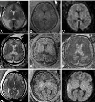
[image:3.594.55.373.42.210.2]observed in all group B fetuses and in only 1 group A fetus (case 11, on-line Table 1).
Vasogenic Edema
We studied and evaluated vasogenic edema on sections stained with H&E in the 2 groups of fetuses. We delineated 3 degrees of edema (see Materials and Methods): “degree 1” (Fig 2A), “degree 2” (Fig 2B), and “degree 3” (Fig 2C). Statistical analysis of the distribution of the 3 degrees of edema in the fetuses of both groups allowed us to find that degrees 2 and 3 edema were significantly more common in group B (7/9, 77.8%) than in group A (2/12, 16.7%) (P⫽.031) (Fig 2D).
Astrogliosis
Using GFAP immunohistochemistry, we delineated 3 degrees of astroglial response: “degree 1” (Fig 3B), “degree 2” (Fig 3C), and “degree 3” (Fig 3D). Normal starry astrocytes were also included (Fig 3A). Increased astrogliosis was observed mainly in group B fetuses. More cases with degree 3 astrogliosis were found in group B (8/9, 88.9%) than in group A (4/12, 33.3%) (P⫽.029) (Fig 3E). Moreover, no degree 1 astrogliosis was observed in group B, but it was found in 5 of 12 fetuses in group A (41.7%).
Microgliosis, Neuronal and Oligodendrocytic
Abnormalities, and Proliferation or Congestion of Vessels Using CD45 immunohistochemical and standard histologic examinations, we defined 3 degrees of histologic appearance according to microgliosis and proliferation or congestion of vessels. The distribution of these degrees was analyzed, but no significant statistical difference was observed between both groups.
Using standard histologic evaluation and NF, MAP2, OLIG2, and MBP immunohistochemical methods, we evalu-ated the number and integrity of interstitial neurons and oli-godendrocytes to take into account cellular loss or morpho-logic changes. No significant difference was observed between both groups (data not shown).
Discussion
The detection of WM lesions in the early stages of hypoxic-ischemic damage, before the establishment of irreversible le-sions, is of paramount importance in the determination of appropriate pregnancy management, or accurate fetal prog-nosis. MR imaging should have a key role in this field because the clinical and biologic contribution is limited and sonogra-phy has a poor sensitivity. In fetuses, some at-risk situations Fig 3.Histologic analysis for astrogliosis. Immunohistochem-ical examination of astrogliosis with a GFAP-specific anti-body (A–D).A,Normal astrocytic population.B,Degree 1 astrogliosis.C,Degree 2 astrogliosis (arrowheadsandarrows
[image:4.594.54.371.38.448.2]may lead to an impairment of brain perfusion and subsequent cerebral ischemia. In the prenatal period, although acute events such as in utero death of 1 monochorionic twin or maternal hypovolemic shock or trauma rarely happen, unlike what is observed in the postnatal period, chronic damage is much more common and may be related mainly to placental insufficiency or maternofetal infection.9MR imaging with T2,
T1, and T2*-weighted sequences has proved to be valuable in the detection of focal ischemic WM lesions but much less ac-curate in the detection of diffuse WM abnormalities.1Indeed,
because of high water content and incomplete myelination of the fetal brain in utero, WM shows a physiologic T1 hypoin-tensity and T2 hyperinhypoin-tensity. So, on the basis of signal inten-sity analysis only, it is very difficult to assert with any certainty the presence of diffuse ischemic lesion lengthening on both T1 and T2 sequences. In our study, there was a good agreement between WM MR imaging signal intensity and abnormalities in the ADCs. In only 1 case (case 11, group A), discussed be-low, ADC was normal despite marked T1 hypointensity and T2 hyperintensity (Figs 1A–C). Evaluation of signal intensity remains subjective and difficult, as described above, and ADC measurement provides a more objective way of analyzing the cerebral parenchyma.
The contribution of DWI in evaluation of perinatal brain injury has been studied widely in neonates, and the DWI pat-terns of lesions reflecting microstructural changes in the WM and basal ganglia have been described.10-12 So far, little is
known about DWI in the fetal brain. Mean normal values of ADC have been reported in 2 small series,7,8but no study has evaluated the contribution of DWI in the detection of WM ischemic damage.
Correlations between DWI and histologic lesions in the ischemic brain have been reported in experimental studies, mainly with animal models of focal ischemic stroke.13In light
of these experiments, it has been stated that acute brain injury is responsible for disruption of water and electrolyte ho-meostasis, ensuing cellular swelling and cytotoxic edema. Therefore, the initial decrease in ADC results from a shift of water from the extracellular to the intracellular space; water in the extracellular space is supposed to have a higher diffusion coefficient than water in the intracellular space. Secondary loss of integrity of the cell membrane and cell lysis are responsible for increases in the volume of the extracellular space, vaso-genic edema, and subsequent increased ADC. During this transition from acute to subacute and chronic injury, a period of DWI pseudonormalization is observed during which the histologic damage persists.14,15Swollen astrocytes rather than
shrunken neurons could be the major contributor to the initial reduction in ADC.13Most experimental studies with animal models are based on an acute external injury and therefore do not reproduce the sequence of events arising in fetuses. In a recently published study,16WM lesions were induced in
neo-natal rats by prolonged exposition under protracted preneo-natal hypoxia, until the day before delivery. Neonatal rat brains were investigated by in vivo DWI and standard histologic and immunohistochemical evaluations from the day of birth to the twenty-first day. Prenatal hypoxia was associated with cystic-like lesions of WM, more numerous macrophages, severe as-trogliosis, increased cell death, and enhanced angiogenesis. These changes were observed mostly before the seventh day
after birth. On this day, ADC values were significantly in-creased compared with the control group, whereas T2 and T1 densities were not significantly different in the control and hypoxic groups. The conditions of this study were very similar to what is observed in fetuses with chronic brain damage and suggest that increased ADC may precede modifications of WM MR imaging signal intensity.
Our study shows a correlation between increased ADC and the presence of vasogenic edema and astrogliosis. These results are in agreement with what has been reminded above (corre-lations between DWI and histologic findings) and with Baud’s experiments.16It is noteworthy that in all cases with increased
ADC, abnormalities in signal intensity of the WM were also present, which is not surprising because vasogenic edema also generates changes in signal intensity of the WM.13In some
cases, abnormalities in WM were marked, whereas in other cases, they were more subtle. Normal variants in the signal intensity of the WM are not well known in fetuses,17so that in
these questioning cases, the use of DWI might be helpful be-cause determination of ADC provides a more objective way of evaluating the integrity of the WM.
In 4 cases (cases 5 and 11, group A; cases 4 and 7, group B), there was a disagreement between MR imaging and histologic results. In case 5, the delay between the 2 examinations was 16 days, and the edema might have developed during the days after MR imaging. In case 11, signal intensity and histologic abnormalities were marked, whereas ADC was within the nor-mal range (Figs 1A–C). As usual, there was no known acute causal event, so that we do not know when the injury started, but an explanation for the disagreement between the 2 meth-ods might be the fact that DWI was performed during the phase of pseudonormalization. We could not find any expla-nation for cases 4 and 7.
Increased microgliosis was observed in 7 fetuses (cases 9, 10 and 11, group A; and cases 2, 3, 4, and 9, group B). In all cases, except for 2 (cases 9 and 10, group A), fetuses presented with intrauterine growth restriction or cytomegalovirus infection. Four fetuses showed increased ADC. These findings correlate well with the results of Baud’s study.16
Conversely, we could not find any correlation between in-creased ADC and neuronal or oligodendrocytic modifications and proliferation or congestion of vessels. However, absence of increased neuronal death is not surprising in a case of chronic injury; indeed, a recent study has shown that under chronic ischemic conditions, neuronal death was prevented by increased connexin-43 expression.18This protein might have
neuroprotective effects and would be produced by astrocytes in response to hypoxic stimulation. Furthermore, oligoden-drocytes are most vulnerable to hypoxic-ischemic brain in-jury, but, in our study, we did not observe any decrease in the oligodendrocytic population. This finding could be explained by oligodendrocytic proliferation as demonstrated by Zaidi et al,19who observed, in rodents, a generation of new oligoden-drocytes after a hypoxic-ischemic brain injury. The absence of vascular changes may also be observed in association with ce-rebral edema, and it has been shown that the blood-brain bar-rier opening was clearly not required for edematous forma-tion.20 Moreover, low vessel density in the deep WM
It is difficult to evaluate the contribution of the process of TOP in neuropathologic lesions. This process could affect the brain, and, for example, if the delay between feticide and de-livery is longer than 48 hours, cellular lysis starts in utero and tissues are injured. However, in our cases this delay did not exceed 24 to 48 hours. Furthermore, the drug (oxytocin) used for inducing delivery generates uterine contractions that may affect the fetal brain (when they are numerous or extensive) by increasing blood pressure and may be responsible for vascular defects in the brain.22Nevertheless, in our study, both groups
had the same constraints and therefore may be compared. Our results may be biased by the small number of fetuses and the delay, sometimes rather long (notably in cases 1, 3, 4, 5, 6, and 10 of group A and cases 3 and 7 of group B) between MR imaging and TOP. This long delay (superior to 2 weeks) may account for some disagreement between the findings on MR imaging and the fetopathologic findings.
Conclusion
Our preliminary study provides very interesting information about the role of DWI in the evaluation of fetuses with diffuse hypoxic-ischemic damage to the WM, which is one of the most challenging situations encountered in prenatal diagno-sis. Evaluation of diffuse WM injuries with MR imaging on the basis of signal intensity abnormalities only is always subjective and difficult. Measurement of ADC provides a more objective way of analyzing the fetal WM. The presence of a strong cor-relation between increased ADC and cerebral edema with as-trogliosis emphasizes the necessity of DWI in the evaluation of diffuse hypoxic-ischemic WM injury in fetuses. The prognosis of WM abnormalities in signal intensity with increased ADC is unknown, and the postnatal follow-up of children presenting with these findings during the prenatal period will undoubt-edly be of paramount importance.
Acknowledgments
We thank Drs M. Bucourt, M. Catala, Y. Huten, Ph. Rouvier, and the late F. Daikha-Dahmane for their contribution to this study. We also thank M.N. Lotiquet and B. Bartolt for their technical assistance, and Vincent Delezoide for correcting the manuscript.
References
1. Garel C, Delezoide AL, Elmaleh-Berges M, et al.Contribution of fetal MR im-aging in the evaluation of cerebral ischemic lesions.AJNR Am J Neuroradiol
2004;25:1563– 68
2. Righini A, Zirpoli S, Mrakic F, et al.Early prenatal MR imaging diagnosis of polymicrogyria.AJNR Am J Neuroradiol2004;25:343– 46
3. Choi DW, Rothman SM.The role of glutamate neurotoxicity in hypoxic-isch-emic neuronal death.Annu Rev Neurosci1990;13:171– 82
4. Hao AJ, Dheen ST, Ling EA.Expression of macrophage colony-stimulating factor and its receptor in microglia activation is linked to teratogen-induced neuronal damage.Neuroscience2002;112:889 –900
5. Landis DM.The early reactions of non-neuronal cells to brain injury.Annu Rev Neurosci1994;17:133–51
6. Matsumoto K, Lo EH, Pierce AR, et al.Role of vasogenic edema and tissue cavitation in ischemic evolution on diffusion-weighted imaging: comparison with multiparameter MR and immunohistochemistry.AJNR Am J Neurora-diol1995;16:1107–15
7. Bui T, Daire JL, Chalard F, et al.Microstructural development of human brain assessed in utero by diffusion tensor imaging.Pediatr Radiol2006;36:1133– 40 8. Righini A, Bianchini E, Parazzini C, et al.Apparent diffusion coefficient deter-mination in normal fetal brain: a prenatal MR imaging study.AJNR Am J Neuroradiol2003;24:799 – 804
9. Girard N, Gire C, Sigaudy S, et al.MR imaging of acquired fetal brain disor-ders.Childs Nerv Syst2003;19:490 –500
10. Hu¨ppi PS, Dubois J.Diffusion tensor imaging of brain development.Semin Fetal Neonatal Med2006;11:489 –97
11. Robertson RL, Ben-Sira L, Barnes PD, et al.MR line-scan diffusion-weighted imaging of term neonates with perinatal brain ischemia.AJNR Am J Neurora-diol1999;20:1658 –70
12. Rutherford M, Srinivasan L, Dyet L, et al.Magnetic resonance imaging in peri-natal brain injury: clinical presentation, lesions and outcome.Pediatr Radiol
2006;36:582–92
13. Rivers CS, Wardlaw JM.What has diffusion imaging in animals told us about diffusion imaging in patients with ischaemic stroke? Cerebrovasc Dis
2005;19:328 –36
14. Neil J, Miller J, Mukherjee P, et al.Diffusion tensor imaging of normal and injured developing human brain—a technical review. NMR Biomed
2002;15:543–52
15. Sotak CH.Nuclear magnetic resonance (NMR) measurement of the apparent diffusion coefficient (ADC) of tissue water and its relationship to cell volume changes in pathological states.Neurochem Int2004;45:569 – 82
16. Baud O, Daire JL, Dalmaz Y, et al.Gestational hypoxia induces white matter damage in neonatal rats: a new model of periventricular leukomalacia.Brain Pathol2004;14:1–10
17. Garel C. New advances in fetal MR neuroimaging. Pediatr Radiol
2006;36:621–25
18. Nakase T, Yoshida Y, Nagata K.Enhanced connexin 43 immunoreactivity in penumbral areas in the human brain following ischemia.Glia2006;54:369 –75 19. Zaidi AU, Bessert DA, Ong JE, et al.New oligodendrocytes are generated after
neonatal hypoxic-ischemic brain injury in rodents.Glia2004;46:380 –90 20. Beaumont A, Marmarou A, Hayasaki K, et al.The permissive nature of blood
brain barrier (BBB) opening in edema formation following traumatic brain injury.Acta Neurochir Suppl2000;76:125–29
21. Miyawaki T, Matsui K, Takashima S.Developmental characteristics of vessel density in the human fetal and infant brains.Early Hum Dev1998;53:65–72 22. Li H, Gudmundsson S, Olofsson P.Acute centralization of blood flow in