genetic variants associated with cardiac
structure and function
Philipp S. Wild, … , Ramachandran S. Vasan, Marcus Dörr
J Clin Invest.
2017;
127(5)
:1798-1812.
https://doi.org/10.1172/JCI84840
.
BACKGROUND.
Understanding the genetic architecture of cardiac structure and function
may help to prevent and treat heart disease. This investigation sought to identify common
genetic variations associated with inter-individual variability in cardiac structure and
function.
METHODS.
A GWAS meta-analysis of echocardiographic traits was performed, including
46,533 individuals from 30 studies (EchoGen consortium). The analysis included 16 traits of
left ventricular (LV) structure, and systolic and diastolic function.
RESULTS.
The discovery analysis included 21 cohorts for structural and systolic function
traits (
n
= 32,212) and 17 cohorts for diastolic function traits (
n
= 21,852). Replication was
performed in 5 cohorts (
n
= 14,321) and 6 cohorts (
n
= 16,308), respectively. Besides 5
previously reported loci, the combined meta-analysis identified 10 additional genome-wide
significant SNPs: rs12541595 near
MTSS1
and rs10774625 in
ATXN2
for LV end-diastolic
internal dimension; rs806322 near
KCNRG
, rs4765663 in
CACNA1C
, rs6702619 near
PALMD
, rs7127129 in
TMEM16A
, rs11207426 near
FGGY
, rs17608766 in
GOSR2
, and
rs17696696 in
CFDP1
for aortic root diameter; and rs12440869 in
IQCH
for Doppler
transmitral A-wave peak velocity. Findings were in part validated in other cohorts and in
GWAS of related disease traits. The genetic loci showed […]
Clinical Medicine
Cardiology
Genetics
Find the latest version:
Large-scale genome-wide analysis identifies genetic
variants associated with cardiac structure and function
Philipp S. Wild,1,2,3 Janine F. Felix,4 Arne Schillert,5,6 Alexander Teumer,7,8 Ming-Huei Chen,9 Maarten J.G. Leening,4,10 Uwe Völker,8,11 Vera Großmann,2 Jennifer A. Brody,12 Marguerite R. Irvin,13 Sanjiv J. Shah,14 Setia Pramana,15 Wolfgang Lieb,16 Reinhold Schmidt,17 Alice V. Stanton,18,19 Dörthe Malzahn,20 Albert Vernon Smith,21,22 Johan Sundström,23 Cosetta Minelli,24 Daniela Ruggiero,25 Leo-Pekka Lyytikäinen,26,27 Daniel Tiller,28 J. Gustav Smith,29,30,31 Claire Monnereau,4,32,33 Marco R. Di Tullio,34 Solomon K. Musani,35 Alanna C. Morrison,36 Tune H. Pers,37,38,39,40 Michael Morley,41 Marcus E. Kleber,42 AortaGen Consortium,43 Jayashri Aragam,44,45 Emelia J. Benjamin,46,47 Joshua C. Bis,12 Egbert Bisping,48 Ulrich Broeckel,49 CHARGE-Heart Failure Consortium,50 Susan Cheng,46,51 Jaap W. Deckers,10 Fabiola Del Greco M,52 Frank Edelmann,53 Myriam Fornage,54 Lude Franke,55 Nele Friedrich,8,56 Tamara B. Harris,57 Edith Hofer,17,58 Albert Hofman,4 Jie Huang,59,60 Alun D. Hughes,61 Mika Kähönen,62,63 KNHI investigators,64 Jochen Kruppa,5,65 Karl J. Lackner,66 Lars Lannfelt,67 Rafael Laskowski,68 Lenore J. Launer,69
Margrét Leosdottir,70 Honghuang Lin,46,71 Cecilia M. Lindgren,72,73 Christina Loley,5 Calum A. MacRae,73,74 Deborah Mascalzoni,52 Jamil Mayet,75,76 Daniel Medenwald,28 Andrew P. Morris,72,77 Christian Müller,78 Martina Müller-Nurasyid,79,80,81 Stefania Nappo,25 Peter M. Nilsson,82,83 Sebastian Nuding,84 Teresa Nutile,25 Annette Peters,80,85 Arne Pfeufer,86 Diana Pietzner,28
Peter P. Pramstaller,52,87,88 Olli T. Raitakari,89,90 Kenneth M. Rice,91 Fernando Rivadeneira,4,32,92 Jerome I. Rotter,93
Saku T. Ruohonen,90 Ralph L. Sacco,94,95,96 Tandaw E. Samdarshi,97 Helena Schmidt,98 Andrew S.P. Sharp,99 Denis C. Shields,100,101 Rossella Sorice,25,102 Nona Sotoodehnia,12,103 Bruno H. Stricker,4,92,104 Praveen Surendran,19,101 Simon Thom,75,76 Anna M. Töglhofer,98 André G. Uitterlinden,4,92 Rolf Wachter,105 Henry Völzke,7,8 Andreas Ziegler,5,6,106,107 Thomas Münzel,3,68 Winfried März,42,108,109 Thomas P. Cappola,41 Joel N. Hirschhorn,37,38,110 Gary F. Mitchell,111 Nicholas L. Smith,112,113,114 Ervin R. Fox,97 Nicole D. Dueker,115 Vincent W.V. Jaddoe,4,32,33 Olle Melander,82,83 Martin Russ,84,116 Terho Lehtimäki,26,27 Marina Ciullo,25,102 Andrew A. Hicks,52 Lars Lind,23 Vilmundur Gudnason,21,22 Burkert Pieske,48,53,117 Anthony J. Barron,75,76 Robert Zweiker,48 Heribert Schunkert,80,118 Erik Ingelsson,119,120 Kiang Liu,14 Donna K. Arnett,13 Bruce M. Psaty,113,121 Stefan Blankenberg,6,78 Martin G. Larson,122,123 Stephan B. Felix,8,124 Oscar H. Franco,4 Tanja Zeller,6,78 Ramachandran S. Vasan,46,47 and Marcus Dörr8,124
1Preventive Cardiology and Preventive Medicine, Department of Medicine 2, and 2Center for Thrombosis and Hemostasis, University Medical Center of the Johannes Gutenberg-University Mainz, Mainz,
Germany. 3DZHK (German Centre for Cardiovascular Research), partner site RhineMain, Mainz, Germany. 4Department of Epidemiology, Erasmus MC, University Medical Center Rotterdam, Rotterdam,
Netherlands. 5Institute for Medical Biometry and Statistics, University Lübeck, University Medical Center Schleswig-Holstein, Lübeck, Germany. 6DZHK, partner site Hamburg/Kiel/Lübeck, Hamburg,
Germany. 7Institute for Community Medicine, University Medicine Greifswald, Greifswald, Germany. 8DZHK, partner site Greifswald, Greifswald, Germany. 9Department of Neurology, Boston University School
of Medicine, Boston, Massachusetts, USA. 10Department of Cardiology, Erasmus MC, University Medical Center Rotterdam, Rotterdam, Netherlands. 11Interfaculty Institute of Genetics and Functional
Genomics, University Medicine Greifswald, Greifswald, Germany. 12Cardiovascular Health Research Unit, Department of Medicine, University of Washington, Seattle, Washington, USA. 13Department of
Epidemiology, School of Public Health, University of Alabama at Birmingham, Birmingham, Alabama, USA. 14Northwestern University Feinberg School of Medicine, Chicago, Illinois, USA. 15Department of
Medical Epidemiology and Biostatistics, Karolinska Institutet, Stockholm, Sweden. 16Institute of Epidemiology and Popgen Biobank, Christian-Albrechts University of Kiel, Kiel, Germany. 17Department of
Neurology, Clinical Division of Neurogeriatrics, Medical University Graz, Graz, Austria. 18Blood Pressure Unit, Beaumont Hospital, Dublin, Ireland. 19Department of Molecular and Cellular Therapeutics, Royal
College of Surgeons in Ireland, Dublin, Ireland. 20Department of Genetic Epidemiology, University Medical Center, Georg-August University, Göttingen, Germany. 21Icelandic Heart Association, Kopavogur,
Iceland. 22Faculty of Medicine, University of Iceland, Reykjavik, Iceland. 23Department of Medical Sciences, Cardiovascular Epidemiology, Uppsala University, Uppsala, Sweden. 24Population Health and
Occupational Disease, National Heart and Lung Institute (NHLI), Imperial College London, London, United Kingdom. 25Institute of Genetics and Biophysics A. Buzzati-Traverso, CNR, Naples, Italy.
26Department of Clinical Chemistry, Fimlab Laboratories, Tampere, Finland. 27Department of Clinical Chemistry, Faculty of Medicine and Life Sciences, University of Tampere, Tampere, Finland. 28Institute of
Medical Epidemiology, Biostatistics, and Informatics, Martin-Luther-University Halle-Wittenberg, Halle (Saale), Germany. 29Department of Cardiology, Lund University and Skåne University Hospital, Lund,
Sweden. 30Program in Medical and Population Genetics, Broad Institute, Cambridge, Massachusetts, USA. 31Center for Human Genetic Research and Cardiovascular Research Center, Massachusetts General
Hospital and Harvard Medical School, Boston, Massachusetts, USA. 32The Generation R Study Group and 33Department of Pediatrics, Erasmus MC, University Medical Center Rotterdam, Rotterdam,
Netherlands. 34Department of Medicine, Columbia University Medical Center, New York, New York, USA. 35Jackson Heart Study, University of Mississippi Medical Center, Jackson, Mississippi, USA.
36Department of Epidemiology, Human Genetics, and Environmental Sciences, University of Texas Health Science Center at Houston, Houston, Texas, USA. 37Medical and Population Genetics Program, Broad
Institute of MIT and Harvard, Cambridge, Massachusetts, USA. 38Division of Endocrinology and Center for Basic and Translational Obesity Research, Boston Children’s Hospital, Boston, Massachusetts, USA.
39Novo Nordisk Foundation Center for Basic Metabolic Research, University of Copenhagen, Copenhagen, Denmark. 40Statens Serum Institut, Department of Epidemiology Research, Copenhagen, Denmark.
41Penn Cardiovascular Institute and Division of Cardiovascular Medicine, Perelman School of Medicine, University of Pennsylvania, Philadelphia, Pennsylvania, USA. 42Vth Department of Medicine, Medical
Faculty Mannheim, Heidelberg University, Mannheim, Germany. 43Members of the AortaGen Consortium and their affiliations are detailed in the Supplemental Acknowledgments. 44Harvard Medical School,
Boston, Massachusetts, USA. 45Veteran’s Administration Hospital, West Roxbury, Boston, Massachusetts, USA. 46National Heart, Lung, and Blood Institute’s and Boston University’s Framingham Heart
Study, Framingham, Massachusetts, USA. 47Sections of Cardiology, Preventive Medicine and Epidemiology, Department of Medicine, Boston University Schools of Medicine and Public Health, Boston,
Massachusetts, USA. 48Department of Cardiology, Medical University Graz, Graz, Austria. 49Medical College of Wisconsin, Milwaukee, Wisconsin, USA. 50Members of the CHARGE-Heart Failure Consortium are
detailed in the Supplemental Acknowledgments. 51Cardiovascular Division, Brigham and Women’s Hospital, Harvard Medical School, Boston, Massachusetts, USA. 52Center for Biomedicine, European
Academy of Bolzano/Bozen, Bolzano, Italy – Affiliated institute of the University of Lübeck, Lübeck, Germany. 53Department of Cardiology, Charité-Universitätsmedizin Berlin, Campus Virchow-Klinikum,
Berlin, Germany. 54University of Texas Health Science Center, Houston, Texas, USA. 55Department of Genetics, University of Groningen, University Medical Centre Groningen, Groningen, Netherlands.
56Institute of Clinical Chemistry and Laboratory Medicine, University Medicine Greifswald, Greifswald, Germany. 57Laboratory of Epidemiology, Demography, and Biometry, National Institute on Aging, NIH,
Bethesda, Maryland, USA. 58Institute for Medical Informatics, Statistics and Documentation, Medical University Graz, Graz, Austria. 59Boston VA Research Institute, Boston, Massachusetts, USA. 60Brigham
and Women’s Hospital Division of Aging, Harvard Medical School, Boston, Massachusetts, USA. 61Institute of Cardiovascular Science, University College London, London, United Kingdom. 62Department of
Introduction
Heart failure (HF) is associated with substantial morbidity, mor-tality, and health care costs, and is increasing in prevalence with the aging of the global population (1). Hence, prevention and treatment of HF by identifying its genetic and environmental determinants is a public health priority. The identification of the
BACKGROUND. Understanding the genetic architecture of cardiac structure and function may help to prevent and treat heart
disease. This investigation sought to identify common genetic variations associated with inter-individual variability in cardiac structure and function.
METHODS. A GWAS meta-analysis of echocardiographic traits was performed, including 46,533 individuals from 30 studies
(EchoGen consortium). The analysis included 16 traits of left ventricular (LV) structure, and systolic and diastolic function.
RESULTS. The discovery analysis included 21 cohorts for structural and systolic function traits (n = 32,212) and 17 cohorts
for diastolic function traits (n = 21,852). Replication was performed in 5 cohorts (n = 14,321) and 6 cohorts (n = 16,308), respectively. Besides 5 previously reported loci, the combined meta-analysis identified 10 additional genome-wide significant SNPs: rs12541595 near MTSS1 and rs10774625 in ATXN2 for LV end-diastolic internal dimension; rs806322 near KCNRG, rs4765663 in CACNA1C, rs6702619 near PALMD, rs7127129 in TMEM16A, rs11207426 near FGGY, rs17608766 in GOSR2, and rs17696696 in CFDP1 for aortic root diameter; and rs12440869 in IQCH for Doppler transmitral A-wave peak velocity. Findings were in part validated in other cohorts and in GWAS of related disease traits. The genetic loci showed associations with putative signaling pathways, and with gene expression in whole blood, monocytes, and myocardial tissue.
CONCLUSION. The additional genetic loci identified in this large meta-analysis of cardiac structure and function provide
insights into the underlying genetic architecture of cardiac structure and warrant follow-up in future functional studies.
FUNDING. For detailed information per study, see Acknowledgments.
Authorship note: P.S. Wild, J.F. Felix, A. Schillert, and A. Teumer, as well as T. Zeller, R.S. Vasan, and M. Dörr, contributed equally to this work.
Conflict of interest: Author conflicts of interest are listed in the supplemental material.
Submitted: October 5, 2015; Accepted: February 16, 2017.
Reference information: J Clin Invest. 2017;127(5):1798–1812. https://doi.org/10.1172/JCI84840.
and their affiliations are detailed in the Supplemental Acknowledgments. University of Veterinary Medicine, Foundation Institute of Veterinary Medicine and Genetics, Hannover, Germany. Institute of
Clinical Chemistry and Laboratory Medicine, University Medical Center Mainz, Mainz, Germany. 67Department of Public Health and Caring Sciences, Geriatrics, Uppsala University, Uppsala, Sweden.
68Department of Medicine 2, University Medical Center Mainz, Mainz, Germany. 69Neuroepidemiology Section, National Institute on Aging, NIH, Bethesda, Maryland, USA. 70Department of Cardiology, Lund
University, and Skåne University Hospital, Malmö, Sweden. 71Section of Computational Biomedicine, Department of Medicine, Boston University School of Medicine, Boston, Massachusetts, USA. 72Wellcome
Trust Centre for Human Genetics, University of Oxford, Oxford, United Kingdom. 73Broad Institute of the Massachusetts Institute of Technology and Harvard University, Cambridge, Massachusetts, USA.
74Brigham and Women’s Hospital, Boston, Massachusetts, USA. 75International Centre for Circulatory Health, Hammersmith Hospital, London, United Kingdom. 76NHLI, Imperial College London, London,
United Kingdom. 77Department of Biostatistics, University of Liverpool, Liverpool, United Kingdom. 78Department of General and Interventional Cardiology, University Heart Center Hamburg, Hamburg,
Germany. 79Department of Medicine I, Ludwig-Maximilians-University Munich, Munich, Germany. 80DZHK, partner site Munich Heart Alliance, Munich, Germany. 81Institute of Genetic Epidemiology,
Helmholtz Zentrum München – German Research Center for Environmental Health, Neuherberg, Germany. 82Department of Clinical Sciences, Lund University, Malmö, Sweden. 83Department of Internal
Medicine, Skåne University Hospital, Malmö, Sweden. 84Department of Medicine III, University Clinics Halle (Saale), Martin-Luther-University Halle-Wittenberg, Halle (Saale), Germany. 85Institute of
Epidemiology II, Helmholtz Zentrum München – German Research Center for Environmental Health, Neuherberg, Germany. 86Institute of Human Genetics, Helmholtz Zentrum München, Neuherberg,
Germany. 87Department of Neurology, General Central Hospital, Bolzano, Italy. 88Department of Neurology, University of Lübeck, Lübeck, Germany. 89Department of Clinical Physiology and Nuclear Medicine,
Turku University Hospital, Turku, Finland. 90Research Centre of Applied and Preventive Cardiovascular Medicine, University of Turku, Turku, Finland. 91Department of Biostatistics, University of Washington,
Seattle, Washington, USA. 92Department of Internal Medicine, Erasmus MC, University Medical Center Rotterdam, Rotterdam, Netherlands. 93Institute for Translational Genomics and Population Sciences,
Los Angeles Biomedical Research Institute and Department of Pediatrics, Harbor-UCLA Medical Center, Torrance, California, USA. 94Department of Neurology and 95McKnight Brain Institute, Miller School of
Medicine, University of Miami, Miami, Florida, USA. 96Departments of Public Health Sciences and Human Genomics, University of Miami, Miami, Florida, USA. 97Division of Cardiology, University of
Mississippi Medical Center, Jackson, Mississippi, USA. 98Institute of Molecular Biology and Biochemistry, Medical University Graz, Graz, Austria. 99Department of Cardiology, Royal Devon and Exeter Hospital
and University of Exeter, Exeter, United Kingdom. 100UCD Conway Institute of Biomolecular and Biomedical Research and 101School of Medicine and Medical Sciences, University College Dublin, Dublin,
Ireland. 102IRCCS Neuromed, Pozzilli, Isernia, Italy. 103Division of Cardiology, University of Washington, Seattle, Washington, USA. 104Inspectorate for Health Care, Utrecht, Netherlands. 105Department of
Cardiology and Pneumology, University Medical Center of Göttingen, Georg-August University, Göttingen, Germany. 106School of Mathematics, Statistics and Computer Science, University of KwaZulu-Natal,
Durban, South Africa. 107Zentrum für Klinische Studien, Universität Lübeck, Lübeck, Germany. 108Synlab Academy, Synlab Services GmbH, Mannheim, Germany. 109Clinical Institute of Medical and Chemical
Laboratory Diagnostics, Medical University of Graz, Graz, Austria. 110Department of Genetics, Harvard Medical School, Boston, Massachusetts, USA. 111Cardiovascular Engineering Inc., Norwood,
Massachusetts, USA. 112Department of Epidemiology, University of Washington, Seattle, Washington, USA. 113Group Health Research Institute, Group Health Cooperative, Seattle, Washington, USA. 114Seattle
Epidemiologic Research and Information Center, Department of Veterans Affairs Office of Research and Development, Seattle, Washington, USA. 115John P. Hussman Institute for Human Genomics, Miller
School of Medicine, University of Miami, Miami, Florida, USA. 116Helios-Amperklinikum Dachau, Dachau, Germany. 117German Heart Institute Berlin DHZB, Department of Internal Medicine/Cardiology, Berlin,
Germany. 118Deutsches Herzzentrum, Technische Universität München, Munich, Germany. 119Department of Medical Sciences, Molecular Epidemiology and Science for Life Laboratory, Uppsala University,
Uppsala, Sweden. 120Department of Medicine, Division of Cardiovascular Medicine, Stanford University School of Medicine, Stanford, California, USA. 121Cardiovacular Health Research Unit, Departments of
Medicine, Epidemiology, and Health Services, University of Washington, Seattle, Washington, USA. 122Biostatistics Department, Boston University School of Public Health, Boston, Massachusetts, USA.
Results
Cohort descriptions and the echocardiographic characteristics are presented in Supplemental Tables 1–5; supplemental mate-rial available online with this article; https://doi.org/10.1172/ JCI84840DS1.
Individual study genomic inflation factors are shown in Sup-plemental Table 6. The meta-analytic genomic inflation factor
(λ) was 1.09 or less for all traits evaluated. The genomic inflation
factors for the traits with significant results (see below) were 1.09 (for aortic root diameter [AoD]) and 1.08 (for LV diastolic inter-nal dimension [LVDD]). To address to what extent the genomic inflation might be due to unaccounted population stratification versus truly associated genetic markers, we applied the recently developed linkage disequilibrium (LD) score regression method to these two traits (10). The genomic inflation factor due to potential confounding bias reduced to 1.05 for AoD and to 1.03 for LVDD, suggesting that our meta-analytic approach accounted for popula-tion stratificapopula-tion reasonably well. Quantile-quantile (Q-Q) plots are shown in Supplemental Figures 1–16.
genetic architecture of HF may be facilitated by evaluating echo-cardiographic traits of left ventricular (LV) structure and func-tion. These heritable, quantitative traits can antedate HF and are more amenable to genetic analysis than more “distal” heart disease traits (2). Initial studies that related common genetic variants to echocardiographic traits and incident HF (2–5) were limited by modest sample size, analysis of only a few echocardio-graphic phenotypes, or evaluation of “all HF,” a phenotypically heterogeneous group (6–9).
[image:4.585.71.506.62.395.2]We conducted a meta-analysis of genome-wide association studies (GWAS) on a comprehensive set of echocardiographic traits in carefully phenotyped individuals primarily of Europe-an Europe-ancestry within the EchoGen consortium (2) comprising 30 studies. We associated our identified genetic loci with echocar-diographic traits in other ethnicities, in populations with relat-ed disease traits. Additionally, we further characterizrelat-ed loci by evaluating putative signaling pathways and examining their association with gene expression in whole blood, monocytes, and cardiac tissue.
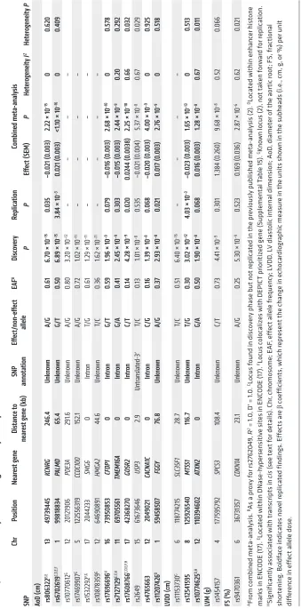
Table 1. Genetic loci associated with echocardiographic traits of LV struc ture and sys tolic func tion with genome -wide significanc e at P < 5.0 × 10
–8 in
the disc over y dataset, replication results, and a meta-analysis combining disc over y and replication data Chr Position Ne ar es t gene Dis tanc e t o ne ar es
t gene (kb
) SNP anno tation Eff ec t/ non-eff ec t allele EAF A Disc ov er y Replication Combined met a-analysis SNP P P Eff ec t ( SEM) P Het erogeneit y I 2 Het erogeneit y P AoD ( cm) rs806322 B,E 13 497 39 445 KCNR G 246. 4 Unknown A/G 0.6 1 6.7
0 × 10
–1 5 0.0 35 –0.0 21 ( 0.00 3)
2.22 × 10
–1 5 0 0.620 rs67 026 19 C,D ,E,F 1 998 188 34 PALMD 65 .4 Unknown G/ T 0.50
6.89 × 10
–1
5
3.84 × 10
–3 0.0 21 ( 0.00 3) <1.
10 × 10
–1 6 0 0. 409 rs10 77 06 12 G 12 20121906 PDE3A 291.6 Unknown A/G 0.8 0
3.20 × 10
–1 2 – – – – rs1 746 990 7 G 5 1225563 19 CCDC100 15 2.1 Unknown A/G 0. 72 1.0
2 × 10
–1 1 – – – – rs1532292 F,G 17 2044233 SMG6 0 Intr on T/ G 0. 61
1.29 × 10
–1 1 – – – – rs108 78 35 9 G 12 646 908 91 HMGA2 44 .6 Unknown T/ C 0. 36
1.62 × 10
–1 1 – – – – rs1 76 966 96 H 16 73 950853 CFDP1 0 Intron G/ T 0.5 9 1.9
6 × 10
–9 0.0 79 –0.016 ( 0.00 3)
2.68 × 10
–1 0 0 0.57 8 rs7 127 129 E,F ,H 11 69 70556 1 TMEM16A 0 Intron G/A 0. 41 2.4
5 × 10
–9 0.30 3 –0.015 ( 0.00 3)
2.44 × 10
–9 0.20 0.292 rs1 7608 766 C,D ,E,F ,H 17 42 36827 0 GOSR2 0 Intron C/ T 0.14
4.28 × 10
–9 0.0 20 0.0 244 ( 0.00 38 ) 2.2
5 × 10
–1 0 0.66 0.0 32 rs264 9 15 616 73646 USP3 2.9 Untr anslated-3 ′ T/ C 0.1 3
1.01 × 10
–8 0.535 –0 .0 21 ( 0.004 ) 5.
37 × 10
–8 0. 67 0.0 29 rs47 65663 12 204 90 21 CA CNA1C 0 Intron C/ G 0.16 1.3
9 × 10
–8 0.068 –0.0 20 ( 0.00 3)
4.00 × 10
–9 0 0.92 5 rs1120 74 26 D 1 594 5850 7 FGG Y 76.8 Unknown A/G 0.3 7 2.9
3 × 10
–8 0.0 21 0.01 7 ( 0.00 3) 2.7
6 × 10
–9 0 0.5 18 LVDD ( cm) rs11153 730 G 6 118 77 4215 SL C35F1 28 .7 Unknown T/ C 0. 51
6.40 × 10
–1 6 – – – – rs12 54 15 95 8 12 5926 540 MTS S1 116.7 Unknown T/ G 0.30 3.0
2 × 10
–1
2
4.0
3 × 10
–3 –0.0 23 ( 0.00 3) 1.6
5 × 10
–1 3 0 0.5 13 rs10 77 462 5 D, H 12 110 39460 2 AT XN2 0 Intron G/A 0.50
1.90 × 10
–8
0.068
0.016 (
0.00
3)
1.28 × 10
–8 0.67 0.011 LVM (g ) rs145 415 7 4 1775 95 792 SPC S3 108 .4 Unknown C/ T 0. 73 4.4
1 × 10
–9 0. 301 1.38 4 ( 0.260 )
9.68 × 10
–8 0. 52 0.066 FS ( % ) rs947 036 1 6 36 73 135 7 CDKN1A 23 .1 Unknown A/G 0.25 5.
30 × 10
–9 0.5 23 0. 16 9 ( 0.036 ) 2.8
7 × 10
–6
0.62
0.0
21
AFrom c
ombined
meta-analysis.
BAs a prox
y f or rs2 76 204 9, R
2 = 1.0, D
′
= 1.0.
CLocus f
ound in disc
over
y phase but not r
eplicated in the pr
eviously published
meta-analysis (
2).
DLocated within enhanc
er hist
one
marks in ENC
ODE (
17
).
ELocated within DNase
-hypersensitive sites in ENC
ODE (
17
).
FLocus c
olocalizes with DEPICT prioritized gene (
Supplemental T
able 15).
GKnown locus (
2), not tak
en f or w ard f or r eplication.
HSignificantly associated with tr
anscripts in cis (see te xt f or details ). Chr
, chromosome; E
AF
, eff
ect allele fr
equenc
y; L
VDD
, L
V diast
olic internal dimension; A
oD
, diameter of the aor
tic root; FS, fr
actional
shor
tening
. Boldf
ac
e indicates no
vel r
eplicated findings. Eff
ects ar
e
β
coefficients, which r
epr
esent the change in echocardiogr
aphic me
asur
e in the units shown in the subhe
ads (i.e., cm, g
, or %
) per unit
diff
er
enc
e in eff
[image:5.585.70.409.58.743.2]an ancestry in the Generation R study (12), and none in Hispanics (Northern Manhattan Study [NOMAS] study) or African Ameri-cans (Jackson Heart Study [JHS] and NOMAS study; Supplemen-tal Table 8). When evaluating associations of the newly discovered SNPs with related disease traits, rs17696696, which was found to be associated with AoD, was also associated with pulse wave velocity in the AortaGen consortium (Supplemental Table 9 and ref. 13). There were no statistically significant associations with incident HF or mortality in HF patients of the CHARGE-Heart Failure (CHARGE-HF) consortium (Supplemental Table 10), or with all-cause mortality, HF, or cardiovascular mortality in the Ludwigshafen Risk and Cardiovascular Health (LURIC) cohort of patients with suspected coronary artery disease (CAD) (Sup-plemental Table 11). In the CARDIOGRAMplusC4D consortium data, rs17696696, rs17608766, and rs10774625 were significant-ly associated with CAD; rs10774625 was also strongsignificant-ly associat-ed with the narrower phenotype myocardial infarction (MI; P =
5.09 × 10⁻¹¹, Supplemental Table 12).
Biological pathways related to echocardiographic traits. In path-way analysis, the observed genetic variants were significantly enriched for canonical pathways that might be involved in the biological regulation of echocardiographic traits: protein kinase A
signaling (P = 5.8 × 10⁻⁶), death receptor signaling (P = 6.9 × 10⁻⁵),
the Wnt/Ca2+ pathway (P = 2.2 × 10⁻⁴), and P2Y purigenic receptor
signaling (P = 4.1 × 10⁻⁴, Supplemental Tables 13 and 14,
Supple-mental Figure 20, and refs. 14–16).
When investigating the potential regulatory effect of the top loci using Encyclopedia of DNA Elements (ENCODE) data (17), 4 SNPs (rs10774625, rs6702619, rs17608766, and rs11207426) were located within enhancer histone marks and 4 (rs806322, rs6702619, rs7127129, and rs17608766) within DNase-hypersensi-tive sites. The literature search tool Snipper revealed no additional information, and no significant direct or indirect protein-protein interactions were found between loci using DAPPLE software (18). No significantly reconstituted gene sets were identified by the DEPICT tool (ref. 19 and Supplemental Table 15). DEPICT pri-oritized (false discovery rate [FDR] <0.05) 10 genes across
associ-ated (P < 1 × 10⁻⁵) loci, including 4 colocalizing with genome-wide
significant loci (Tables 1 and 2, and Supplemental Table 15). Analyses of expression quantitative trait loci and gene expression in whole blood, monocytes, and myocardial tissue. Our data showed 4 SNPs that were significantly associated with cis transcripts in both datasets (whole blood and monocytes, Supplemental Table 16):
rs10774625 with SH2B adaptor protein 3 (SH2B3, P = 8.15 × 10⁻²⁰
and P = 1.83 × 10⁻⁴), rs17696696 with craniofacial development
protein 1 (CFDP1, P = 6.21 × 10⁻¹¹ and P = 7.59 × 10⁻⁵), rs7127129
with Fas-associated death domain–containing protein (FADD,
P = 1.61 × 10–37 and P = 2.71 × 10⁻⁴), and rs1532292 with serine
race-mase (SRR, P = 3.40 × 10⁻²⁰ and P = 4.63 × 10⁻¹⁰).
We also examined the associations of our top loci with gene expression in human LV tissue provided by the Myocardial Applied Genomics Network consortium (MAGNet consortium; unpub-lished data). Two SNPs were significantly associated with LV gene expression: rs12541595 showed cis-association with metastasis
suppressor 1 (MTSS1, P = 1.25 × 10⁻¹⁹), with the effect allele T
asso-ciated with lower MTSS1 expression; rs1532292 showed again a
cis-association with SRR (P = 2.62 × 10⁻⁴), with the effect allele T
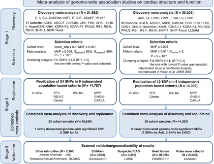
Single nucleotide polymorphisms related to cardiac structure and function (stage 1). We applied a two-stage design proposed by Skol et al. (11), including an additional stage for assessing the gener-alizability of the find, with details on samples and single nucleo-tide polymorphisms (SNPs) for each stage given in Figure 1. The meta-analysis of LV cardiac structure and systolic function traits included data from 21 cohorts with up to 30,201 individuals. For LV diastolic function, data were available from 17 cohorts with up to 21,852 individuals. We identified genome-wide significant
associations (all P < 5 × 10⁻⁸) of: 1 locus with LV mass (LVM), 3
with LVDD, 12 with AoD, 1 with LV fractional shortening (LVFS). Additionally, the following associations were observed at a higher
P value threshold (all P < 1 × 10⁻⁶): 2 with the peak velocity of the
transmitral E-wave (Mv-E), 5 with the peak velocity of the trans-mitral A-wave (Mv-A), 5 with the ratio of Mv-E to Mv-A (E/A), 2 with deceleration time of Mv-E (DecTime), 4 with isovolumetric relaxation time (IVRT), 1 with the peak velocity of the excursion of the lateral mitral annulus in the early diastolic phase (E′), 3 with the ratio of Mv-E to E′ (E/E′), 1 with asymptomatic LV diastolic dysfunction with preserved ejection fraction (DDpEF), and 2 with HF with preserved ejection fraction (HFpEF). Using pre-defined selection criteria (Figure 1) and excluding known loci from our previous report (2), 12 SNPs for traits of cardiac structure and LV systolic function (Table 1) and 24 SNPs for traits of LV diastolic function (Table 2) were considered for additional analysis detailed
in stage 2 below. Full results for all SNPs with P < 1 × 10⁻⁴ are shown
in Supplemental Table 7.
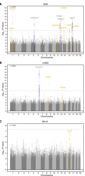
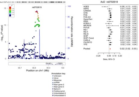
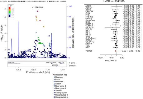
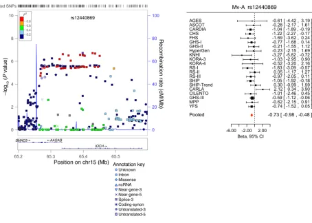
Replication and combined meta-analysis (stage 2). SNPs taken forward for stage 2 replication were analyzed in 5 cohorts (n = 14,002; 2 with in silico GWAS data, 3 with de novo genotyping) for cardiac structure and LV systolic function; and in 6 cohorts (n = 14,787; 3 with in silico GWAS data, 3 with de novo geno-typing) for LV diastolic function (Figure 1). A final combined meta-analysis of discovery and replication data from overall 30 cohort samples included 44,203 individuals with data on cardiac structure and systolic function, and 36,639 individuals with data on LV diastolic function. The investigation revealed 10 SNPs with genome-wide significance: rs10774625 and rs12541595 for LVDD; rs806322, rs4765663, rs6702619, rs7127129, rs11207426, rs17608766, and rs17696696 for AoD; and rs12440869 for Mv-A (Tables 1 and 2). Manhattan plots for these 3 traits are present-ed in Figure 2. Forest plots for the most significantly associat-ed SNPs for AoD (rs6702619), LVDD (rs12541595), and Mv-A (rs12440869) with the corresponding regional plots including functional annotation are presented in Figures 3, 4, and 5. The plots for the other genome-wide significant loci are shown in Supplemental Figures 17 and 18. Funnel plots for the significantly associated SNPs are shown in Supplemental Figure 19. All known and novel loci combined explained 1.7%, 0.5%, and 0.2% of the phenotypic variance in AoD, LVDD, and Mv-A, respectively, in a combined analysis of 3 of the larger cohorts.
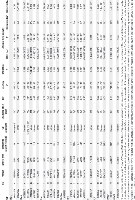
Europe-Table 2. Genetic loci associated with echocardiographic traits of dias tolic func tion with P < 1.0 × 10
–6 in
the disc over y dataset, replication results, and a meta-analysis combining disc over y and replication data Chr Position Ne ar es t gene Dis tanc e t o ne ar es
t gene (kb
) SNP anno tation Eff ec t/ non-eff ec t allele EAF A Disc ov er y Replication Combined met a-analysis SNP P P Eff ec t ( SEM) P Het erogeneit y I 2 Het erogeneit y P Mv -E rs46 908 79 4 158215 162 GLRB 1.6 Ne ar -gene -5 ′ T/ C 0. 81
6.45 × 10
–7 0.550 0.639 ( 0. 156 )
4.45 × 10
–5 0.64 0.016 rs12564392 1 2108 4215 2 ATF3 0 Intr on A/C 0. 02 6.4
1 × 10
–7 0. 162 –0 .994 ( 0. 38 9) 0.011 0. 78 4,6
7 × 10
–4 Mv -A rs10396 92 B 6 547 38825 FAM8 3B 80 .7 Unknown G/A 0.06 8.
14 × 10
–6 0.258 –0 .5 96 ( 0.23 7) 0.012 0.85 2.7
2 × 10
–5 rs1158 947 9 1 153299932 AD AM15 0 Coding -synon ym A/G 0.1 7 6.
18 × 10
–8 0.1 85 0.5 49 ( 0. 133)
3.88 × 10
–5
0.
72
3.5
4 × 10
–3 rs12 44086 9 C 15 653 98005 IQCH 0 Intron T/ A 0.26
1.90 × 10
–7
9.04 × 10
–3 –0.7 26 ( 0.128 ) 1.3
1 × 10
–8 0.54 0.054 rs1447563 2 464 90868 EP AS1 23 .5 Unknown C/A 0.4 7
5.13 × 10
–7 0.046 –0 .586 ( 0. 114 ) 2.7
6 × 10
–7 0.1 8 0.299 rs6 905862 6 3123558 1 TCF19 0 Intr on A/G 0.4 2
2.90 × 10
–7 0.958 0. 455 ( 0. 120 )
1.46 × 10
–4
0.
65
0.015
E/A rs6
79104 9 3 59 728 311 FHIT 0 Intr on T/ C 0.06
1.14 × 10
–7 0. 87 1 –0 .019 ( 0.005 )
1.05 × 10
–4
0.68
8.7
4 × 10
–3 rs18 39005 14 412366 93 LRFN5 0 Intr on C/A 0.21 1.6
5 × 10
–7 0.85 8 0.011 ( 0.003) 8.
47 × 10
–5 0.62 0.0 21 rs7 904368 B 10 168 98 593 RSU1 0 Intr on T/ C 0. 79 8.
31 × 10
–7
0.
397
0.012 (
0.003)
8.92 × 10
–6 0.62 0.0 22 rs12534 994 7 4662 70 74 TNS3 65 4. 2 Unknown C/ T 0.20
6.94 × 10
–7 0. 862 –0 .010 ( 0.003)
2.11 × 10
–4 0.66 0.011 rs18 91293 10 104 991 78 7 INA 35. 1 Unknown A/G 0.55
2.01 × 10
–7 0.947 –0 .009 ( 0.00 2) 3.7
6 × 10
–5
0.
59
0.032
DecTime rs1455
795 8 760866 55 CRISPLD1 0 Intr on A/G 0.1 0
2.82 × 10
–7 0.4 73 0.003 ( 0.001) 1.0
2 × 10
–5 0.5 0.091 IVR T rs4 96 125 2 8 1421 74 126 DENND3 33. 8 Unknown A/G 0.62 4.
31 × 10
–7 0.013 –0 .00 2 ( 0.0004 )
5.12 × 10
–8 0 0. 474 rs6860194 5 68 12 7986 SL C30A5 29 7.6 Unknown G/C 0. 24
1.44 × 10
–7 0. 87 5 –0 .00 2 ( 0.0004 )
1.94 × 10
–6 0.4 6 0. 115 rs926 138 7 6 3016 9340 TRIM31 9. 3 Unknown C/ T 0.92 3.5
7 × 10
–7 0. 347 0.003 ( 0.001) 6.9
7 × 10
–7 0 0.5 14 rs1 7868 16 7 B 2 516 16 109 NRXN1 507 Unknown A/C 0.03
1.26 × 10
–6 0. 78 5 0.004 ( 0.001) 6.4
9 × 10
–6 0.4 9 0.095 rs77 29095 5 944 14226 MCTP1 0 Intr on G/C 0.8 0
3.18 × 10
–7 0.7 95 –0 .00 2 ( 0.0004 )
9.14 × 10
–6 0.4 9 0. 100 E
′ rs1048
4775 6 15096 708 1 PLEKHG1 0 Intr on T/A 0.50
3.66 × 10
–7 0.89 7 0.055 ( 0.0 23) 0.014 0. 81 3.6
1 × 10
–4 E/E ′ rs71398 72 13 199 75 134 CR YL1 0 Intr on G/A 0.99
3.28 × 10
–7 0. 724 –0 .768 ( 0. 18 4)
3.13 × 10
–5 0. 61 0.03 7 rs1939680 11 115 23 792 7 CADM1 35 7.6 Unknown C/G 0.55 6.
30 × 10
–7 0.5 22 –0 .043 ( 0.0 24 ) 0.068 0.83
9.95 × 10
–5 rs12068 977 1 69455 98 5 LRR C7 542.5 Unknown C/G 0.01 7.7
9 × 10
–7 0.211 0.038 ( 0.06 9) 0.58 5 0. 87
4.12 × 10
–6
DDpEF rs136
77 2 22 48 186471 FLJ4438 5 14 1.9 Unknown A/G 0.90
7.44 × 10
–7 0.686 0.001 ( 0.010 ) 0.904 0.89
7.43 × 10
–6 HF pEF rs12304309 12 52 49356 7 CAL COC O1 86. 1 Unknown C/ T 0.98
2.34 × 10
–7 0. 348 –0 .03 7 ( 0.0 29 ) 0.20 7 0.90
1.60 × 10
–6
AFrom c
ombined
meta-analysis.
BProx
y SNP f
or le
ad SNP of the locus.
CSignificantly associated with tr
anscripts in cis (see te xt f or details ). Chr
, chromosome; E
AF
, eff
ect allele fr
equenc
y; Mv-E, pe
ak velocit y of the e arly diast olic tr ansmitr al inflow ; Mv-A , pe ak velocit
y of the tr
ansmitr
al inflow c
orr
esponding t
o atrial c
ontr
action; E
/A
, r
atio of the mitr
al E
- and A
-w
ave; DecTime, dec
eler
ation time of the mitr
al E -w ave; IVR T, iso volumetric r ela
xation time; E
′
, pe
ak velocit
y of the e
xcursion of the later
al mitr
al annulus in the e
arly diast
olic phase; E
/E
′
, r
atio of E and E
′
; DDpEF
, diast
olic dysfunction with pr
eser ved EF ; HFpEF , HF with pr eser ved EF . Boldf ac
e indicates no
vel r
eplicated findings. Eff
ects ar
e
β
coefficients, which r
epr
esent the change in echocardiogr
aphic me
asur
e in the units shown in the subhe
ads (i.e., cm, g
, or %
) per unit
diff
er
enc
e in eff
[image:7.585.77.533.66.740.2]associated with lower SRR expression. Both expression quantita-tive trait locus (eQTL) associations from the LV tissue were also supported by the GTEx database (http://gtexportal.org/home/). The association with SRR expression for rs1532292 had the same direction of effect in different tissues, with the T allele generally
associated with lower gene expression levels, e.g., in the aorta and in blood cells. Additionally, the follow-ing eQTLs with genes from the reference sequence database (RefSeq; https://www.ncbi.nlm.nih.gov/ refseq/) in the aorta or heart tissue were found for the replicated SNPs in the GTEx database: rs17696696 (BCAR1), rs12541595 (LINC00964), and rs11153730 (SSXP10). Detailed GTEx results are given in Supple-mental Table 17.
Discussion
In the present investigation, we identified 7 genetic loci associated with aortic root size and confirmed the associations of 4 other loci previously reported (2). These 11 variants explained 1.7% of the inter-individ-ual variation in aortic root size (Supplemental Table 18). However, use of genome-wide complex trait analysis (GCTA) software in one of the larger cohorts (Study of Health in Pomerania [SHIP]) as an illus-trative example demonstrated that common genetic variation explains about 30% of the variation in AoD (Supplemental Table 19), underscoring the potential for more, as-yet-undiscovered, loci. Additionally, we observed three genetic loci that were associated with LV diastolic dimensions (including one previously reported; see below) and one locus that was associat-ed with the transmitral A-wave velocity.
[image:8.585.39.331.48.648.2]Among the SNPs identified in our study as being associated with LVDD, one was rs12541595 close to MTSS1, which interacts with cytoplasmic actin near the cell surface and modulates intercellular connec-tions in the kidney and metastatic potential in tumors (20, 21). When investigating our top loci for cis- associations with gene expression in human LV myo-cardial tissue (MAGNet consortium, unpublished data) and the GTEx database, rs12541595 showed a significant association with MTSS1 expression, with the LVDD-lowering allele (T) associated with lower MTSS1 expression in this tissue (Supplemental Table 9). We spec-ulate that a reduction in MTSS1 may promote favorable LV remod-eling, perhaps by affecting cell junctions. The other novel variant associated with LVDD, rs10774625, was associated with expres-sion of SH2B3 in eQTL analysis and lies in ATXN2 (ataxin 2),
tissue (unpublished data from the MAGNet consortium; GTEx database, see Supplemental Table 9).
One of the SNPs associated with AoD in our meta-analysis was also associated with AoD in children in the Generation R Study. Additionally, one SNP was associated with pulse wave velocity. Two SNPs associated with AoD and one SNP associated with LVDD were also significantly associated with CAD, the LVDD SNP also with MI in the CARDIOGRAMplusC4D consortium. These associations strengthen the evidence of involvement of these loci in echocar-diographic traits. However, given the sample sizes of cohorts with different ethnicities as well as the SNP allele frequencies, and tak-ing the effect sizes into account, the power was not more than 35% to reveal a statistically significant association of select SNPs with traits in “look-up” exercises. Therefore, some of the null results for the assessment of the generalizability of observed associations to non-European samples should be interpreted with care.
Pathway analysis suggested enrichment of the Wnt/Ca2+
canonical pathway among the genetic variants associated with echocardiographic traits. These observations are consistent with the known effects of this pathway on myocardial biology (35). The
Wnt/Ca2+ pathway connects to the nuclear factor of activated T
cells (NFAT) transcription factor (14, 15) and gene expression via calcineurin. Interestingly, both calcineurin and its target NFAT are involved in cardiac hypertrophy (16).
The association of our findings with expression data from human blood revealed 4 genes with potential functional signif-which is adjacent to SH2B3, previously associated with retinal
venular diameter, CAD, and arterial hypertension in separate reports (22–26). For LVDD, we also replicated the previously iden-tified SLC35F1 locus (soluble transporter membrane protein) adja-cent to the phospholamban (PLN) locus (protein inhibiting cardiac
muscle sarcoplasmic reticulum Ca++-ATPase) (2).
[image:9.585.69.531.55.377.2]Three loci associated with AoD have been linked previous-ly to blood pressure as well as MI (GOSR2, Golgi SNAP recep-tor complex member 2; refs. 24, 27), blood pressure response to treatment (CACNA1C, calcium channel, voltage-dependent, L type, alpha 1C subunit; ref. 28), and carotid intimal-medial thickness, as well as with CAD (CFDP1; refs. 29, 30). The oth-er novel AoD-associated genetic loci woth-ere in or close to PALMD (palmdelphin, a paralemmin-related cytosolic protein; ref. 31), KCNRG (soluble protein with regulatory function in voltage- gated potassium channels; ref. 32), FGGY (carbohydrate kinase domain–containing protein, phosphorylates carbohydrates; ref. 33), and in TMEM16A (transmembrane member 16A, protein involved in transepithelial anion transport and smooth muscle contraction; ref. 34). We also replicated in our discovery sample 4 loci associated with aortic diameter from our previous report (2): SMG6 (Smg-6 homolog, nonsense-mediated mRNA decay factor), CCDC100 (centrosomal protein 120kDa), HMGA2 (high-mobility group AT-hook 2), and PDE3A (phosphodies-terase 3A, cGMP-inhibited). The effect allele of rs1532292 was associated with lower SRR expression in human LV myocardial
likely have further improved diagnosis and classification of LV dia-stolic dysfunction in our study if this method had been available in more cohorts. Likewise, as noted above, several of the LV diastolic filling measures are notoriously susceptible to variation in ven-tricular loading conditions (38). The genetic variants identified in our study have small effect sizes and explain a relatively small percentage of the variance in the echocardiographic phenotypes. Larger studies with more detailed reference panels, as well as more detailed functional studies and studies into the interactions of the variants found with factors such as hypertension, will likely shed further light on the molecular mechanisms underlying these complex traits. Furthermore, alterations of the transmitral A wave velocity are challenging to interpret alone, without consideration of other measures of LV diastolic function and filling patterns. The transmitral A wave velocity reflects the late diastolic phase of the LV filling, i.e., the phase of atrial contraction. Thus, in theory this single measure provides important information about active atrial function. Yet in practice, this measure changes variably and in a complex manner with the progression of LV diastolic dysfunction: Increasing impaired ventricular relaxation is at first accompanied by a decrease in E-wave with a compensatory increase in A-wave, resulting in a “relaxation abnormality” pattern; it results in the further, continuous decrease in A-wave velocity, reflecting a pro-gressive deterioration of the contractility of the left atrium, and also changes in LV compliance (39, 40). These pathophysiological considerations underline the importance of the active contraction icance (Supplemental Table 8). Of these, rs7127129 is located
within TMEM16A, but its eQTL FADD has been shown to be associated with myocardial ischemia/reperfusion injury in an HF mouse model (36).
[image:10.585.63.522.57.377.2]Our study is strengthened by the large sample size, the use of standardized echocardiographic techniques with adequate quali-ty, and a harmonization of phenotypic data. Nonetheless, several limitations must be acknowledged. We did not observe any associ-ation of common genetic variants with the other echocardiograph-ic measurements studied, e.g., LA size, LV wall thechocardiograph-ickness (LVWT), LVM, LV systolic dysfunction (LVSD), and most measures of LV diastolic function, with the exception of the transmitral A-wave velocity. In particular, we did not find any statistically significant associations for HFpEF, although we included only carefully phe-notyped individuals in our study to reduce the phenotypic hetero-geneity (37). The lack of association of select echocardiographic traits with common genetic variation is intriguing. It is likely that heterogeneity in both phenotypic assessment and study design and modest statistical power may have limited our ability to detect modest genetic associations, and associations with rare variants could not be assessed by design. A proportion of the intra-individ-ual variability of functional traits might have been influenced by physiological factors (e.g., posture, state of hydration, heart rate, or medication use) (38). In this context, it should be noted that some echocardiographic measures may be imprecise, e.g., analy-sis of tissue Doppler imaging (TDI) of the mitral annulus would
For analysis of LV diastolic dysfunction, we excluded individuals with reduced ejection fraction (EF) (defined as <50%, LVFS <29% or poor/ impaired LV systolic function by visual estimation).
Strategy for analysis
For the identification of genetic variants associated with cardiac struc-ture and function, we followed a 3-stage analysis plan (Figure 1). First, a discovery meta-analysis of up to 21 population- and hospital-based GWAS was performed (stage 1). Second, replication of the findings from stage 1 was performed in up to 6 independent cohort studies (3 with in silico data and 3 with de novo genotyping), and a combined meta-analysis of discovery and replication data was carried out (stage 2). In stage 3, SNPs that were genome-wide significant in the combined meta-analysis were investigated for the generalizability of the observed associations in a cohort of white children of European ancestry (the Generation R study), in two cohorts of other ethnicities (Hispanic in the NOMAS Study and African American in the JHS and in the NOMAS study), and in associations with related disease traits (data from the AortaGen and CHARGE-HF consortia, and the LURIC study).
Echocardiographic methods
Detailed echocardiographic methods used and distributions of traits in each cohort study are reported in Supplemental Methods and Sup-plemental Tables 3 and 4.
The present investigation focused on 5 traits of cardiac struc-ture: LVM, LVDD, LVWT, AoD, and left atrial size (LA). Additionally, of the left atrium. Last, we did not directly assess the functional
significance of all the associated SNPs or perform mechanistic studies, other than for the MTSS1 locus associated with LVDD (unpublished data from the MAGNet consortium).
To conclude, we report the largest genetic association study to our knowledge of a comprehensive set of LV echocardiographic traits. The large number of interesting genetic loci identified for AoD and LV diastolic dimensions, and the biological pathways enriched within our association results provide new insights into the biology of cardiac remodeling. Additional studies are warrant-ed to further evaluate experimentally the functional significance of the reported genetic variants and loci.
Methods
EchoGen consortium
[image:11.585.74.528.52.376.2]The EchoGen consortium was initiated in 2007 and has grown to a consortium of 30 studies with population-based and hospital-based cohorts primarily of European ancestry, and additionally including two cohorts of African American and one of Hispanic individuals. For the present investigation, we applied harmonized phenotype defi-nitions, covariate selection, and genotyping protocols and the same statistical analysis plan across all cohorts. For traits of cardiac struc-ture and systolic function, individuals with a history of MI, clinical diagnoses of HF, or valve disease were excluded if this information was known or recorded during the echocardiographic examination.
Map (https://www.genome.gov/10001688/international-hapmap-
project/) release 28 CEU dataset using PLINK (settings r2 > 0.2, 1 Mb
distance) (44). For each identified independent locus, the SNP with the lowest P value was defined as the lead SNP and taken forward for replication. SNPs representing loci identified and replicated in our previously published report (2) were not taken forward for replication.
Replication and combined meta-analysis (stage 2). In stage 2, SNPs
were related to echocardiographic traits in 6 cohort samples (Figure 1). We chose proxies for 4 of the top SNPs, as no reliable assays were available for wet lab replication of the originally identified SNPs:
rs1039692 was used as a proxy for rs949796 (Mv-A, P = 6.60 × 10⁻⁷,
R2 = 1.0), rs7904368 as a proxy for rs7074647 (E/A, P = 8.30 × 10⁻⁷,
R2 = 0.95), rs17868167 as a proxy for rs17862703 (IVRT, P = 9.70 × 10⁻⁷,
R2 = 1.0), and rs806322 as a proxy for rs2762049 (AoD, P = 3.85 × 10⁻¹⁵,
R2 = 1.0). The dbSNP (https://www.ncbi.nlm.nih.gov/projects/SNP/)
identifiers of the proxies are reported in the final results.
For the combined meta-analysis of discovery and replication
cohorts, SNPs with a P value of <5 × 10⁻⁸ in the combined meta-
analysis were considered to be significantly associated with their respective outcomes, as the overall sample size of the replication cohorts was very small. Genome-wide significant association signals were deemed novel for the corresponding traits if they were >500 kb away from the lead SNPs reported in our previous study (2) and not in
high LD with them (R2 < 0.5).
Look-up in other cohorts to test for generalizability of findings For the genome-wide significant SNPs representing novel loci, we per-formed “look-ups” in relation to the corresponding echocardiographic traits in children (the Generation R study), Hispanics (NOMAS), and African Americans (meta-analysis of data from JHS and NOMAS). Additionally, we evaluated associations of these SNPs with traits of interest: SNPs for aortic root diameter with pulse wave velocity in the AortaGen consortium (45, 46); and all newly identified SNPs with incident HF and mortality in the CHARGE-HF consortium (3), with all-cause, cardiovascular, and HF mortality in the LURIC study (a cohort of patients with suspected CAD), as well as with MI and CAD in the CARDIOGRAMplusC4D consortium data (47). Further details for the look-up investigations are presented in Supplemental Methods.
Proportion of trait variance explained
The proportion of variance in echocardiographic traits explained by the significantly associated SNPs from our meta-analyses was estimat-ed in 3 of the larger cohorts (Rotterdam study [RS], SHIP, and FHS).
Within each cohort, R2 values of two models were compared for each
trait: one model including the covariates (age, sex, height, and weight) only; and one model additionally including the new and known loci. The proportion of the sex-, age-, height-, and weight-adjusted vari-ance explained by all common (MAF >0.01) autosomal genotyped SNPs for each trait was calculated in the SHIP sample using the REML method of GCTA software version 1.24.4 (48).
Known associations of genome-wide significant SNPs
We combined a manual review of the literature with the use of the tool Snipper version 1.2 (http://csg.sph.umich.edu/boehnke/snipper/), which conducts an automated search of the published literature using speci-fied search terms and the putative SNP to evaluate previously reported disease/trait associations for the genome-wide significant SNPs. we evaluated 2 traits of systolic cardiac function (LVFS and LVSD)
and 9 traits of LV diastolic function: Mv-A, Mv-E, E/A, E′, the ratio E/E′ as a surrogate for LV end-diastolic pressure, DecTime, and IVRT, as well as DDpEF and HFpEF (41). Measurements were based on the European and American guidelines for the echocardiographic assessment of the LV (42).
Genotyping methods and imputation
Details on genotyping, imputation, and quality control are presented in Supplemental Table 5. Population stratification as well as family structure, if applicable, was accounted for in each individual cohort’s analysis. For replication, 3 of the 6 cohorts (Gutenberg Health Study III [GHS-III]; Cardiovascular Risk Factors, Living and Ageing in Halle [CARLA] study; and Malmö Preventive Project [MPP] study)
underwent de novo genotyping using 5′ nuclease assays on 384-well
plates. For quality control, genotypes were validated in 10% of the samples for all SNPs.
Definition of traits and statistical methods
Discovery (stage 1). All traits were analyzed as continuous traits, with
the exception of LVSD, DDpEF, and HFpEF. LVSD was defined as an EF <50%, fractional shortening (FS) <29% or reduced (poor or impaired) EF by visual estimation. Aggregate binary phenotypes were defined for asymptomatic participants with echocardiographic evi-dence of LV DDpEF and for those with HFpEF based on information on classes of HF according to the New York Heart Association (NYHA) and medication for HF in addition to echocardiography.
Stage 1 analyses were first performed separately at the individual cohort level for each trait (Figure 1). Continuous echocardiographic traits were related to genotype dosage (0–2 copies of the effect allele) for each autosomal SNP using linear regression assuming additive genetic models adjusted for age, sex, height, weight, and study site (only applicable for the Cardiovascular Health Study [CHS] and Anglo-Scandinavian Cardiac Outcomes Trial [ASCOT]). For bina-ry traits, we used logistic regression models with the same adjust-ments. In the Framingham Heart Study (FHS), linear mixed-effects models were applied to account for familial correlations. The asso-ciations of genotypes with echocardiographic traits were quantified by beta estimates, SEM, and P values. After verifying strand align-ment across studies and applying genomic control to each study, we performed inverse variance–weighted fixed-effects meta-analysis across the discovery cohorts with METAL (43) for the structural and the systolic function traits and with the R package MetABEL (http:// www.r-project.org) for the diastolic traits. After the meta-analysis, we excluded SNPs with a minor allele frequency (MAF) below 0.5% for diastolic function traits and below 1% for structural traits, and FS and below 3% for LVSD.
We used an a priori P value threshold of <5 × 10⁻⁸ to indicate
genome-wide statistical significance in the discovery meta-analysis for the selection of SNPs taken forward to the next stage. As no SNP reached genome-wide significance in the analysis of diastolic
func-tion traits, SNPs with P < 1 × 10⁻⁶ were taken forward for replication as
Hap-OTR, PMN, PS, PSW, RL, RLS, RM, R. Schmidt, R. Sorice, RSV, RW, RZ, SB, SC, SJS, SKM, S. Nappo, ST, STR, TBH, TES, TL, TM, TN, TZ, V. Großmann, and V. Gudnason. Genotyping: AAH, AGU, AMT, A. Pfeufer, APM, CML, DR, EI, ERF, FR, H. Schmidt, JGS, JH, JIR, JS, KJL, KL, L. Lind, LPL, MC, MEK, MF, MM, MRI, OM, PSW, R. Sorice, SB, SKM, TES, TL, TN, TPC, TZ, UB, UV, and V. Großmann. Statistical analysis: AS, AT, CL, C. Minelli, C. Monnereau, C. Müller, DCS, D. Malzahn, D. Medenwald, DP, DR, DT, EH, FDGM, GFM, HL, JAB, JCB, JFF, JGS, JK, JNH, JS, KMR, LF, LPL, MC, MEK, MF, MGL, MHC, MM, MMN, MRI, NDD, PS, PSW, R. Sorice, SJS, SKM, S. Nuding, SP, and THP. Interpretation of data: ACM, ADH, AGU, AH, AJB, AS, ASPS, AT, A.V. Smith, A.V. Stanton, AZ, BHS, BP, CAM, DKA, EB, FR, GFM, HL, H. Schmidt, H. Schunkert, JFF, JGS, JM, JNH, JWD, KL, LF, LJL, MD, MF, MGL, MJGL, ML, MM, NS, OHF, OM, PMN, PS, PSW, R. Schmidt, RSV, SBF, SJS, ST, TBH, THP, TPC, TZ, V. Gudnason, VWVJ, WL, and WM. Drafting the manuscript: AS, AT, JFF, MD, PSW, RSV, and TZ. Revising manuscript critically for important intellectual content: all authors.
Funding summary and Acknowledgments
This work was supported by a grant from the US National Heart, Lung, and Blood Institute (N01-HL-25195; R01HL 093328 to RSV), a MAI-FOR grant from the University Medical Center Mainz, Germany (to PSW), the Center for Translational Vascular Biology (CTVB) of the Johannes Gutenberg-University of Mainz, and the Federal Ministry of Research and Education, Germany (BMBF 01EO1003 to PSW). This work was also supported by the research project Greifswald Approach to Individualized Medicine (GANI_MED). GANI_MED was funded by the Federal Ministry of Education and Research and the Ministry of Cultural Affairs of the Federal State of Mecklenburg, West Pomerania (contract 03IS2061A). We thank all study participants, and the col-leagues and coworkers from all cohorts and sites who were involved in the generation of data or in the analysis. We especially thank Andrew Johnson (FHS) for generation of the gene annotation database used for analysis. We thank the German Center for Cardiovascular Research (DZHK e.V.) for supporting the analysis and publication of this project. RSV is a member of the Scientific Advisory Board of the DZHK. Data on CAD and MI were contributed by CARDIoGRAMplusC4D inves-tigators. See Supplemental Acknowledgments for consortium details. PSW, JFF, AS, AT, TZ, RSV, and MD had full access to all of the data in the study and take responsibility for the integrity of the data and the accuracy of the data analysis.
Study-specific Acknowledgments, funding, and ethics statements The following list provides the study-specific names and funding sources with grant numbers only. Details on the study acronyms, study-specific Acknowledgments, funding, support for researchers, and ethics statements are provided in the supplemental material.
AortaGen
AGES. NIH N01-AG-1-2100, National Institute on Aging (NIA)
Intra-mural Research Program, Hjartavernd (the Icelandic Heart Associa-tion), Althingi (the Icelandic Parliament).
ASCOT. Pfizer, New York; Servier Research Group, Paris, and
Leo Laboratories, Copenhagen; partial funding: Barts and the London School of Medicine and Dentistry, Centre Nationale de Genotypage Paris, and Irish Research Council GREP award.
cis eQTL analysis
To evaluate the potential functional significance of our findings, we related each replicated SNP to the expression levels of genes in three sets of tissues: human whole blood samples from n = 5,311 individuals evaluated by Westra et al. (49), human monocytes from n = 1,372 par-ticipants in the GHS (50), and LV free-wall tissue from n = 313 patients with HF undergoing transplantation and from unused donor hearts from the MAGNet consortium (http://www.med.upenn.edu/magnet). Further details are presented in Supplemental Methods. To evaluate possible cis eQTLs across multiple tissues, an additional look-up was performed in the GTEx database for the new findings.
Pathway analysis
The collective effects of multiple genetic variants on biological sys-tems were investigated by pathway analysis, first for the 7 structural and systolic traits combined, and then for the 9 combined diastolic traits and for all 16 echocardiographic traits combined (for details, see Supplemental Methods).
To identify whether any of the associated SNPs fall within regula-tory regions of the genome, we evaluated data from ENCODE (17). We compared the expected overlap of the putative SNPs with functional domains due to chance with the actual observed overlap by creating a permuted set of non-associated SNPs that were evaluated for overlap with the functional domains. We also used the DEPICT tool to further explore functionality of the identified SNPs (19). In addition, variants
with P < 5 × 10⁻⁷ were used as the input for the DAPPLE software (18),
which then built both direct and indirect interaction networks from seed genes near the top loci.
Statistics
If not specified otherwise, a Wald test statistic was calculated by divid-ing the estimated effect size by its standard error and compardivid-ing them with a normal distribution (2-tailed) with mean zero. In the GWAS,
P < 5 × 10⁻⁸ for the combined stage 1 and 2 analysis was deemed
sig-nificant (11), which corresponds to a significance level of 0.05 after correcting for 1 million independent SNPs (51). For pathway analyses, a FDR was applied as multiple testing correction with a cutoff-value <0.05 for statistical significance.
Study approval
All study protocols of participating cohorts were reviewed and approved by a local ethics committee and followed the recommen-dations of the Declaration of Helsinki. All subjects in the cohorts provided informed written consent prior to their participation in the study. Therefore, no specific approval was required for this meta-analysis of human data. The institutional review boards are listed in the supplemental material.
Author contributions
JHS. NHLBI and National Institute on Minority Health and
Health Disparities (NIMHD) (HSN268201300046C,
HHSN268-201300047C, HHSN268201300048C, HHSN2682013 00049C,
HHSN268201300050C).
KNHI. National Genome Research Network (NGFN), German
Federal Ministry of Education and Research (BMBF; 01GS0837 and 01GS0422); Competence Network for Heart Failure Germany.
KORA-F3 and KORA-F4. Helmholtz Zentrum München, German
Research Center for Environmental Health, Neuherberg, Germany, BMBF; partial funding: NGFN.
LURIC. European Union 7th Framework Program (AtheroRemo,
grant agreement 201668, and RiskyCAD, grant agreement 305739), INTERREG-IV-Oberrhein-Program (project A28, Genetic Mecha-nisms of Cardiovascular Diseases), European Regional Development Fund (ERDF), Wissenschaftsoffensive TMO.
MAGNet. NIH R01HL105993.
MICROS. Ministry of Health and Department of Educational
Assistance, University and Research of the Autonomous Province of Bolzano, the South Tyrolean Sparkasse Foundation, and European Union framework program 6 EUROSPAN project (contract LSHG-CT-2006-018947).
MPP. Swedish Heart-Lung Foundation, Swedish Research
Coun-cil, European Research CounCoun-cil, Faculty of Medicine of Lund Universi-ty, Skåne University Hospital, and the Crafoord Foundation.
NOMAS. NINDS, NIH (R37 NS2993 and R01 NS36286).
PIVUS. Swedish Foundation for Strategic Research (project grant
ICA08-0047), Swedish Research Council (project grant 2012-1397), Swedish Heart-Lung Foundation (project grant 20120197), Swedish Society of Medicine, and Uppsala University.
RS-I, RS-II, RS-III. Netherlands Organisation of Scientific Research
(NWO) investments (no. 175.010.2005.011, 911-03-012), Research Institute for Diseases in the Elderly (014-93-015; RIDE2), Netherlands Genomics Initiative (NGI)/NWO project 050-060-810, Erasmus MC and Erasmus University, Rotterdam, Netherlands Organization for Health Research and Development (ZonMw), Research Institute for Dis-eases in the Elderly (RIDE), Ministry of Education, Culture and Science, Ministry for Health, Welfare and Sport, European Commission (DG XII), and Municipality of Rotterdam. Partial funding: NWO investments (no. 175.010.2005.011, 911-03-012), Research Institute for Diseases in the Elderly (014-93-015; RIDE2), NGI/NWO project 050-060-810.
SHIP, SHIP-Trend. Federal Ministry of Education and Research
(grants 01ZZ9603, 01ZZ0103, and 01ZZ0403), Ministry of Cultur-al Affairs, SociCultur-al Ministry of the FederCultur-al State of Mecklenburg–West Pomerania. Partial funding: Competence Network Heart Failure of the Federal Ministry of Education and Research, Federal Ministry of Education and Research (grant 03ZIK012) and a joint grant from Sie-mens Healthcare, Erlangen, Germany and the Federal State of Meck-lenburg-West Pomerania.
ULSAM. Swedish Foundation for Strategic Research (project grant
no. ICA08-0047), Swedish Research Council (project grant 2012-1397), Swedish Heart-Lung Foundation (project grant 20120197), Swedish Society of Medicine, and Uppsala University; partial
fund-ing: SNP&SEQ Technology Platform in Uppsala, Uppsala University,
Uppsala University Hospital, Science for Life Laboratory – Uppsala, and the Swedish Research Council (contracts 80576801 and 70374401).
YFS. Academy of Finland: grants 286284, 134309 (Eye), 126925,
121584, 124282, 129378 (Salve), 117787 (Gendi), and 41071 (Skidi); the
ASPS. Austrian Science Fund Project P20545_P05 Genetics of
Cerebral Small Vessel Disease.
CARDIA. National Heart, Lung, and Blood Institute in
col-laboration with the University of Alabama at Birmingham (HHSN268201300025C and HHSN268201300026C), Northwest-ern University (HHSN268201300027C), University of Minnesota (HHSN268201300028C), Kaiser Foundation Research Institute (HHSN268201300029C), and Johns Hopkins University School of Medicine (HHSN268200900041C); partial funding: Intramural Research Program of the NIA, Gene Environment Association Studies (GENEVA) through grants HG004729, HG04424, and U01-HG004446 from the National Human Genome Research Institute.
CARLA. Deutsche Forschungsgemeinschaft (DFG) as part of
Collaborative Research Center 598 ‘‘Heart Failure in the Elderly — Cellular Mechanisms and Therapy,’’ Medical Faculty, Martin-Luther- University Halle-Wittenberg; Wilhelm-Roux Programme, Martin- Luther-University Halle-Wittenberg; Federal Employment Office; and Ministry of Education and Cultural Affairs of Saxony-Anhalt.
CHARGE-HF
CHS. National Heart, Lung, and Blood Institute (NHLBI)
HHSN268201200036C, HHSN268200800007C, N01HC55222, N01HC85079, N01HC85080, N01HC85081, N01HC85082, N01HC85083, N01HC85086, N01HC85084, N01HC35129; and NHLBI grants U01HL080295, R01HL087652, R01HL105756, R01HL103612, R01HL120393, and HL130114, with additional con-tribution from the National Institute of Neurological Disorders and Stroke (NINDS), and R01AG023629 from the NIA; partial funding: National Center for Advancing Translational Sciences, CTSI grant UL1TR000124, and National Institute of Diabetes and Digestive and Kidney Disease Diabetes Research Center (DRC) grant DK063491 to the Southern California Diabetes Endocrinology Research Center.
Cilento. Italian Ministry of Universities and CNR (Interomics
Flagship Project, PON03PE_00060_7), Assessorato Ricerca Regione Campania, Fondazione con il SUD (2011-PDR-13), and Istituto Banco di Napoli – Fondazione to MC.
DEPICT. Danish Council for Independent Research Medical
Sci-ences (FSS), Alfred Benzon Foundation.
FHS1, FHS2, FHS3. National Heart, Lung and Blood Institute’s
Framingham Heart Study (N01-HC-25195 and HHSN268201500001I), research grants (5R01HL107385-04, 1R01HL126136-01A1, R00-HL107642, R01HL131532), Affymetrix Inc. for genotyping services (contract N02-HL-6-4278).
Generation R. Erasmus Medical Center, Rotterdam, Erasmus
Uni-versity Rotterdam, Netherlands Organization for Health Research and Development (ZonMw), Netherlands Organisation for Scientific Research (NWO), Ministry of Health, Welfare and Sport and the Min-istry of Youth and Families.
GHS-I, GHS-II, GHS-III. Government of Rhineland-Palatinate
(“Stiftung Rheinland-Pfalz für Innovation,” contract AZ 961-386261/733), research programs “Wissen schafft Zukunft” and “Center for Transla-tional Vascular Biology (CTVB)” of the Johannes Gutenberg-University of Mainz, Boehringer Ingelheim, and PHILIPS Medical Systems.
HyperGEN. HyperGEN echocardiography ancillary study: NIH
Center of the Johannes Gutenberg-University Mainz, Langen-beckstr. 1, 55131 Mainz, Germany. Phone: 49.6131.17.7163; E-mail: [email protected]. Or to: Janine F. Felix, Depart-ment of Epidemiology, room Na-2906, Erasmus MC, University Medical Center Rotterdam, P.O. Box 2040, 3000 CA Rotterdam, Netherlands. Phone: 31.10.70.43997; E-mail: j.felix@erasmusmc. nl. Or to: Ramachandran S. Vasan, Sections of Preventive cine and E