ISSN Print: 2168-5452
DOI: 10.4236/ijohns.2019.84013 Jun. 14, 2019 113 Int. J. Otolaryngology and Head & Neck Surgery
Pathogenesis and Microbiology of Otitis Media
with Effusion in Children
Tahia Hashem Saleem
1,
Essam A. Abo Elmagd
2*
, Mahmud E. Khalefa
2, Bahaa Elhawary
21Faculty of Medicine, Assiut University, Assiut, Egypt 2Faculty of Medicine, Aswan University, Aswan, Egypt
Abstract
Objective: To detect different etiological factors of otitis media with effusion (OME) and different types of microorganisms in middle ear fluids. Methods: This prospective study included 60 patients with otitis media with effusion diagnosed at the otorhinolaryngology (ENT) outpatient clinic with age ranged from 2 to 16 years, 36 males and 24 females. Results: Predisposing factors of OME were rhinosinusitis in 58.3% of cases, adenoid in 20% of cas-es, adenotonsillitis in 16.7% of cases and tonsillitis in 5% of cases. Microor-ganisms in middle ear fluids were negative in 70% of cases, isolation of strep-tococcus pneumonia in 16.7% of cases, Haemophilus influenzae 6.7% and Moraxella catarrhalis 6.7%. Conclusion: Rhinosinusitis was the most fre-quent predisposing factor of cases of OME. Positive bacterial culture was found in 30% of cases.
Keywords
Otitis Media with Effusion, Microbiology, Pathogenesis
1. Introduction
OME is characterized by a non-purulent effusion of the middle ear that may be either mucoid or serous without acute symptoms. It is one of the common caus-es of deafncaus-ess among children. When inadequately treated, it may lead to major functional limitations like hearing loss and impairment in development of speech and language [1]. The reason for treatment failure is probably due to par-tial knowledge of etiopathological mechanisms responsible for the beginning and the course of the disease in the mucous membrane of the middle ear [2]. OME includes inflammation of the tubotympanum and an accumulation of fluid How to cite this paper: Saleem, T.H.,
Elmagd, E.A.A., Khalefa, M.E. and Elha-wary, B. (2019) Pathogenesis and Microbi-ology of Otitis Media with Effusion in Children. International Journal of Otola-ryngology and Head & Neck Surgery, 8, 113-120.
https://doi.org/10.4236/ijohns.2019.84013
Received: May 7, 2019 Accepted: June 11, 2019 Published: June 14, 2019
Copyright © 2019 by author(s) and Scientific Research Publishing Inc. This work is licensed under the Creative Commons Attribution International License (CC BY 4.0).
DOI: 10.4236/ijohns.2019.84013 114 Int. J. Otolaryngology and Head & Neck Surgery within the middle ear. The disturbance of the excretory function is due to me-chanical obstruction of the Eustachian tube (ET) and/or mucociliary dysfunction of the tubotympanum. Mechanical obstruction has been emphasized for a long time, but recent laboratory investigations have established the critical impor-tance of mucociliary function in the tubotympanum. However, the pathogenesis is not fully understood. It is unclear whether the cilia functions normally throughout the full length of the ET in the chronic phase of OME [3]. OME was previously considered to be bacteriologically sterile. However positive bacterial cultures have been demonstrated in 40% of middle ear fluid; Streptococcus pneu-moniae and Haemophilus influenza account for the majority of cases [4][5][6].
2. Patients and Methods
This prospective study included 60 patients with otitis media with effusion di-agnosed at ENT outpatient clinic, South Valley university hospital during the period from December 2015 to December 2016. All patients gave written in-formed consent before entering the study and the study protocol was approved by the ethical committee of the faculty of Medicine, South Valley University.
The diagnosis of otitis media with effusion was made in our study group on the basis of the following clinical findings in the form of dull tympanic mem-brane, loss of con light, loss of landmarks of the eardrum, blue drum, and/or al-teration in the mobility of tympanic membrane. Every patient had complete ear, nose and throat examination. All cases had detailed assessments aided by X-ray of soft tissue neck (lateral view) for adenoidal enlargement and an audiological assessment. All patients were subjected to tympanometric screening (Immit-tancemeter-Interacoustics-Automatic AZ26, Denmark).
3. For All Patients Included in the Study
Patients subjected to surgical management in the form of myringotomy and ventilation tube insertion (grommet), myringotomy and adenoidectomy or my-ringotomy and adenotonsillectomy according to the predisposing factor. Sam-ples of middle ear effusions were collected using sterile syringe during the puncture of tympanum or tympanostomy tube placement Sample was sent for culture and sensitivity. For all samples, culture done using CLED media, incuba-tion at 37 for 48 hours, for the negative results the time extended 24 hours more. Sensitivity for the positive results applied on neutral agar using the antibiotic discs and according to the diameter of inhibition, the results were recorded.
4. Statistical Analysis
DOI: 10.4236/ijohns.2019.84013 115 Int. J. Otolaryngology and Head & Neck Surgery significant when P < 0.05.
5. Results
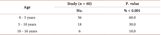
A total of included 60 patients with otitis media with effusion with age ranged from 2 to 16 years, 60% were less 5 years (Table 1), 36 males and 24 females
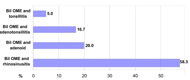
(Table 2). Bilateral OME with rhinosinusitis diagnosed in 58.3%, with adenoid
enlargement in 20%, with adenotonsillitis in 16.7% and with tonsillitis in 5%
(Figure 1). Bilateral myringotomy and grommet tube insertion only was done in
[image:3.595.208.540.300.373.2]58.3% of cases, with adenoidectomy in 20% of cases, with adenotonsillectomy in 16.7% and with tonsillectomy in 5% of cases (Table 3). Culture sensitivity results were negative in 70% of cases while positive culture in 30% of cases (Strepto-coccus pneumonia 16.7%, Haemophilusinfluenzae 6.7, Moraxellacatarrhlis 6.7%) (Table 4).
Table 1. Age distribution of the studied groups.
Age Study (n = 60) P. value
No. % < 0.001
0 - 5 years 36 60.0
5 - 10 years 18 30.0
10 - 16 years 6 10.0
Table 2. Sex distribution of the studied groups.
Sex Study (n = 60) P. value
No. 0.121%
Male 36 60.0
Female 24 40.0
Table 3. Operations done.
Operations done No. (n = 60) % P. value
Bilmyringotomy and grommet tube
insertion and adenoidectomy 12 20.0
<0.001 Bilmyringotomy and grommet tube
insertion and adenotonsillectomy 10 16.7
Bilmyringotomy and grommet tube
insertion and tonsillectomy 3 5.0
Bilmyringotomy and grommet tube
insertion 35 58.3
Table 4. Culture.
Diagnosis No. (n = 60) % P. value
Streptococcus pneumonia 10 16.7
<0.001
Haemophilus influenzae 4 6.7
Moraxella catarrhalis 4 6.7
DOI: 10.4236/ijohns.2019.84013 116 Int. J. Otolaryngology and Head & Neck Surgery
Figure 1. Predisposing factors of OME in study group.
6. Discussion
Otitis media with effusion (OME) is the presence of effusion within the middle ear cleft. It is one of the most common diseases of early childhood, 60% to 80% of children will have at least one episode during their first year of life [7][8] 10 - 11. Microorganisms that locally colonize the adenoids and epithelium of the up-per respiratory tract are the originator of inflammation process, which lead to mucous secretion in the middle ear. Identification of cytokines in secretion of the middle ear of the patients with OME indicates that the inflammatory media-tors play a role in pathogenesis of OME [2].
In our prospective study, we included 60 patients with otitis media with effu-sion. patients subjected to surgery, suction of the effusion fluid was done and the sample sent for culture and sensitivity.
In our study, we found that the preschool age group (≤5 years) represented 60% of our patients which is in agreement with most of the published studies [2]. Li et al., 2016 [9] reported that 60% to 80% of children will have at least one episode during their first year of life.
otopa-DOI: 10.4236/ijohns.2019.84013 118 Int. J. Otolaryngology and Head & Neck Surgery thogenic bacteria live in a specialized structure, called “biofilm” [16]. Regardless of the cause of acute otitis media, eustachian tube dysfunction is nearly universal in otitis media with effusion. As further evidence, ligation of the eustachian tube in animals invariably leads to the formation of a persistent middle ear effusion. Once the acute inflammation and bacterial infection have resolved, a failure of the middle ear clearance mechanism allows middle ear effusion to persist. Many factors have been implicated in the failure of the clearance mechanism, includ-ing ciliary dysfunction; mucosal edema; hyperviscosity of the effusion; and, pos-sibly, an unfavorable pressure gradient [13]. Otitis media with effusion does not necessarily follow acute otitis media. Theories to explain the development of middle ear effusion in this case include the secretion of fluid from inflamed middle ear mucosa. This theory proposes that the middle ear mucosa is sensi-tized by previous exposure to bacteria, and continued antigenic challenge from occasional reflux induces the production of the effusion. On the contrary mul-tiple studies have revealed that the same flora of bacteria is present in otitis me-dia with effusion as in acute otitis meme-dia; these findings indicate that this effu-sion is not sterile [17]. Okomoto et al. 1993 [18] reported that adeno and rhino-viruses of upper respiratory tract may invade the middle ear mucosa and stimu-late it to increase secretory activity. Tran 2005 [19] reported that inadequate an-tibiotic therapy in acute suppurative otitis media lead to low grade infection which act as stimulus for mucosa to secrete more fluid.
7. Conclusion
Of the whole patients of OME, 60% were under school age and only 10% of pa-tients were above 10 years. Rhinosinusitis was the most frequent predisposing factor of cases (58.3%), followed by adenoids enlargement (20%) then adenoton-sillitis (16.7%). Positive bacterial culture was found in 30% of cases.
Conflicts of Interest
The authors declare no conflicts of interest regarding the publication of this pa-per.
References
[1] Siddartha, B.V., Bhandary, S.K., Shenoy, V. and Rashmi (2012) Otitis Media with Effusion in Relation to Socio Economic Status: A Community Based Study. Indian Journal of Otolaryngology and Head & Neck Surgery, 64, 56-58.
https://doi.org/10.1007/s12070-011-0163-4
[2] Kubba, H., Pearson, J.P. and Birchall, J.P. (2000) The Aetiology of Otitis Media with Effusion: A Review. Clinical Otolaryngology, 25, 181-194.
https://doi.org/10.1046/j.1365-2273.2000.00350.x
[3] Zhu, Z.H., Shan, Y.J., Han, Y., et al. (2013) Pathological Study of OME after Treat-ment with Intrnasal Pulmonary Surfactant. Laryngoscope, 123, 3148-3155.
https://doi.org/10.1002/lary.24166
DOI: 10.4236/ijohns.2019.84013 119 Int. J. Otolaryngology and Head & Neck Surgery
Media with Effusion. The Journal of Laryngology & Otology, 103, 253-256.
https://doi.org/10.1017/S0022215100108631
[5] Balram, G., Rani, R.G., Mansour, S.Y. and Jafar, A. (2001) Medical Management of Otitis Media with Effusion. Kuwait Medical Journal, 33, 317-319.
[6] Khan, F., Asif, M., Farooqi, G.H., Ali Shah, S. and Sajid, T. (2006) Management Outcome of Secretory Otitis Media. Journal of Ayub Medical College Abbottabad, 18, 55-58.
[7] Coticchia, J.M., Chen, M., Sachdeva, L. and Mutchnick, S. (2013) New Paradigms in the Pathogenesis of Otitis Media in Children. Frontiers in Pediatrics, 1, 52.
https://doi.org/10.3389/fped.2013.00052
[8] Halken, S. (2004) Prevention of Allergic Disease in Childhood: Clinical and Epide-miological Aspects of Primary and Secondary Allergy Prevention. Pediatric Allergy and Immunology, 15, 9-32.https://doi.org/10.1111/j.1399-3038.2004.0148b.x
[9] Li, L., Guo, X., et al. (2015) Expression of Surfactant Protein—A during LPS-Induced Otitis Media with Effusion in Mice. Otolaryngology—Head and Neck Surgery, 153, 433-439.https://doi.org/10.1177/0194599815587699
[10] Rosenfeild, R.M., Culpepper, L., Doyle, K.J. and Grundfast, K.M. (2004) Clinical Practice Guideline: Otitis Media with Effusion. Otolaryngology—Head and Neck Surgery, 130, S95-S118.https://doi.org/10.1016/j.otohns.2004.02.002
[11] Erdivanli, O.C., Coskun, Z.O., Kazikdas, K.C. and Demirci, M. (2012) Prevalence of Otitis Media with Effusion among Primary School Children in Eastern Black Sea, in Turkey and the Effect of Smoking in the Development of Otitis Media with Effu-sion. Indian Journal of Otolaryngology and Head & Neck Surgery, 64, 17-21.
https://doi.org/10.1007/s12070-011-0131-z
[12] Joshua, T. (2008) Prognosis in Children with Otitis Media with Effusion.
http://d-scholarship.pitt.edu/10153
[13] Damoiseaux, R.A.M.J., Rovers, M.M., Van Balen, F.A.M., Hoes, A.W. and de Melk-er, R.A. (2006) Long-Term Prognosis of Acute Otitis Media in Infancy: Determi-nants of Recurrent Acute Otitis Media and Persistent Middle Ear Effusion. Family Practice, 23, 40-45.https://doi.org/10.1093/fampra/cmi083
[14] Tawfik, S.A., Ibrahim, A.A., Talaat, I.M., El-Alkamy, S.S. and Youssef, A. (2016) Role of Bacterial Biofilm in Development of Middle Ear Effusion. European Arc-hives of Oto-Rhino-Laryngology, 273, 4003-4009.
https://doi.org/10.1007/s00405-016-4094-2
[15] Saafan, M.E., Ibrahim, W.S. and Tomoum, M.O. (2013) Role of Adenoid Biofilm in Chronic Otitis Media with Effusion in Children. European Archives of Oto-Rhino-Laryngology, 270, 2417-2425.
https://doi.org/10.1007/s00405-012-2259-1
[16] Van Hoecke, H., De Paepe, A.S., Lambert, E., Van Belleghem, J.D., Cools, P., Van Simaey, L., Deschaght, P., Vaneechoutte, M. and Dhooge, I. (2016) Haemophilus Influenzae Biofilm Formation in Chronic Otitis Media with Effusion. European Archives of Oto-Rhino-Laryngology, 273, 3553-3560.
https://doi.org/10.1007/s00405-016-3958-9
[17] Higgins, T.S., Talavera, F. and Meyers, A.D. (2015) Otitis Media with Effusion.
http://emedicine.medscape.com/article/858990-overview
Dis-DOI: 10.4236/ijohns.2019.84013 120 Int. J. Otolaryngology and Head & Neck Surgery eases, 168, 1277-1281.https://doi.org/10.1093/infdis/168.5.1277