FINE NEEDLE ASPIRATION CYTOLOGY OF PEDIATRIC CERVICAL LYMPHADENOPATHY
1, *
Dr. Namita Pandey and
1
Department of Pathology, Northern Railway Central Hospital, New
2Incharge Histopathology,
ARTICLE INFO ABSTRACT
Background:
particularly in children. Underlying cause can be evaluated by using various diagnostic modalities. FNAC is a simple, quick and inexpensive method for evaluation of enlarg
FNAC in children is less popular as compared to its use in adults. Objective:
Methods:
2015 to December 2016. 115
were included in the study. FNAC was used for making diagnosis. Results:
cytomorphological finding being reactive hyperplasia. Most of the etiologies were benign. Conclusion:
safe and reliable diagnostic method in children obviating the need for excision biopsy.
Copyright © 2018, Dr. Namita Pandey and Dr. Seema Chadhha
which permits unrestricted use, distribution, and reproduction
INTRODUCTION
Lymphadenopathy is defined as enlargement of lymph nodes. This process is often secondary to infection and is frequently benign and self limited. Viral or bacterial infections lead to localized responses from lymphocytes and macrophages, leading to enlargement of nodes. There may also be localized infilteration by inflammatory cells in response to an infection of the nodes themselves. This is known as lymphadenitis. Finally, it is crucial to rule out rarer, more serious causes such as lymphomas or leukemias, which are due to proliferation of
neoplastic lymphocytes or macrophages.
lymphadenopathy is common in childhood. The difficult to ascertain because it is usually caused respiratory tract infection and is self-limited. C cervical lymphadenopathy in children are listed
Infectious: Most common cause in children.
Viral upper respiratory infection, infectious
(EBV, CMV), group A Streptococcal pharyngitis, bacterial lymphadenitis (e.g staphylococcus
catscratch disease, toxoplasmosis, tuberculosis, mycobacteria.
*Corresponding author: Dr. Namita Pandey,
Department of Pathology, Northern Railway Central Hospital, New Delhi India.
DOI: https://doi.org/10.24941/ijcr.30879.07.2018
ISSN: 0975-833X
Article History: Received 27th April, 2018
Received in revised form 19th May, 2018
Accepted 20th June, 2018
Published online 31st July, 2018
Citation: Dr. Namita Pandey and Dr. Seema Chadhha
Journal of Current Research, 10, (07), 71835-71839. Key words: FNAC, Cervical, Lymphadenopathy, Children.
RESEARCH ARTICLE
FINE NEEDLE ASPIRATION CYTOLOGY OF PEDIATRIC CERVICAL LYMPHADENOPATHY
Dr. Namita Pandey and
2Dr. Seema Chadhha
Department of Pathology, Northern Railway Central Hospital, New Delhi
Incharge Histopathology, Northern Railway Central Hospital, New Delhi
ABSTRACT
Background: Cervical lymphadenopathy is a very common problem encountered in hospital settings, particularly in children. Underlying cause can be evaluated by using various diagnostic modalities. FNAC is a simple, quick and inexpensive method for evaluation of enlarg
FNAC in children is less popular as compared to its use in adults.
Objective: To determine the utility of FNAC in diagnosing pediatric cervical lymphadenopathy. Methods: The study was conducted at Northern Railway Central Hospital, New Delhi, from January 2015 to December 2016. 115 patients presenting with cervical lymphadenopathy, up to the age of 10, were included in the study. FNAC was used for making diagnosis.
ts: Cervical lymphadenopathy was quite common in children, with most common cytomorphological finding being reactive hyperplasia. Most of the etiologies were benign.
Conclusion: Different etiologies play role in development of cervical lymphadenopathy. FNA safe and reliable diagnostic method in children obviating the need for excision biopsy.
Dr. Namita Pandey and Dr. Seema Chadhha. This is an open access article distributed under the reproduction in any medium, provided the original work is properly cited.
Lymphadenopathy is defined as enlargement of lymph nodes. This process is often secondary to infection and is frequently benign and self limited. Viral or bacterial infections lead to localized responses from lymphocytes and macrophages, leading to enlargement of nodes. There may also be localized y inflammatory cells in response to an infection of the nodes themselves. This is known as lymphadenitis. Finally, it is crucial to rule out rarer, more serious causes such as lymphomas or leukemias, which are due to proliferation of
macrophages. Cervical
childhood. The incidence is caused by viral upper limited. Causes of listed as under:
infectious mononucleosis pharyngitis, acute staphylococcus aureus), rubella, tuberculosis, atypical
Department of Pathology, Northern Railway Central Hospital, New Delhi,
Noninfectious: Noninfectious causes of cervical lymphadenopathy in children are less common, but always should be considered in the differential diagnosis.
tissue disorders, kawasaki disease
node > 1.5 cm), periodic fever, aphthous pharyngitis, adenitis (PFAPA),
Neoplastic: Malignant childhood
and neck in ¼ of cases. Neuroblastoma, Hodgkins, and rhabdomyosarcoma
< 6 years old. In older children, lymphoma are more common.
lymphadenopathy in children begins with thorough history and physical examination, which are crucial in narrowing the differential diagnoses. Diagnostic te
important role as well. Histopathology is considered the gold standard for pathological diagnosis. Though
considered diagnostic, but it is a safe, minimally invasive and rapid procedure with good patient acceptance and l
morbidity.FNAC is a popular initial investigation
adults since long, but there are limited studies in the past regarding its utility in pediatric population. In our study, we used FNAC to assess the cyto
enlarged cervical lymph nodes in 115 children upto age of 10 years, in our set up.
International Journal of Current Research Vol. 10, Issue, 07, pp.71835-71839, July, 2018
Dr. Namita Pandey and Dr. Seema Chadhha, 2018. “Fine needle aspiration cytology of pediatric cervical lymphadenopathy
Available online at http://www.journalcra.com
FINE NEEDLE ASPIRATION CYTOLOGY OF PEDIATRIC CERVICAL LYMPHADENOPATHY
Delhi, India
Northern Railway Central Hospital, New Delhi, India
Cervical lymphadenopathy is a very common problem encountered in hospital settings, particularly in children. Underlying cause can be evaluated by using various diagnostic modalities. FNAC is a simple, quick and inexpensive method for evaluation of enlarged cervical lymph nodes.
To determine the utility of FNAC in diagnosing pediatric cervical lymphadenopathy. The study was conducted at Northern Railway Central Hospital, New Delhi, from January
patients presenting with cervical lymphadenopathy, up to the age of 10, were included in the study. FNAC was used for making diagnosis.
Cervical lymphadenopathy was quite common in children, with most common cytomorphological finding being reactive hyperplasia. Most of the etiologies were benign.
Different etiologies play role in development of cervical lymphadenopathy. FNAC is a safe and reliable diagnostic method in children obviating the need for excision biopsy.
Creative Commons Attribution License, cited.
Noninfectious causes of cervical
lymphadenopathy in children are less common, but always should be considered in the differential diagnosis. Connective tissue disorders, kawasaki disease (unilateral cervical lymph periodic fever, aphthous stomatitis,
kikuchi disease, medications.
childhood tumours develop in the head Neuroblastoma, leukemia, non-rhabdomyosarcoma are most common in those
children, Hodgkin’s and non-Hodgkin’s common. Evaluation of cervical lymphadenopathy in children begins with thorough history and physical examination, which are crucial in narrowing the differential diagnoses. Diagnostic tests and imaging play an important role as well. Histopathology is considered the gold standard for pathological diagnosis. Though FNAC is not considered diagnostic, but it is a safe, minimally invasive and rapid procedure with good patient acceptance and low FNAC is a popular initial investigation method in adults since long, but there are limited studies in the past regarding its utility in pediatric population. In our study, we used FNAC to assess the cyto-morphological spectrum of vical lymph nodes in 115 children upto age of 10
INTERNATIONAL JOURNAL OF CURRENT RESEARCH
MATERIALS AND METHODS
A total of 115 pediatric patients aged 0-10 years with palpable cervical lymphadenopathy were included in the study period, extending from January 2015 to December 2016. The cases included both inpatients and outpatients, referred to the cytology section at our hospital. Brief history was taken and clinical examination was done to note the site, number, size and consistency of enlarged lymph nodes, presence of tenderness or other signs of inflammation and other relevant findings, to provide diagnostic clues. Routine FNA procedure was done using 21-24 guage needle, attached to 10 ml syringe. Wet fixed smears in absolute alcohol were stained by Hematoxylin and Eosin and examined microscopically. Special stains were used whenever required and histopathological correlation was done when possible.
RESULTS
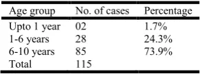
[image:2.595.328.543.53.200.2]Out of 115 cases, 85 cases were in the age group of 6-10 years (73.9%), followed by 28 cases in the age group of 1-5 years (24.3% ) and only 2 were infants (1.7%) (Table 1). Most cases (54.7%) presented with swelling in the neck region without any symptom. Symptomatic patients presented with throat pain (18.2%), painful neck swelling with fever (9.5%), fever and weight loss (7.8%), ear discharge (6.9%) and fever and loss of appetite (2.6%) (Table 2).
Table 1 . Age-wise distribution of cases
Age group No. of cases Percentage
Upto 1 year 02 1.7%
1-6 years 28 24.3%
6-10 years 85 73.9%
Total 115
[image:2.595.328.544.258.402.2]It was observed that most of the children with cervical lymphadenopathy were in the age range of 6-10 years.
Table 2. Symptom-wise distribution of cases
Symptoms No. of cases Percentage
Neck swelling only 63 54.7%
Throat pain 21 18.2%
Painful neck swelling with fever 11 9.5%
Fever and weight los 09 7.8%
Ear discharge 08 6.9%
Fever and loss of appetite 03 2.6%
Total 115
It was observed that the most common presentation was asymptomatic neck swelling.
Table 3. Sites of lymphadenopathy
Site No. of cases Percentage
Lateral cervical 44 38.2%
Bilateral multiple 26 22.6%
Submental 13 11.3%
Submandibular 11 9.5%
Post- auricular 06 5.2%
Pre-auricular 04 3.4%
Occipital 02 1.7%
Part of generalised lymphadenopathy 09 7.8%
Total 115
It was observed that the most common site of cervical lymphadenopathy was lateral cervical.
[image:2.595.89.235.400.454.2]Here we see, polymorphous population of lymphocytes with small mature lymphocytes (black arrows) admixed with large reactive lymphocytes ( white block arrows). (H & E, 40x)
Figure 1. Reactive hyperplasia lymph node.
[image:2.595.326.541.442.603.2]Here we see, polymorphs, lymphocytes and macrophages in a necrotic background with cellular debris. ( H & E, 40x).
Figure 2. Acute suppurative lymphadenitis
Here we see, well defined epithelioid granuloma. ( H & E, 40x)
Figure 3. Tubercular lymphadenitis
Here we see, pink rod like structures in a blue background. Slide positive for AFB.(ZN stain, 100x)
Figure 4. AFB
[image:2.595.57.267.521.602.2] [image:2.595.329.543.634.774.2] [image:2.595.50.276.668.769.2]Here we see, classical RS cell (arrow) is visible in a background of histiocytes and RBCs. ( H & E , 40 x)
Figure 5. HL – MC type
[image:3.595.57.271.51.188.2]Here we see, Hodgkin cells/ popcorn cells (arrow), in a background of lymphocytes.(H & E,40x)
Figure 6. HL – NLP type
Here we see, atypical large lymphoid cells with high N/C
cytoplasm, fine chromatin and inconspicuous nucleoli. Very few mature small lymphocytes and polymorphs seen admixed with the atypical cells.( H & E, 40x)
Figure 7. NHL, of DLBCL subtype
Here we see, large pleomorphic atypical lymphoid cells with irregular nuclei, moderate amount of basophilic cytoplasm, fine vesicular chromatin and inconspicuous nucleoli. Mature small lymphocytes and polymorphs are seen in the background. ( H & E, 40x)
Figure 8. NHL, of ALCL subtype
Here we see, classical RS cell (arrow) is visible in a background of lymphocytes,
Here we see, Hodgkin cells/ popcorn cells (arrow), in a background of
Here we see, atypical large lymphoid cells with high N/C ratio, scant cytoplasm, fine chromatin and inconspicuous nucleoli. Very few mature small lymphocytes and polymorphs seen admixed with the atypical cells.( H & E,
NHL, of DLBCL subtype
cells with irregular nuclei, moderate amount of basophilic cytoplasm, fine vesicular chromatin and inconspicuous nucleoli. Mature small lymphocytes and polymorphs are seen in the background. ( H & E, 40x)
NHL, of ALCL subtype
[image:3.595.59.273.229.380.2]Here we see, clusters of histiocytes with spindled to oval nuclei, abundant pale basophilic cytoplasm and few with lipofuschin pigments ( arrow ), admixed with mature small lymphocytes. ( H & E, 40 x)
Figure 9. Non-specific chronic lym
Clinical examination revealed that out of 115, most of the patients 44 (38.2 %) had lateral cervical lymph node enlargement, followed by bilateral multiple enlarged nodes (mostly small and non tender) in 26 cases (22.6%), submental region in 13 cases (11.3%), submandibular region in 11 cases (09.5%), post auricular region in 6 cases
region in 4 cases (3.4%) and occipital region in 2 cases (01.7%). 9 cases (7.8%) had cervical lymph node involvement as a part of generalised lymphadenopa
100 cases, FNAC results were inconclusive due to inadequate smear in 3 cases( 3% ). Reactive lymphadenitis
suppurative lymphadenitis (20%) and tubercular lymphadenitis (8.6%) were the commonest lesions noted on FNAC findi (Table 4). Comparision of cytological and histological diagnosis was possible in only 3 cases of lymphomas
NHL), the cytological diagnosis was concordant with the histopathological diagnosis in all 3 of them. AFB staining (ZN) was done in cases
lymphadenitis, and 4 out of 6 cases showed AFB positivity. Rest 2 cases were advised for excisional biopsy.
DISCUSSION
Cervical lymphadenopathy is a common problem in pediatric
clinic. Park, in his study, found that around 90% of
aged 4-8 years old have cervical lymphadenopathy
1995). Cervical lymphadenopathy includes bot
(enlarged and tender) and non-
lymph nodes. It can be a part of more generalized lymphadenopathy, defined as enlargement of two or more non contiguous lymph node regions.
Definition(Pediatric Cervical Lymphadenopathy, 2009
Pathologic Lymph Node - > 2 cm in pediatric patients is considered abnormal
Acute Lymphadenopathy - < 2 weeks’ in duration. Sub acute Lymphadenopathy
-Chronic Lymphadenopathy - > 6 weeks’ in duration.
Classification of Cervical Lymphadenopathy Based on
Clinical Presentation
1. Acute Unilateral: This is the most common type of cervical lymphadenopathy. This is usually reactive and secondary to upper respiratory tract infection (URTI), skin infection, or dental infection.
ters of histiocytes with spindled to oval nuclei, abundant pale basophilic cytoplasm and few with lipofuschin pigments ( arrow ), admixed with mature small lymphocytes. ( H & E, 40 x)
specific chronic lymphadenitis (sinus histiocytosis)
Clinical examination revealed that out of 115, most of the patients 44 (38.2 %) had lateral cervical lymph node enlargement, followed by bilateral multiple enlarged nodes (mostly small and non tender) in 26 cases (22.6%), submental %), submandibular region in 11 cases 09.5%), post auricular region in 6 cases (05.2%), pre-auricular region in 4 cases (3.4%) and occipital region in 2 cases (01.7%). 9 cases (7.8%) had cervical lymph node involvement as a part of generalised lymphadenopathy (Table 3). Out of 100 cases, FNAC results were inconclusive due to inadequate smear in 3 cases( 3% ). Reactive lymphadenitis (60%), acute 20%) and tubercular lymphadenitis were the commonest lesions noted on FNAC findings Comparision of cytological and histological diagnosis was possible in only 3 cases of lymphomas (HL and NHL), the cytological diagnosis was concordant with the histopathological diagnosis in all 3 of them. AFB staining ZN) was done in cases diagnosed as Tubercular lymphadenitis, and 4 out of 6 cases showed AFB positivity. Rest 2 cases were advised for excisional biopsy.
Cervical lymphadenopathy is a common problem in pediatric , in his study, found that around 90% of children 8 years old have cervical lymphadenopathy (Park, Cervical lymphadenopathy includes both inflamed inflamed (enlarged, non-tender) lymph nodes. It can be a part of more generalized ed as enlargement of two or more non- contiguous lymph node regions.
Pediatric Cervical Lymphadenopathy, 2009)
> 2 cm in pediatric patients is
< 2 weeks’ in duration. - 2-6 weeks’ in duration. > 6 weeks’ in duration.
of Cervical Lymphadenopathy Based on
[image:3.595.59.270.415.550.2] [image:3.595.57.271.605.752.2]Other rare causes are Kawasaki, cat scratch disease
(Bartonella) and Kikuchi-Fujimoto disease (histolytic
necrotising lymphadenitis).
2. Acute Bilateral: This type of lymphadenitis occurs secondary to viral URTI, Epstein-Barr virus (EBV), and cytomegalovirus (CMV).
3. Sub-acute: The common cause for this is Mycobacterium tuberculosis.
4. Chronic: This can be reactive in process secondary to neoplasia, lymphoma, leukemia, or soft tissue tumours.
Cervical lymph nodes include preauricular, parotid, jugulo-digastric, submental, submandibular, posterior cervical, superficial cervical, deep cervical, occipital and posterior auricular (mastoid ) lymph nodes.
In our study, out of 115 children, most of them had enlarged lateral cervical lymph nodes (38.2%). Most of these cases were incidentally dicovered by the parents and the children had no symptoms related to the lymphadenopathy. Other common sites were bilateral multiple (22.6%) and submental (11.3%). Generally, the cause of lymph node swelling in young patients – from babies to infants and adolescents – is benign in over 80% of cases. This figure decreases considerably with age, to the extent that a malignant cause is found in over 60% of
patients age 50 and above (Isselbacher et al., 1995). However,
if there is a suspicion of malignancy in a young patient, the most frequent causes in children under 6 years of age are acute leukemia, neuroblastoma, rhabdomyosarcoma and non-Hodgkin lymphoma.
Between the ages of 7 and 13, non-Hodgkin lymphoma and
Hodgkin’s lymphoma are roughly equal, with
rhabdomyosarcoma and thyroid cancer occurring more rarely. From the age of 13, Hodgkin’s disease is the leading malignant cause of neck masses during childhood and adolescence
(Brown and Azizkhan, 1998). In our study, recative
lymphadenopathy (60%) was the most common cytological diagnosis followed by acute suppurative lymphadenopathy (20%) and tubercular lymphadenopathy (8.6%). Malignancy was not a common underlying cause in this age group (5.2%). Malignant enlarged cervical lymph nodes showed HL (2.6%) and NHL (1.7%). Our findings were consistent with the above studies. Reactive hyperplasia is diagnosed on FNAC by a polymorphous population of lymphocytes and tingible body macrophages. Tubercular lymphadenitis are diagnosed on smears by presence of epitheloid granulomas, in a background of necrosis, lymphocytes and presence of acid fast bacilli on Ziehl Nelson (ZN) stain. Cytology of acute suppurative lymphadenitis consists of acute inflammatory infilterate in a necrotic background.
Chronic non specific lympadenitis includes follicular hyperplasia or sinus histiocytosis, which on FNA smears shows aggregates of macrophages/histiocytes, sdmixed with lymphocytes. In our study, the diagnoses were made based on these cytomorphological criteria (Figure 1-9). In 6 cases showing granulomatous lymphadenitis, ZN stain was used for confirming tubercular etiology, which showed AFB positivity in 4/6 cases (3.4% ) and negative in 2/6 cases (1.7%). The
latter were given the diagnosis of granulomatous
lymphadenitis and were advised for excisional biopsy for further confirmation. NHLs in children are almost always one of the three types: lymphoblastic, small non- cleaved type (Burkitt and non- Burkitt) and large cell type (ALCL and DLBCL ). These are high grade lymphomas. In our study, we diagnosed 2 cases of NHL, both as high grade, large cell type (Figure 7 & 8). Histopathological examination confirmed the findings. In children diagnosed with HL, MC, NLPHL, and
NS are the subtypes more commonly seen (Thomas et al.,
2002). In our study, we diagnosed 03 cases of HL, 2 MC and 1
NLPHL subtype (Figure 5 & 6).2 cases were confirmed by
HPE. 1 case was lost to follow-up. Primary diagnostic evaluation of childhood peripheral lymphadenopathy is mainly based on group of lymph nodes involved and FNA. A careful history and thorough physical examination are the first steps in establishing the cause of a neck mass. Location, size, consistency, and mobility of the mass provide clues and are useful for comparison during follow-up. Amongst diagnostic modalities, FNA is a rapid, simple, accurate diagnostic procedure and important initial step in the evaluation and management of enlarged cervical lymph nodes in children. It is very well tolerated by children, and there were no complications.
Conclusion
Cervical lymph node enlargement is a common clinical problem in the pediatric population, reactive, pyogenic and granulomatous enlargement being important causes. Since the diagnosis varies from a simple infection to malignancy, this can be a matter of anxiety for both the family as well as the treating doctor. FNAC can be recommended as a first line of investigation in the diagnosis of cervical lymphadenopathy in paediatric age group.
Abbreviations
AFB – Acid fast bacilli.
ALCL – Anaplastic large cell lymphoma. CMV – Cytomegalovirus.
DLBCL – Diffuse large B cell lymphoma. EBV – Ebstein Bar Virus.
[image:4.595.90.498.78.175.2]FNAC – Fine needle aspiration cytology.
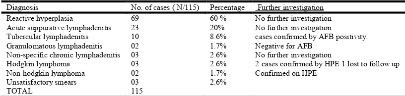
Table 4. Cytological diagnosis of 115 cases of cervical lymphadenopathy
Diagnosis No. of cases ( N/115) Percentage Further investigation
Reactive hyperplasia 69 60 % No further investigation
Acute suppurative lymphadenitis 23 20% No further investigation
Tubercular lymphadenitis 10 8.6% cases confirmed by AFB positivity.
Granulomatous lymphadenitis 02 1.7% Negative for AFB
Non-specific chronic lymphadenitis 03 2.6% No further investigation
Hodgkin lymphoma 03 2.6% 2 cases confirmed by HPE 1 lost to follow up
Non-hodgkin lymphoma 02 1.7% Confirmed on HPE
Unsatisfactory smears 03 2.6%
TOTAL 115
It was observed that most common cause of cervical lymohadenopathy in children, in our study, was reactive hyperplasia. Acute suppurative and tubercular lymphadenitis were other common causes.
HL – Hodgkins Lymphoma.
HPE – Histopathological examination. NHL – Non-Hodgkins Lymphoma. NS – Nodular sclerosis.
ZN – Ziehl Neelsen.
Acknowledgement: I am sincerely grateful to Dr. Seema Chaddha (Incharge Histopathology, Sr. DMO, NRCH, New Delhi ) for her constant support and encouragement.
Conflict of interest statement: No conflict of interest.
Funding statement: Self
REFERENCES
Brown RL, Azizkhan RG. 1998. Pediatric head and neck
lesions. Pediatr Clin North Am., 45(4):889–905. doi:
10.1016/S0031-3955(05)70052-3.
Cummings: Otolaryngology: Head and Neck Surgery, 4th ed. Isselbacher KJ, Braunwald E, Wilson JD, Martin JB, Fauci
AS, Kasper DL. 1995. Harrisons Innere Medizin.13th ed. Wien: Blackwell.
Paediatric Guidelines 2013-2014.
Park YW. 1995. Evaluation of neck masses in children. Am.
Family Physician, 51:1904-1912.
Pediatric Cervical Lymphadenopathy, 2009. Grand Rounds Presentation, The University of Texas Medical Branch, Department of Otolaryngology.
Thomas RK, Re D, Zander T, et al. 2012. Epidemiology and
etiology of Hodgkin's lymphoma. Ann Oncol., 13 Suppl
4:147–152.