Published Ahead of Print 21 November 2012.
2013, 195(3):545. DOI: 10.1128/JB.01980-12.
J. Bacteriol.
Minton, Davide Serruto and Meera Unnikrishnan
Roberto Adamo, Sarah A. Kuehne, Maria Scarselli, Nigel P.
Tanja Ðapa, Rosanna Leuzzi, Yen K. Ng, Soza T. Baban,
Clostridium difficile
Formation by the Anaerobic Pathogen
http://jb.asm.org/content/195/3/545
Updated information and services can be found at:
These include:
SUPPLEMENTAL MATERIAL
Supplemental material
REFERENCES
http://jb.asm.org/content/195/3/545#ref-list-1
at:
This article cites 65 articles, 25 of which can be accessed free
CONTENT ALERTS
more»
articles cite this article),
Receive: RSS Feeds, eTOCs, free email alerts (when new
http://journals.asm.org/site/misc/reprints.xhtml
Information about commercial reprint orders:
http://journals.asm.org/site/subscriptions/
To subscribe to to another ASM Journal go to:
on November 18, 2013 by Univ of Nottingham
http://jb.asm.org/
Downloaded from
on November 18, 2013 by Univ of Nottingham
http://jb.asm.org/
Pathogen
Clostridium difficile
Tanja Ðapa,aRosanna Leuzzi,aYen K. Ng,bSoza T. Baban,bRoberto Adamo,aSarah A. Kuehne,bMaria Scarselli,aNigel P. Minton,b
Davide Serruto,aMeera Unnikrishnana
Novartis Vaccines and Diagnostics, Siena, Italya
; Clostridia Research Group, School of Molecular Medical Sciences, Centre for Biomolecular Sciences, University of Nottingham, Nottingham, United Kingdomb
Bacteria within biofilms are protected from multiple stresses, including immune responses and antimicrobial agents. The bio-film-forming ability of bacterial pathogens has been associated with increased antibiotic resistance and chronic recurrent infec-tions. Although biofilms have been well studied for several gut pathogens, little is known about biofilm formation by anaerobic gut species. The obligate anaerobeClostridium difficilecausesC. difficileinfection (CDI), a major health care-associated prob-lem primarily due to the high incidence of recurring infections.C. difficilecolonizes the gut when the normal intestinal micro-flora is disrupted by antimicrobial agents; however, the factors or processes involved in gut colonization during infection remain unclear. We demonstrate that clinicalC. difficilestrains, i.e., strain 630 and the hypervirulent strain R20291, form structured biofilmsin vitro, with R20291 accumulating substantially more biofilm. Microscopic and biochemical analyses show multiple layers of bacteria encased in a biofilm matrix containing proteins, DNA, and polysaccharide. Employing isogenic mutants, we show that virulence-associated proteins, Cwp84, flagella, and a putative quorum-sensing regulator, LuxS, are all required for maximal biofilm formation byC. difficile.Interestingly, a mutant in Spo0A, a transcription factor that controls spore formation, was defective for biofilm formation, indicating a possible link between sporulation and biofilm formation. Furthermore, we demonstrate that bacteria in clostridial biofilms are more resistant to high concentrations of vancomycin, a drug commonly used for treatment of CDI. Our data suggest that biofilm formation byC. difficileis a complex multifactorial process and may be a crucial mechanism for clostridial persistence in the host.
B
iofilms are sessile surface-associated microbial communities, encapsulated within self-produced polymeric matrices (1). Biofilms represent the predominant state of bacteria in nature; only a small fraction of bacteria in natural ecosystems are believed to exist planktonically (2). Bacteria in biofilms are known to be more resistant to different environmental stresses, including an-tibiotics (2). The human large intestine is a good example of an extremely complex ecosystem, home to numerous bacterial spe-cies of microflora, which play an important role in protection against gut diseases (3). Various gut pathogens, including entero-toxigenicEscherichia coli,Salmonella,Yersinia, etc., can alter the dynamics of the gut due to their highly adhesive and invasive properties (4) and thus establish infections. Biofilm formation by gut pathogens such as enteroaggregativeE. colihas been well stud-ied bothin vitroandin vivo(5,6). Several bacterial factors such as adhesins and pili, which mediate biofilm formation, have been implicated in bacterial colonization and virulence (7).Biofilm formation has been characterized for very few individ-ual bacterial gut species of the numerous anaerobic species that populate the gut. This could be attributed to difficult cultivation and genetic manipulation of such bacteria.Clostridium difficileis a spore-forming, Gram-positive anaerobic bacillus that can causes severe gastrointestinal infections in humans (8).C. difficile infec-tion (CDI) is usually associated with antimicrobial therapy since it results in disruption of the normal microbiota. The clinical symp-toms of CDI range from mild or severe diarrhea to serious inflam-matory conditions, including pseudomembranous colitis (8). CDI is one of the predominant nosocomial infections worldwide today. The population with the largest risk for CDI is the elderly, although CDI in younger patients is on the rise (9). The
transmis-sion of clostridial disease occurs through spores, as demonstrated recently using a murine model forC. difficileinfection (10,11).
CDI is primarily a toxin-mediated disease. The two large clos-tridial toxins A (TcdA) and B (TcdB) are the best-characterized virulence factors ofC. difficile(12–15).C. difficiletoxins have been shown to have many effects, including disorganization of the cell actin cytoskeleton and tight junctions, induction of apoptosis, fluid accumulation, and destruction of the epithelium (14,15). Although the toxins are key virulent factors, a role for bacterial colonization has been highlighted in recent years. Mice immu-nized withC. difficilesurface proteins demonstrated reduced in-testinal colonization withC. difficile(16). Recently, the fibronec-tin-binding adhesin protein A was shown to play a role inC. difficilecolonization (17). The high- and low-molecular-weight surface layer proteins (SLPs) are predicted to be involved in ad-herence ofC. difficileto host cells during the infection (18,19). Cell wall proteins (CWPs) such as Cwp66 and Cwp84 have been shown to be important in the adherence and degradation of the extracellular matrix (20,21).
Received9 October 2012Accepted16 November 2012 Published ahead of print23 November 2012
Address correspondence to Meera Unnikrishnan, meera.unnikrishnan @novartis.com.
Supplemental material for this article may be found athttp://dx.doi.org/10.1128 /JB.01980-12.
Copyright © 2013, American Society for Microbiology. All Rights Reserved.
doi:10.1128/JB.01980-12
on November 18, 2013 by Univ of Nottingham
http://jb.asm.org/
Recurrent bacterial infections have been associated with the ability to form resilient biofilms for various pathogens (22). Per-sistent recurringC. difficileinfections have presented a major hur-dle in the treatment of CDI (23). The ability to form biofilms has been demonstrated recently for related clostridial species and for
C. difficilein relation to other intestinal species (24,25). However, biofilm formation byC. difficile, or the mechanisms involved in this process, has not yet been characterized. We describe biofilm formation by clinicalC. difficile strainsin vitrousing multiple techniques. Using isogenic mutants of proteins associated with clostridial pathogenesis we have identified surface and regulatory proteins that are required for biofilm development by the hyper-virulent strain R20291. We also report a role forC. difficile bio-films in resistance to antibiotics commonly used for the treatment of CDI.
MATERIALS AND METHODS
Bacterial strains and media.TwoC. difficilestrains, 630 and strain B1/ NAP1/027 R20291 (isolated from the Stoke Mandeville outbreak in 2004 and 2005), were used in our studies.C. difficilestrains were grown at 37°C in an anaerobic workstation (Don Whitley, United Kingdom) in BHIS, brain heart infusion (Bacto, USA) supplemented withL-cysteine (0.1% (wt/vol), and yeast extract (5 mg/ml; Bacto, USA). Details about antibi-otics are described in the supplemental material. Table S1 in the supple-mental material lists the strains and plasmids used in the present study.
Biofilm formation assay.For the generation of biofilms, overnight cultures ofC. difficilewere diluted 1:100 into fresh BHIS, containing 0.1 M glucose or 0.3 M NaCl, and incubated in the tissue culture treated 24-well polystyrene plates (Costar, USA), 1 ml per well, in anaerobic conditions at 37°C for 1 h to 120 h. Twenty-four-well cell culture plates were preincu-bated in anaerobic conditions for 48 h prior to use. To prevent evapora-tion of liquid, the plates were wrapped with parafilm.
Measurement of biofilm biomass.Measurement of biofilm biomass with crystal violet (CV) was done as described in previously published methods (24). After the required incubation, wells of the 24-well plate were gently washed twice with sterile phosphate-buffered saline (PBS) and then allowed to dry for 10 min. The biofilm was stained with 1 ml of filter-sterilized 0.2% CV and incubated for 30 min at 37°C in anaerobic conditions. CV was removed from the wells, followed by two washes with sterile PBS. The dye was extracted by adding 1 ml of methanol to each well, followed by incubation for 30 min at room temperature in aerobic con-ditions. The methanol-extracted dye was diluted 1:1, 1:10, or 1:100 and
A570was measured with spectrophotometer Ultrospec 500pro
(Amer-sham Biosciences).
For bacterial cell counts from biofilms, the planktonic phase was first removed and wells were washed twice with PBS. The adherent biofilms were then detached by scraping with a sterile pipette tip, washed in PBS, and plated on BHIS for determination of the number of CFU present in the biofilm.
Enzymatic inhibition of biofilms.To study whether enzymes affect the biofilm formation, 0.1 mg of proteinase K/ml and 2 U of DNase I/ml was added to biofilms at time zero. To see whether these enzymes affect preformed biofilms, biofilms were allowed to form in 24-well plate as described above, washed twice with sterile PBS. Fresh BHIS plus 0.1 M Glc with 0.1 mg of proteinase K/ml or 2 U of DNase I/ml was added, and the biofilms were incubated for a further 24 h, followed by staining with 0.2% CV as described above.
Confocal microscopy analysis of biofilm formation. C. difficile
strains were grown in 4-well glass chamber slides (BD Falcon, USA) in BHIS plus 0.1 M glucose at 37°C in anaerobic conditions for 24 or 72 h and then stained with various staining reagents. For biofilm staining with the fluorescent BacLight Live/Dead stain mixture (Molecular Probes/Invitro-gen), which contains the nucleic acid stains Syto 9 and propidium iodide, wells were gently washed twice with PBS– 0.1% saponin to remove
unat-tached cells, followed by incubation with dye for 15 min at 37°C. Before removal from the anaerobic chamber, the bacteria were fixed with 4% paraformaldehyde (PFA) for 15 min.
For immunofluorescent staining bacteria were fixed with 4% PFA for 15 min, washed three times with 2% bovine serum albumin (BSA)–PBS, and blocked with 2% BSA and 0.1% saponin in PBS for 10 min. The samples were then incubated with mouse serum against fixed R20291 strain or against a syntheticC. difficile PSII polysaccharide (synthetic phosphorylated hexasaccharide repeating unit) (26) diluted 1:1,000 for 1 h, followed by incubation with Alexa Fluor 568 goat anti-mouse antibod-ies diluted 1:500 for 1 h.
Biofilm matrix was labeled by SYPRO Ruby biofilm matrix stain (Mo-lecular Probes/Invitrogen), which stains matrices of biofilms (labels most classes of proteins, including glycoproteins, phosphoproteins, lipopro-teins, calcium-binding prolipopro-teins, and fibrillar proteins). After two washes with sterile PBS– 0.1% saponin sample were incubated for 30 min with 1 ml of Ruby biofilm matrix stain at room temperature and then washed with distilled water.
All chamber slides were mounted with ProLong Gold Antifade reagent (Molecular Probes/Invitrogen) and analyzed with a Zeiss Observer LSM 710 confocal scanning microscope.
Construction ofC. difficilemutants and complemented strains.
Mutations in the genessleC,spo0A,fliC, andluxSwere generated using the insertional inactivation system, ClosTron, as previously described (27–
30). Construction of thefliCandluxSmutants is described in the supple-mental material. The positions of in-frame deletions and ClosTron inser-tions are reported in Table S1 in the supplemental material. To generate thecwp84mutant, the 3=end of thecwp84gene was deleted by using allelic exchange as described elsewhere (Y. K. Ng et al. unpublished data). Com-plementation studies are described in the supplemental material.
Measurement ofC. difficilespores in biofilm.C. difficilespores were measured as previously described (27). At day 1, 3, and 5 of biofilm for-mation samples were removed from the anaerobic chamber and heated at 80°C for 25 min to kill the vegetative cells but not the spores. Serially diluted were then plated onto BHI agar and BHI supplemented with 0.1% taurocholate (Sigma-Aldrich, USA). The plates were incubated for 24 to 48 h.
Biofilm resistance to antibiotics.To determine the resistance capa-bilities of planktonic and biofilm cells, we used antibiotic vancomycin (Sigma-Aldrich) at 100 times the MIC (the MIC was determined to be 0.2
g/ml for R20291 [data not shown]). Resistance was determined to both sessile and planktonic populations from the sameC. difficileculture. The supernatant (planktonic fraction) from 1- or 3-day biofilm cultures in six-well plates was removed, washed once in sterile PBS, resuspended in BHIS– 0.1 M glucose with 20g of vancomycin (Sigma-Aldrich)/ml, and incubated in fresh six-well plates. In parallel, to six-well plate where su-pernatants were removed, fresh BHIS– 0.1 M glucose containing 20g of vancomycin/ml was added to the adherent biofilms (sessile part). Treat-ment with vancomycin was performed for 6 or 24 h. CFU from planktonic and adherent phases were determined by plating dilutions on BHIS agar. Biofilm cell counts were determined as described above. The percent sur-vival was calculated by dividing the number of CFU posttreatment by the initial number of CFU.
To investigate whether the protection from vancomycin is a result of biofilm structure or a physiological attribute of the cells in the biofilm, we perform experiment as described above. The sessile part of biofilm was disrupted gently by pipetting and incubated with BHIS with 0.1 M Glc and 20g of vancomycin/ml. The planktonic growth from the same well was washed and incubated with BHIS with 0.1 M Glc and 20g of vancomy-cin/ml. CFU counts were determined after 6 and 24 h, and counts before and after different times of treatment were compared.
Statistical analyses.All biofilm experiments were performed in trip-licates and at least three independent experiments were performed. A paired Studentttest was performed to determine whether the differences
on November 18, 2013 by Univ of Nottingham
http://jb.asm.org/
between two groups was significant.Pvalues of⬍0.05 were considered statistically significant.
RESULTS
C. difficileforms time- and strain-dependent biofilmsin vitro.
In order to investigate biofilm formation byC. difficile, we studied two strains,C. difficile630, a commonly studied clinical strain, and the hypervirulent strain R20291. Sessile biomass (biofilm) was measured by staining the biofilms with CV, as described in Mate-rials and Methods. We tested biofilm formation in several media (chemically defined medium, rich medium TYM, and BHIS) sup-plemented with combinations of sodium chloride and glucose (data not shown). Both strains form higher amounts of biofilm in rich medium BHIS supplemented with 0.1 M glucose. In the ab-sence of glucose, strain 630 forms significantly lower amounts of biofilm compared to that seen in the presence of 0.1 M glucose. However, the presence of glucose does not significantly affect bio-film formation for the strain R20291. The presence of 0.3 M so-dium chloride inhibits biofilm formation in both strains (Fig. 1A). Biofilms formed in all conditions tested were adherent in na-ture and were found to form only on the bottoms of the wells (of 24-well plates) and not on the surface of the culture medium. Biofilm formation in BHIS containing 0.1 M glucose was moni-tored over a time period of 1 to 120 h and quantitated by both CV staining and bacterial counts.C. difficile630 biofilm accumulation appears to be maximal at day 5 (120 h) (Fig. 1B). Strain R20291 starts to form biofilm after 6 h of incubation, with maximum biofilm formation after 24 h, as measured by CV staining (Fig. 1C). Biofilm formed by the strain R20291 uniformly covers the entire surface area of the bottom of the wells (24-well plate) at 24 h (Fig. 1D), while at the same time point for strain 630 (Fig. 1D) we saw much less biofilm formation. At 72 h (Fig. 1E), we ob-served relatively more uniform biofilms for strain 630 and uneven biofilms for strain R20291 (Fig. 1E). Bacterial counts from bio-films indicated a similar trend of increasing bacterial numbers until 24 h and a decrease after this period for both strains. How-ever, it is clear that R20291 biofilms have significantly higher number of bacteria as well as higher amounts of biofilms as quan-titated by CV, compared to 630, under the conditions tested.
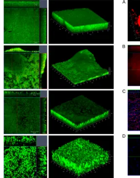
C. difficilebiofilms are multilayered and encased in a thick matrix.To examine biofilm formation by strains R20291 and 630 further, biofilms were allowed to form on glass slides in BHIS containing 0.1 M glucose for one or 3 days. Live/Dead staining (with Syto 9 and propidium iodide dyes staining live bacteria green and dead bacteria red, respectively) was used in order to evaluate the bacterial viability and biofilm thickness. We found that the majority of bacteria in both R20291 (Fig. 2AandB) and 630 (Fig. 2CandD) biofilms were alive with a minor number of dead cells after 1 day (Fig. 2AandC) and 3 days (Fig. 2BandD). Z-stack acquisitions revealed multiple layers of bacteria in the biofilm underneath a dense layer of material (Fig. 2AtoD, right panels). Three-dimensional (3D) images show the presence of uniformly spread biofilms for R20291 on day 1 (Fig. 2A), with uneven secondary structures by day 3 (Fig. 2B), and with a thick-ness ranging from 30 to 45m. For strain 630, we observe that the Live/Dead staining of biofilms was not homogeneous as seen for R20291 after day 1 (Fig. 2C) or day 3 (Fig. 2D). The maximum thickness of the biofilm was 30m (72 h). Thus, microscopic analysis suggests thatC. difficilebiofilms are structured, with sev-eral layers of largely live bacteria encased within a dense matrix.
Composition of the C. difficile biofilm matrix. Since the R20291 biofilms were more robust and reproducible, we exam-ined the R20291 biofilms further using multiple staining to eval-uate the extracellular matrix. Interestingly, antibodies against
FIG 1C. difficilebiofilm formationin vitro. (A) Biofilm formation by strain 630 and hypervirulentstrainR20291,inBHISsupplementedwith0.1Mglucoseor0.3MNaCl, for 3 days at 37°C in anaerobic conditions. Biofilm formation was measured by CV staining. (B and C) Time course for biofilm formation for strains 630 (B) and R20291 (C) measured by CV staining (bars) and colony counts (CFU/ml, line). The results are presented in log scale, and the error bars represent standard deviations (P⬍0.05). The data are representative of at least three independent experiments, each performed in triplicates. (D and E) Photographs of biofilms formed on a 24-well plate for strains 630 and R20291 on day 1 (D) and day 3 (E) are shown.
on November 18, 2013 by Univ of Nottingham
http://jb.asm.org/
whole bacteria were able to stain a complex biofilm matrix and some superficial individual bacteria (Fig. 3A). The formation of a thick mature biofilm was visualized by a biofilm matrix tracer Ruby stain, which labels a range of protein classes including gly-coproteins, lipoproteins, and phosphoproteins (Fig. 3B). In addi-tion, when using staining with an antibody against a syntheticC. difficilePSII polysaccharide (synthetic phosphorylated hexasac-charide repeating unit) (26), we observed staining of fiber-like structures on biofilms (Fig. 3C). To confirm whether proteins are part of biofilm matrix, we treated biofilms with 0.1 mg of protei-nase K/ml. Although planktonic growth was not inhibited by pro-teinase K (see Fig. S1 in the supplemental material), incubation of the bacterial cultures with the enzyme resulted in a significant decrease in biofilm formation on days 1 and 3, with protease (Fig. 3E, dark-shaded bars). One-day-old biofilms were also disrupted when treated with proteinase K (Fig. 3E, light-shaded bars). We also performed similar experiments with DNase I treatment and found that DNase I also inhibited biofilm formation (Fig. 3F, dark-shaded bars) and is able to reduce preformed biofilms
(Fig. 3F, light-shaded bars), although to a lesser degree compared to proteinase K. These results indicate that bacterial proteins, DNA, and surface polysaccharide are components of the biofilm matrix formed byC. difficile in vitro.
An intact S layer and flagella are important inC. difficile
biofilm formation. Numerous cell surface proteins and surface
FIG 2Confocal microscopy analysis of biofilms formed byC. difficile. Live/ Dead staining shows dead (red) and live (green) bacteria (propidium io-dide and Syto 9, respectively) in strains R20291 (A and B) and 630 (C and D) biofilms after incubation for 1 day (A and C) and 3 days (B and D). 3D images of biofilms depicting biofilm thickness inm are shown in the left panels.
FIG 3Characterization ofC. difficilebiofilm matrix. (A) shows 3D confocal microscopy images of staining of R20291 biofilms with murine anti-R20291 (left panel) and mouse preimmune serum (right panel) after 3 days of incuba-tion. (B) Biofilms stained with Ruby matrix stain after 3 days of incubaincuba-tion. Biofilms were stained with antibodies to a syntheticC. difficilePSII polysac-charide (red) and DAPI (blue), which stains the bacterial DNA (C), or with the control mice preimmune serum and DAPI (D). Biofilms were incubated with proteinase K (E) or DNase I (F) as described in Materials and Methods. The dark gray bars represent data from treatment of either enzyme at the start of incubation (inhibition of biofilm formation), and the light gray bars represent data from incubating preformed 1-day-old biofilms with either enzyme (dis-ruption of biofilms). The data shown are representative of at least two inde-pendent experiments performed in triplicates (P⬍0.05).
on November 18, 2013 by Univ of Nottingham
http://jb.asm.org/
[image:5.594.50.337.68.431.2] [image:5.594.293.533.69.514.2]structures such as flagella and pili have been shown to be important for biofilm formation in Gram-positive bacteria. Cwp84 is a key sur-face protease involved in maturation of the S layer ofC. difficile. We compared biofilm formation of a strain with a deletion of thecwp84
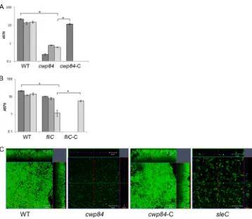
gene, CdiR20291⌬cwp84(⌬cwp84), with the wild type (WT) using multiple methods. Although there was no defect in planktonic growth (see Fig. S2 in the supplemental material), we saw a dramatic decrease in biofilm accumulation forcwp84strain, as measured by CV staining (Fig. 4A). Thecwp84mutant showed a more dramatic defect in biofilm formation on day 1 compared to days 3 and 5. Mi-croscopic analysis showed a single layer of bacteria forcwp84strain (Fig. 4C, panel 2) compared to the WT (Fig. 4C, panel 1). The biofilm defect for thecwp84strain was fully complemented by restoring the WT gene on the chromosome CdiR20291⌬cwp8-C (referred to here ascwp84-C, where the suffix “-C” indicates that the gene is comple-mented).
To examine the role of flagella, a mutant in the flagellin gene,
fliC, CRG3351 (fliC), was tested for biofilm formation. We ob-served a significant decrease in biofilm accumulation for thefliC
mutant compared to the WT on day 5 but not at earlier times using CV staining (Fig. 4B) and microscopic analysis (Fig. 4C, panel 4). This was reversed upon expressing the FliC protein epi-somally from its native promoter (fliC-C) (Fig. 4B). No differ-ences in planktonic growth were observed between these strains (data not shown). These data may indicate that while a mature S
layer is important in early biofilm stages such as adhesion, flagella may be more important for later stages in biofilm formation.
A putative role for quorum sensing in biofilm formation.
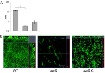
Quorum sensing plays a vital part in biofilm formation, and the involvement of quorum sensing regulators such asluxShas been demonstrated for various other bacteria (31–33). We tested a mu-tant of aluxS, homologue inC. difficile, by homology to other Gram-positive bacteria. A dramatic defect in biofilm formation was observed for theluxSmutant, CRG1183 (luxS), both by CV staining (Fig. 5A) and by microscopy (Fig. 5B, center panel), with no significant differences at different times after incubation (data not shown). Examination of theluxSmutant showed that it is unable to form even a bacterial monolayer on glass surface. Bio-film defects of this mutant were complemented by episomal ex-pression of the full-length gene under the control of the native promoter (luxS-C). Although the complementation did not com-pletely restore the WT phenotype, we were able to detect several layers of bacteria for the complemented strain (Fig. 5B, right panel). Although the growth curves for mutant and comple-mented strains were similar (see Fig. S3 in the supplemental ma-terial), it is possible that expressingluxSepisomally may be toxic for the bacteria, since we observed a smaller colony size for this strain on plates (data not shown).
Sporulation/germination pathway mutants are defective in biofilm formation.Sporulation is a key pathway that is initiated
FIG 4Role of S layer and flagella in biofilm formation. (A) Biofilm formation by WT R20291, acwp84mutant (⌬cwp84) for days 1, 3, and 5 and a complemented strains (cwp84-C) for day 1in vitroas measured by CV staining. (B) Biofilm formation by WT R20291,fliCmutant (fliC) for days 1, 3, and 5 and complemented strainsfliC(fliC-C) for day 5in vitroas measured by CV staining. (C) Confocal microscopy images of Live/Dead staining of biofilms formed by the WT,⌬cwp84,
cwp84-C, andfliC. The results are presented in log scale, and the error bars represent standard deviations (P⬍0.05). Biofilm assays were performed in triplicates, and data are representative of at least three independent experiments.
on November 18, 2013 by Univ of Nottingham
http://jb.asm.org/
[image:6.594.113.471.62.374.2]byC. difficileunder conditions of stress. Regulators of sporulation such as Spo0A are also involved in formation of biofilms in other Gram-positive bacteria (34). We first studied whether spore for-mation occurs inC. difficilebiofilms in our growth conditions, for adherent biofilms and planktonic phase from the same well of the 24-well plate (Fig. 6A). We found that there are very few spores in the biofilm (0.0001%) and in planktonic phases (none detectable) on day 3 and day 5 (0.0001%, data not shown). However, in the control, which was bacteria cultured in a tube (where biofilm for-mation did not occur), spores were formed by day 3 (40 to 50%) (Fig. 6A). Nevertheless, aspo0Amutant CRG1375 (spo0A), which is defective in sporulation (see Fig. S5A in the supplemental ma-terial), demonstrated significantly lesser biofilm formation com-pared to WT both by CV staining (Fig. 6B) and microscopy (Fig. 6D, panel 2) at day 1 and days 3 and 5 (data not shown). We also tested a mutant for a protein involved inC. difficile germina-tion, CRG1166 (sleC) (Fig. S5B). ThesleCmutant is able to form biofilm-like structures (Fig. 6C), but the biofilm is uneven and the thickness of biofilm produced by this strain is never more than 20
m (Fig. 6D, panel 4). The cellular morphology of the bacteria in these biofilms is different to wild-type, and appears to form fila-mentous structures. Biofilm defects of both mutants were com-plemented by episomal expression of genesspo0AorsleC(spoA-C andsleC-C) under the control of their respective native promoters (Fig. 6BtoD).
In addition to the ClosTron mutants described above (fliC,
luxS,spo0A, andsleC), other mutants in the binary toxins,cdtA
andcdtB, or germination-specific N-acetylmuramoyl-L-alanine amidase,cwlD(generated using ClosTron) were also tested, but did not display significant defects in biofilm formation (data not shown).
Effects of vancomycin onC. difficilebiofilms.Since biofilms
are known to be a means by which bacteria protect themselves from antibiotics, we studied whetherC. difficilebiofilms are im-portant in mediating antibiotic resistance. We tested the resis-tance of bacteria in biofilms to the antibiotic vancomycin, which is commonly used for the treatment of CDI.In vitro, vancomycin, has excellent activity againstC. difficile; an MIC of 0.75 to 2.0
g/ml is sufficient to inhibit 90% of strains (23). Both sessile and planktonic phases, from the same C. difficile biofilm culture, 1-day-old (see Fig. S6A in the supplemental material) and 3-day-old biofilms (see Fig. S6B in the supplemental material) were ex-posed to 20g of vancomycin/ml (100 times the MIC of strain R20291) for 6 and 24 h as described in Materials and Methods. The percentages of surviving bacteria after treatment with antibiotics for 24 h for 1-day-old and 3-day-old biofilms are presented inFig. 7A. Bacteria in 1-day-old and 3-day-old biofilms survived 5- and 12-fold more, respectively, than planktonic bacteria. These data support a role forC. difficilebiofilms in resisting antibiotics.
To try and understand whether resistance to vancomycin is due to protection conferred by biofilm matrix structure or an inherent property of the bacteria in biofilm, we studied the effect of vancomycin on adherent biofilms that were disrupted by pipetting (Fig. 7B). Disrupted sessile biofilm and the planktonic phase from 1-day-old biofilm were incubated for 6 and 24 h with 20g of vancomycin/ml. Bacteria from the disrupted adherent biofilms were not more resistant to high concentrations of antibi-otics of biofilm compared to bacteria from the planktonic phase. Although the bacteria from disrupted biofilms do not form new adherent biofilms after the incubation with antibiotics even after 48 h of incubation (data not shown), we observe unstable, thread-like structures in the wells (which were disrupted by pipetting before performing CFU counts). The lack of antibiotic resistance by disrupted biofilm bacteria may indicate a lack of genetic
FIG 5Potential role for quorum sensing inC. difficilebiofilm formation. (A) Biofilm formation by WT R20291, putative quorum-sensing geneluxSmutant (luxS), and complemented strains (luxS-C) as measured by CV staining. (B) Confocal microscopy analysis of WT,luxS, and complemented strainluxS-C. The results are presented in log scale, and the error bars represent the standard deviations (P⬍0.05). The data from biofilm assays are representative of at least three independent experiments performed in triplicates.
on November 18, 2013 by Univ of Nottingham
http://jb.asm.org/
[image:7.594.111.474.63.316.2]changes in the bacteria within resistant biofilms and suggest that the biofilm matrix and/or other epigenetic mechanisms are in-volved in mediating vancomycin resistance.
Biofilm formation, on the other hand, has been reported to be stimulated by subinhibitory concentrations of antibiotics (35). To study whether vancomycin stimulates the biofilm formationin vitro, bacteria were treated with a range of concentrations of van-comycin (0 to 0.5g/ml), both lower and higher than the tube
growth MIC (0.2g/ml), and biofilm formation was measured at day 1 and day 3. No significant induction of biofilm was observed for any of the vancomycin concentrations after 1 day. Inhibition of biofilm formation was evident for concentrations of vancomy-cin ofⱖ0.5g/ml (Fig. 7C). Interestingly, after 3 days incubation, a significant induction of biofilms was observed with 0.5g of vancomycin/ml and to a lesser extent with a subinhibitory con-centration (0.25g/ml) of vancomycin (Fig. 7C). These results
FIG 6Sporulation/germination proteins affectC. difficilebiofilm formation. (A) Quantitation of the number of spores present in the adherent, planktonic phases of biofilm and in planktonic tube culture, in brain heart infusion media (BHI) with sodium taurocholate (BHI⫹T) and heat treatment (80°C), as described in Materials and Methods. (B and C) Biofilm formation by WT R20291, sporulation transcription factorspo0Amutant (spo0A) and complementedspo0Amutant (spo0A-C) after 1 day (B) and WT R20291,sleCmutant (sleC) and complementedsleCmutant (sleC-C) after 3 days (C). (D) Confocal microscopy analysis of WT and mutantsspo0A, complementedspo0Amutant (spo0A-C), andsleC. The results are presented in log scale, and the error bars represent standard deviations (P⬍0.05). Both biofilm and spore quantitation experiments were performed in triplicates, and the data shown are representative of at least three independent experiments.
on November 18, 2013 by Univ of Nottingham
http://jb.asm.org/
[image:8.594.72.509.62.540.2]suggest that exposure to inhibitory vancomycin concentrations can stimulate biofilm formationin vitro.
DISCUSSION
Biofilms are the most representative form of growth of bacteria in the large intestine (4). The ability to form biofilms is known to influence the virulence and also the transmission of intestinal pathogenic bacteria such asVibrio cholerae(36–38). In the present study, we report for the first time the ability of two clinically rele-vantClostridium difficilestrains, “nonepidemic” strain 630 and “epidemic” hypervirulent strain R20291, to form structured bio-filmsin vitro. Furthermore, we identify several genes that may influenceC. difficilebiofilm development.
The ability to adhere and form biofilms influences the ability of pathogens to colonize and establish an infection (39,40). Our data show that R20291 forms more biofilm in all tested conditionsin vitro. It has been reported that strain R20291, a hypervirulent strain, produces higher levels of toxinin vitrocompared to 630 (41). Although colonization of these strains has yet to be examined carefullyin vivo, higher biofilm formation by this strain could indicate better colonizationin vivo. While for the strain R20291 addition of glucose does not increase the levels of biofilm forma-tion, it is an important factor for biofilm formation for strain 630. It is well known that different carbohydrates can modulate biofilm formation. Carbohydrates induce biofilm formation in Strepto-coccus gordonii(42), while inBacillus subtilisthe CcpA protein
represses formation of biofilm in medium with high levels of glu-cose (43). It is possible thatin vivothe nutritional environment in the gut modulates colonization ofC. difficile.
The biofilm matrix is known to protect bacteria during infec-tions by providing an enclosed environment to escape immune responses (5,36). Similar to many clinically relevant biofilm form-ers such asPseudomonas aeruginosaandStaphylococcus aureus,C. difficilealso appears to form a complex matrix comprising of pro-teins, DNA and polysaccharide (44–46). Staining with antibodies againstC. difficilesuggests that this compact biofilm may com-prise surface-associated or secreted bacterial components and may be impenetrable to antibodies. Furthermore, it is interesting that we observe biofilm matrix staining for a synthetic derivative of theC. difficilesurface PSII polysaccharide, which was recently reported to be immunogenic (26). The presence of a complex and perhaps impermeable matrix may protectC. difficilefrom unfa-vorable agentsin vivoin the gut.
Regulation of biofilm formation in Gram-positive bacteria in-volves multiple factors including adhesins, surface structures such as flagella and pili (47). TheC. difficileS layer is composed of S-layer proteins (SLPs) that are present as heterodimeric complex (48). The signal peptide of protein precursor SlpA is removed by proteolytic cleavage. Additional cleavage is essential for matura-tion of SLPs, which is mediated by the cysteine protease Cwp84 (49,50). A mutant incwp84has previously been shown to be
FIG 7Effect of antibiotics onC. difficilebiofilms. (A)Clostridium difficile1- to 3-day-old biofilms and the corresponding planktonic growth were exposed to 20
g of vancomycin/ml (100 times the MIC) for 24 h. The data are presented as percentage of surviving bacteria after treatment with antibiotics for 24 h for 1- and 3-day-old biofilms. (B) Bacterial counts from disrupted 1-day-old biofilms were incubated with 20g of vancomycin/ml for 6 h or 24 h. (C) Biofilm formation measured by CV staining at days 1 and 3 after treatment with subinhibitory and inhibitory concentration of antibiotic vancomycin (MIC for R20291 was 0.2
g/ml). The results are presented in log scale, and the error bars represent standard deviations (P⬍0.05). An asterisk (*) denotes significant differences compared to biofilm formation in the absence of vancomycin (0g/ml).
on November 18, 2013 by Univ of Nottingham
http://jb.asm.org/
[image:9.594.138.450.66.361.2]defective in the S-layer synthesis (50). This protease is also impor-tant for the degradation of extracellular matrix proteins such as fibronectin, laminin, and vitronectin (21). Our data prove that a mature S layer is essential forC. difficilebiofilm formation, per-haps due to the fact that this layer may be essential for anchoring various cell wall associated proteins, which may be required for the early steps such as adhesion during biofilm formation. A role for specific CWPs in biofilms remains to be investigated. Further-more, the observation that antibodies raised against fixed whole bacteria recognize complex structures on biofilms may indicate that surface components such as CWPs may compose the biofilm matrix.
Bacterial flagella are known to modulate attachment, the first step in biofilm formation in motile bacteria, however, the precise role varies between species. For Gram-positive bacteria such asB. subtilisthe presence of flagella is important but not essential for formation of biofilms (51), while forListeria monocytogenes, mo-tility is essential for mature biofilm formation (52). In our exper-iments a mutant in flagellin, a principal component of flagella, clearly affects biofilmsin vitro, indicating that motility ofC. diffi-cileis key in formation of biofilms. Recently, strain 630C. difficile
flagellar mutants were reported to have better adherence in anin vitromodel (53). Our data further suggest that flagella are impor-tant at later biofilm stages, as the muimpor-tant does not display defects in ourin vitroassays at earlier time points.
Cell-cell communication is crucial in a complex structure like biofilms where bacteria are in strict contact with one another. Quorum sensing has an important role in bacterial biofilm forma-tion (33,54). The enzyme LuxS, that synthesizes autoinducer-2 (AI-2), is one of the major modulators of QS and is largely con-served across bacterial species (55). It was demonstrated thatC. difficilegenome carries a 453-bp gene that encodes a protein which shares 40% identity to theV. harveyiLuxS protein and is respon-sible for AI-2 production (56). The role ofluxSin toxin produc-tion is not clear as there are conflicting reports in the literature (56,
57). While precise mechanisms by whichluxSfunctions inC. dif-ficileis unclear at present, our data suggest a role for putative
luxS-encoded molecules in formation of biofilmsin vitroand may indicate that aluxS-mediated quorum-sensing system exists inC. difficile.
Sporulation is a critical pathway in bacterial responses to envi-ronmental stresses. Spo0A is the main response regulator which controls entry into sporulation and is well conserved inBacillus
andClostridiumspecies (58,59). Spo0A is also known to regulate a range of regulatory factors, thus affecting pathways unrelated to sporulation (60).C. difficile spo0Amutants have been reported to modulate toxin production, although there are contradictory data about the nature of this regulation (11,59). Although we do not know yet if there is a link between toxin production and biofilms, clearlyC. difficile spo0Ais involved in the formation of biofilmsin vitro. Biofilm environments have been shown to be optimal for spore formation and spores are part of biofilms for many spore-forming bacteria under nutrient-starved conditions (47). How-ever, we find extremely low numbers of spores in biofilms (adher-ent and planktonic phases of biofilms) under our conditions. Previously, Hamon et al. demonstrated that aB. subtilismutant of
spo0Awas defective for biofilm formation, and similar to our ob-servation, they did not detect spores inBacillusbiofilmsin vitro
(34). We hypothesize thatspo0AinC. difficileacts as a switch between different stress-related pathways such as sporulation,
biofilm formation, and toxin production. Our observation that bacteria sporulate during tube culture, but not in biofilms, when incubated under similar conditions, may support this hypothesis. Recently,spo0Amutants were shown to be defective in persistence and transmission in a murine infection model, primarily due to the inability of thespo0Amutant strain to form spores (11). In addition to production of spores, the formation of biofilmsin vivo
may also account for the persistence defects observed for the
spo0Astrain. Indeed, under otherin vitroconditions orin vivo, spores may also form part of biofilms.
Interestingly, we find that a mutant lacking SleC, a protein recently reported to be specifically involved in germination ofC. difficilespores (27), is defective for biofilm formation. A role for spore germination in biofilms has not been well studied; however, in our conditions given the lack of spores, it is unlikely that SleC is involved in germination of spores. Given that thesleCmutant biofilms shows strikingly different cellular morphologies, it is pos-sible that SleC has other functions such as in hydrolysis of vegeta-tive cell peptidoglycans.
The relevance of biofilm formation in the context of infection and treatment has been widely studied. The role of biofilms in mediating resistance to antibiotics had been well studied for sev-eral bacteria, e.g., methicillin-resistantS. aureus(61). Resistance to antibiotics in biofilm can increase from 10- to 1,000-fold more compared to planktonic bacteria (62). The rising incidence of re-sistance to antibiotic treatments for nosocomial pathogens such as
S. aureusandC. difficilehas been well documented in recent years (63). Highly antibiotic-resistantC. difficilestrains and treatment of recurring clostridial infections have been the major challenges for managing CDI (23). Our findings indicate thatC. difficile bio-films are more resistant to the antibiotic vancomycin, which is commonly used for treatment of CDI. Furthermore, our initial studies on the mechanisms involved in antibiotic resistance show that the bacteria from resistant biofilms do not appear to carry inheritable changes and may suggest a role for theC. difficile bio-film matrix structure in antibiotic penetration, as seen for other bacteria (62). It is also possible that the physiological state of bac-teria within biofilms is important in mediating resistance (64). Interestingly, lower concentrations of vancomycin induce biofilm formation. A role for antibiotics in stimulating biofilm formation has been examined previously for other bacteria and second mes-senger signaling involving c-di-GMP has been implicated in this process (35,65). It is believed that such an induction of biofilms could be clinically relevant when there is exposure to low doses of antibiotics, e.g., the beginning and end of antibiotic therapy, and could perhaps explain ineffective treatment (65). In CDI, the es-tablishment of persistent biofilmsin vivo, in addition to the for-mation of spores, could potentially explain the occurrence of re-current infections.
In conclusion, we demonstrate that clinically relevant strains of the anaerobic gut pathogenC. difficileare able to form complex biofilmsin vitro. C. difficile biofilm formation appears to be a multifactorial process with a role for proteins that are important in different aspects of bacterial physiology. Indeed, the details of the precise roles of each of these proteins/pathways and their reg-ulation remain to be studied. A possible model of infection is that
C. difficilecolonizes the colon via formation of microcolonies or biofilms, followed by toxin production. Formation of biofilmsin vivoperhaps provides the bacterium with a mechanism to protect itself from the cellular immune responses invoked by the toxins, in
on November 18, 2013 by Univ of Nottingham
http://jb.asm.org/
addition to a mechanism of persistence in the presence of antibi-otics. Investigation ofC. difficilebiofilm development during in-fection and factors controlling it could give us a better insight into their role inC. difficilepathogenesis.
ACKNOWLEDGMENTS
The research leading to these results received funding from the European Community’s Seventh Framework Programmes CLOSTNET (PEOPLE-ITN-2008-237942). The funder had no role in study design, data collec-tion and analysis, decision to publish, or preparacollec-tion of the manuscript.
REFERENCES
1.Branda SS, Vik S, Friedman L, Kolter R. 2005. Biofilms: the matrix revisited. Trends. Microbiol.13:20 –26.
2.Davey ME, O’Toole AG.2000. Microbial biofilms: from ecology to mo-lecular genetics. Microbiol. Mol. Biol. Rev.64:847– 867.
3.Cummings JH, Antoine JM, Azpiroz F, Bourdet-Sicard R, Brandtzaeg P, Calder PC, Gibson GR, Guarner F, Isolauri E, Pannemans D, Shortt C, Tuijtelaars S, Watzl B.2004. PASSCLAIM: gut health and immunity. Eur. J. Nutr.43(Suppl 2):II118 –II173.
4.Macfarlane S, Dillon JF.2007. Microbial biofilms in the human gastro-intestinal tract. J. Appl. Microbiol.102:1187–1196.
5.Beloin C, Roux A, Ghigo JM.2008.Escherichia colibiofilms. Curr. Top. Microbiol. Immunol.322:249 –289.
6.Tzipori S, Montanaro J, Robins-Browne RM, Vial P, Gibson R, Levine MM.1992. Studies with enteroaggregativeEscherichia coliin the gnotobi-otic piglet gastroenteritis model. Infect. Immun.60:5302–5306. 7.Kline KA, Falker S, Dahlberg S, Normark S, Henriques-Normark B.
2009. Bacterial adhesins in host-microbe interactions. Cell. Host Microbe
5:580 –592.
8.Rupnik M, Wilcox MH, Gerding DN.2009.Clostridium difficile infec-tion: new developments in epidemiology and pathogenesis. Nat. Rev. Mi-crobiol.7:526 –536.
9.Freeman J, Bauer MP, Baines SD, Corver J, Fawley WN, Goorhuis B, Kuijper EJ, Wilcox MH.2010. The changing epidemiology ofClostridium difficileinfections. Clin. Microbiol. Rev.23:529 –549.
10. Lawley TD, Clare S, Walker AW, Goulding D, Stabler RA, Croucher N, Mastroeni P, Scott P, Raisen C, Mottram L, Fairweather NF, Wren BW, Parkhill J, Dougan G.2009. Antibiotic treatment of clostridium difficile carrier mice triggers a supershedder state, spore-mediated transmission, and severe disease in immunocompromised hosts. Infect. Immun.77: 3661–3669.
11. Deakin LJ, Clare S, Fagan RP, Dawson LF, Pickard DJ, West MR, Wren BW, Fairweather NF, Dougan G, Lawley TD.2012.Clostridium difficile spo0Agene is a persistence and transmission factor. Infect. Immun.80: 2704 –2711.
12. Kuehne SA, Cartman ST, Heap JT, Kelly ML, Cockayne A, Minton NP.
2010. The role of toxin A and toxin B inClostridium difficileinfection. Nature467:711–713.
13. Lyras D, O’Connor JR, Howarth PM, Sambol SP, Carter GP, Phu-moonna T, Poon R, Adams V, Vedantam G, Johnson S, Gerding DN, Rood JI.2009. Toxin B is essential for virulence ofClostridium difficile.
Nature458:1176 –1179.
14. Voth DE, Ballard JD.2005.Clostridium difficiletoxins: mechanism of action and role in disease. Clin. Microbiol. Rev.18:247–263.
15. Shen A.2012.Clostridium difficiletoxins: mediators of inflammation. J. Innate Immun.4:149 –158.
16. Pechine S, Janoir C, Boureau H, Gleizes A, Tsapis N, Hoys S, Fattal E, Collignon A. 2007. Diminished intestinal colonization byClostridium difficileand immune response in mice after mucosal immunization with surface proteins ofClostridium difficile.Vaccine25:3946 –3954. 17. Barketi-Klai A, Hoys S, Lambert-Bordes S, Collignon A, Kansau I.2011.
Role of fibronectin-binding protein A inClostridium difficileintestinal colonization. J. Med. Microbiol.60:1155–1161.
18. Calabi E, Calabi F, Phillips AD, Fairweather NF. 2002. Binding of
Clostridium difficilesurface layer proteins to gastrointestinal tissues. In-fect. Immun.70:5770 –5778.
19. Karjalainen T, Waligora-Dupriet AJ, Cerquetti M, Spigaglia P, Mag-gioni A, Mauri P, Mastrantonio P.2001. Molecular and genomic analysis of genes encoding surface-anchored proteins fromClostridium difficile.
Infect. Immun.69:3442–3446.
20. Waligora AJ, Hennequin C, Mullany P, Bourlioux P, Collignon A, Karjalainen T.2001. Characterization of a cell surface protein of Clostrid-ium difficilewith adhesive properties. Infect. Immun.69:2144 –2153. 21. Janoir C, Pechine S, Grosdidier C, Collignon A.2007. Cwp84, a
surface-associated protein ofClostridium difficile, is a cysteine protease with de-grading activity on extracellular matrix proteins. J. Bacteriol.189:7174 – 7180.
22. Hall-Stoodley L, Stoodley P.2009. Evolving concepts in biofilm infec-tions. Cell. Microbiol.11:1034 –1043.
23. Surawicz CM, Alexander J.2011. Treatment of refractory and recurrent
Clostridium difficileinfection. Nat. Rev. Gastroenterol. Hepatol.8:330 – 339.
24. Varga JJ, Therit B, Melville SB.2008. Type IV pili and the CcpA protein are needed for maximal biofilm formation by the gram-positive anaerobic pathogenClostridium perfringens.Infect. Immun.76:4944 – 4951. 25. Donelli G, Vuotto C, Cardines R, Mastrantonio P. 2012.
Biofilm-growing intestinal anaerobic bacteria. FEMS Immunol. Med. Microbiol.
65:318 –325.
26. Adamo R, Romano MR, Berti F, Leuzzi R, Tontini M, Danieli E, Cappelletti E, Cakici OS, Swennen E, Pinto V, Brogioni B, Proietti D, Galeotti CL, Lay L, Monteiro MA, Scarselli M, Costantino P. 2012. Phosphorylation of the synthetic hexasaccharide repeating unit is essential for the induction of antibodies toClostridium difficilePSII cell wall poly-saccharide. ACS Chem. Biol.7:1420 –1428.
27. Burns DA, Heap JT, Minton NP.2010. SleC is essential for germination ofClostridium difficilespores in nutrient-rich medium supplemented with the bile salt taurocholate. J. Bacteriol.192:657– 664.
28. Heap JT, Pennington OJ, Cartman ST, Carter GP, Minton NP.2007. The ClosTron: a universal gene knockout system for the genus Clostrid-ium. J. Microbiol. Methods70:452– 464.
29. Kuehne SA, Heap JT, Cooksley CM, Cartman ST, Minton NP.2011. ClosTron-mediated engineering ofClostridium. Methods Mol. Biol.765: 389 – 407.
30. Heap JT, Kuehne SA, Ehsaan M, Cartman ST, Cooksley CM, Scott JC, Minton NP.2010. The ClosTron: mutagenesis inClostridiumrefined and streamlined. J. Microbiol. Methods80:49 –55.
31. Lombardia E, Rovetto AJ, Arabolaza AL, Grau RR. 2006. A LuxS-dependent cell-to-cell language regulates social behavior and develop-ment inBacillus subtilis.J. Bacteriol.188:4442– 4452.
32. Ohtani K, Hayashi H, Shimizu T.2002. TheluxSgene is involved in cell-cell signalling for toxin production inClostridium perfringens.Mol. Microbiol.44:171–179.
33. Vendeville A, Winzer K, Heurlier K, Tang CM, Hardie KR. 2005. Making ‘sense’ of metabolism: autoinducer-2, LuxS, and pathogenic bac-teria. Nat. Rev. Microbiol.3:383–396.
34. Hamon MA, Lazazzera BA.2001. The sporulation transcription factor Spo0A is required for biofilm development inBacillus subtilis.Mol. Mi-crobiol.42:1199 –1209.
35. Hoffman LR, D’Argenio DA, MacCoss MJ, Zhang Z, Jones RA, Miller SI.2005. Aminoglycoside antibiotics induce bacterial biofilm formation. Nature436:1171–1175.
36. Watnick PI, Lauriano CM, Klose KE, Croal L, Kolter R. 2001. The absence of a flagellum leads to altered colony morphology, biofilm devel-opment and virulence inVibrio choleraeO139. Mol. Microbiol.39:223– 235.
37. Faruque SM, Biswas K, Udden SM, Ahmad QS, Sack DA, Nair GB, Mekalanos JJ.2006. Transmissibility of cholera:in vivo-formed biofilms and their relationship to infectivity and persistence in the environment. Proc. Natl. Acad. Sci. U. S. A.103:6350 – 6355.
38. Zhu J, Mekalanos JJ.2003. Quorum sensing-dependent biofilms enhance colonization inVibrio cholerae.Dev. Cell5:647– 656.
39. Nobbs AH, Lamont RJ, Jenkinson HF.2009.Streptococcusadherence and colonization. Microbiol. Mol. Biol. Rev.73:407– 450.
40. Allsopp LP, Totsika M, Tree JJ, Ulett GC, Mabbett AN, Wells TJ, Kobe B, Beatson SA, Schembri MA.2010. UpaH is a newly identified auto-transporter protein that contributes to biofilm formation and bladder colonization by uropathogenicEscherichia coliCFT073. Infect. Immun.
78:1659 –1669.
41. Warny M, Pepin J, Fang A, Killgore G, Thompson A, Brazier J, Frost E, McDonald LC.2005. Toxin production by an emerging strain of Clostrid-ium difficileassociated with outbreaks of severe disease in North America and Europe. Lancet366:1079 –1084.
42. Gilmore KS, Srinivas P, Akins DR, Hatter KL, Gilmore MS. 2003.
on November 18, 2013 by Univ of Nottingham
http://jb.asm.org/
Growth, development, and gene expression in a persistentStreptococcus gordoniibiofilm. Infect. Immun.71:4759 – 4766.
43. Stanley NR, Britton RA, Grossman AD, Lazazzera BA.2003. Identifi-cation of catabolite repression as a physiological regulator of biofilm for-mation byBacillus subtilisby use of DNA microarrays. J. Bacteriol.185: 1951–1957.
44. Mann EE, Wozniak DJ.2012.Pseudomonasbiofilm matrix composition and niche biology. FEMS Microbiol. Rev.36:893–916.
45. Mann EE, Rice KC, Boles BR, Endres JL, Ranjit D, Chandramohan L, Tsang LH, Smeltzer MS, Horswill AR, Bayles KW.2009. Modulation of eDNA release and degradation affectsStaphylococcus aureusbiofilm mat-uration. PLoS One4:e5822. doi:10.1371/journal.pone.0005822. 46. Colvin KM, Gordon VD, Murakami K, Borlee BR, Wozniak DJ, Wong
GC, Parsek MR.2011. The Pel polysaccharide can serve a structural and protective role in the biofilm matrix ofPseudomonas aeruginosa.PLoS Pathog.7:e1001264. doi:10.1371/journal.ppat.1001264.
47. Abee T, Kovacs AT, Kuipers OP, van der Veen S.2011. Biofilm forma-tion and dispersal in Gram-positive bacteria. Curr. Opin. Biotechnol.22: 172–179.
48. Fagan RP, Albesa-Jove D, Qazi O, Svergun DI, Brown KA, Fairweather NF.2009. Structural insights into the molecular organization of the S-layer fromClostridium difficile.Mol. Microbiol.71:1308 –1322. 49. Calabi E, Ward S, Wren B, Paxton T, Panico M, Morris H, Dell A,
Dougan G, Fairweather N.2001. Molecular characterization of the sur-face layer proteins fromClostridium difficile.Mol. Microbiol.40:1187– 1199.
50. Kirby JM, Ahern H, Roberts AK, Kumar V, Freeman Z, Acharya KR, Shone CC.2009. Cwp84, a surface-associated cysteine protease, plays a role in the maturation of the surface layer ofClostridium difficile.J. Biol. Chem.284:34666 –34673.
51. Kobayashi K.2007. Gradual activation of the response regulator DegU controls serial expression of genes for flagellum formation and biofilm formation inBacillus subtilis.Mol. Microbiol.66:395– 409.
52. Lemon KP, Freitag NE, Kolter R.2010. The virulence regulator PrfA promotes biofilm formation byListeria monocytogenes.J. Bacteriol.192: 3969 –3976.
53. Dingle TC, Mulvey GL, Armstrong GD.2011. Mutagenic analysis of the
Clostridium difficileflagellar proteins, FliC and FliD, and their contribu-tion to virulence in hamsters. Infect. Immun.79:4061– 4067.
54. Hammer BK, Bassler BL.2003. Quorum sensing controls biofilm forma-tion inVibrio cholerae.Mol. Microbiol.50:101–104.
55. Hardie KR, Heurlier K. 2008. Establishing bacterial communities by ‘word of mouth’: LuxS and autoinducer 2 in biofilm development. Nat. Rev. Microbiol.6:635– 643.
56. Carter GP, Purdy D, Williams P, Minton NP.2005. Quorum sensing in
Clostridium difficile: analysis of aluxS-type signaling system. J. Med. Mi-crobiol.54:119 –127.
57. Lee AS, Song KP.2005. LuxS/autoinducer-2 quorum sensing molecule regulates transcriptional virulence gene expression inClostridium difficile.
Biochem. Biophys. Res. Commun.335:659 – 666.
58. Lopez D, Kolter R.2010. Extracellular signals that define distinct and coexisting cell fates inBacillus subtilis.FEMS Microbiol. Rev.34:134 –149. 59. Underwood S, Guan S, Vijayasubhash V, Baines SD, Graham L, Lewis RJ, Wilcox MH, Stephenson K.2009. Characterization of the sporulation initiation pathway ofClostridium difficileand its role in toxin production. J. Bacteriol.191:7296 –7305.
60. Molle V, Fujita M, Jensen ST, Eichenberger P, Gonzalez-Pastor JE, Liu JS, Losick R.2003. The Spo0A regulon ofBacillus subtilis.Mol. Microbiol.
50:1683–1701.
61. Olson KM, Starks CM, Williams RB, O’Neil-Johnson M, Huang Z, Ellis M, Reilly JE, Eldridge GR.2011. Novel pentadecenyl tetrazole enhances susceptibility of methicillin-resistantStaphylococcus aureusbiofilms to gentamicin. Antimicrob. Agents Chemother.55:3691–3695.
62. Mah TF, O’Toole GA.2001. Mechanisms of biofilm resistance to antimi-crobial agents. Trends. Microbiol.9:34 –39.
63. French GL.2010. The continuing crisis in antibiotic resistance. Int. J. Antimicrob. Agents36(Suppl 3):S3–S7.
64. Lewis K.2008. Multidrug tolerance of biofilms and persister cells. Curr. Top. Microbiol. Immunol.322:107–131.
65. Boehm A, Steiner S, Zaehringer F, Casanova A, Hamburger F, Ritz D, Keck W, Ackermann M, Schirmer T, Jenal U.2009. Second messenger signaling governsEscherichia colibiofilm induction upon ribosomal stress. Mol. Microbiol.72:1500 –1516.