PATHOGENICITY REDUCTION OF RHIZOCTONIA BY
PHYLLOSPHERE MODIFICATION OF ADHATODA VASICA LEAVES
Debojit Saha*, Rishika Chakraborty, Arpan Majumdar, Joyce D. John and Kasturi Biswas, Arup Kumar Mitra
Department of Microbiology, St. Xavier’s College (Autonomous) 30, Mother Teresa Sarani, Kolkata –700016.
ABSTRACT
Adhatoda vasica is an important medicinal plant, commonly found in Asia. The plant contains a quinazoline alkaloid, Vasicine in all its parts. The Adhatoda vasica leaves that were collected from a garden near Hari Singh Marg (Jammu) had lesions which were yellow and irregular in nature. Inoculation of the infected parts of the leaves on Potato Dextrose Agar slants, followed by incubation, produced profuse black mycelial growth. The fungus causing the infection was identified to be Rhizoctonia spp on the basis of spore morphology. Inductive coupled plasma mass spectroscopic analysis reported the presence of Lead while Cadmium was found in negligible amount. In-vivo test applying Koch’s postulates was performed which showed that percentage germination of the spore had decreased in presence of lead (88.89%) and fungicide Carbendazim (94.4%). The in-vitro slide bio assay test substantiated the above result. Both Lead (10ppm) and Carbendazim (1500ppm) was found to reduce the fungal biomass by 45.29% and 99.4% respectively. On performing HPLC and Vasicine spectral assay with fresh and infected leaves, it was found that Vasicine content had reduced by a substantial amount (60%) in the infected leaf. Hence, Rhizoctonia leaf spot constitutes a potential threat to this important medicinal plant. An increase in metal concentration resulted in a decrease in fungal biomass, elucidating the possible use of metal stress as a controlling agent against the infection. As the vasicine content is greatly reduced in infected Adhatoda vasica leaves, its medicinal value is affected. This can simply be controlled by Carbendazim spray and Lead salt dusting.
Volume 4, Issue 2, 549-559. Research Article ISSN 2277– 7105
Article Received on 01 Dec 2014,
Revised on 26 Dec 2014, Accepted on 21 Jan 2015
*Correspondence for
Author
Debojit Saha
Department of
Microbiology, St. Xavier’s College
(Autonomous) 30,
Mother Teresa Sarani,
KEYWORDS: Rhizoctonia spp, Adhatoda vasica, lead ,Carbendazim, Koch’s postulates, Vasicine.
INTRODUCTION
Adhatoda vasica plant, commonly known as Malabar Nut, is a perennial shrub with a high medicinal value used in traditional as well as modern medicines to treat several diseases. The
Adhatoda vasica plants growing in a garden near Hari Singh Marg at a high altitude showed a characteristic lesion of some infection on their leaves. The total disease incidence on the leaf surface was found to be 15% to 20%. The fungus causing the infection was isolated and identified as Rhizoctonia spp based on spore morphology and cultural characteristics (Verma, Singh and Sharma, 2006). Inductive coupled plasma spectroscopy as referred in APHA 22nd edition indicated the presence of lead as phyllosphere pollutant. It was followed by the in-vitro test using Koch’s postulates where it was seen that the diseased lesion area of the leaf decreased by 2.15 mm2 and spore frequency was reduced by 88.89% in presence of lead which revealed an antagonistic metal-spore interaction. The fungicide biomass reduction assay results revealed that 1500 ppm of Carbendazim was most effective in controlling the spore germination (Reddy, Bharathudu, 1978). In-vitro studies substantiated this result as Carbendazim was able to reduce the spore frequency maximally (by 94.4%). HPLC studies showed the infection causes substantial decrease in total Vasicine content (by 60%). Hence it can be concluded that the interaction adversely affects the medicinal value of the plant. Our study is also indicative of the fact that Carbendazim spray and lead salt dusting can be used to control the infection, thus keeping the medicinal properties intact.
MATERIALS AND METHODS Collection of infected leaves
During December 2013, a leaf spot disease was found to be present on most of the Adhatoda vasica plantations located in the garden near Hari Singh Marg, Jammu. Leaves of all ages showed the presence of the disease but were more severe on older leaves. Symptoms such as round, light brown spots along with some irregular shaped dark brown spots were observed, all over the leaf lamina.
Isolation of associated organism
pH, electrical conductivity and TDS of the distilled water with which the leaf was washed, was calculated.
The diseased part of the leaves, which were surface-sterilized in mercuric chloride for two minutes and then washed with sterile water for 3 min, were aseptically inoculated on sterilized potato dextrose agar slants (PDA), supplemented with Streptomycin. This was followed by incubation for five days at 25
Determination of phyllosphere contaminant of the leaves
High TDS value of the water with which the leaf was washed indicated the possible presence of potential phyllosphere contaminants on the leaf surface. Hence acidulation of the water was done with 10% HNO3, followed by heating in a porcelain crucible for 2 min. Possible
presence of Lead (Pb) and Cadmium (Cd) was detected using inductive coupled plasma mass spectroscopy as reported in APHA 22nd edition. This is achieved by ionizing the sample with inductively coupled plasma and then using a mass spectrometer to separate and quantify those ions.
Amount of Cd present was exceedingly small and hence, Cd was not considered as a phyllosphere contaminant and was not further used for the following analysis.
Preparation and maintenance of pure culture and identification
Reinoculation of the hyphal tips coming out of the diseased leaves after incubation was performed again on Potato Dextrose Agar slants and pure culture was prepared.5 day old culture showed the presence of dark brown to blackish profuse mycelial growth. Based on the cultural characters and morphology of the spores, the fungus was identified as Rhizoctonia spp.
Pathogenecity test
1. Fungal spore suspension(FSS) + Sugar solution(SS) 2. Fungal spore suspension (FSS)+ Blitox(Fungicide 1) 3. Fungal spore suspension(FSS) + Bavistin(Fungicide 2)
4. Fungal spore suspension(FSS) + Sugar solution(SS) + Blitox(Fungicide 1)
5. Fungal spore suspension(FSS) + Sugar solution(SS) + Carbendazim (Fungicide 2) 6. Fungal spore suspension(FSS)+ Lead
After incubation for 3 days the presences of lesions were observed. Tissue sectioning of the lesions and further microscopic analysis followed by quantification of the spores were done.
Slide bio assay
The pathogenecity was checked in vitro using slide bio assay technique. Slides were made clean and on it fungal spore suspension was added along with different components like sugar, fungicide and metal with the help of a micro tip and was placed aseptically in petri dishes lined with filter paper, wetted with distilled water. A control was prepared by only adding fungal suspension and distilled water on it. Following incubation for 3 days microscopic analysis of the samples was done and quantification and germination of spores were noted.
Fungal biomass reduction assay
The isolated fungus was grown on Potato Dextrose Agar plates and following incubation, profuse mycelial growth was observed. Then the growth was transferred to Potato Dextrose Broth (PDB) kept in a conical flask with the help of a cork borer. The PDB was supplemented with three different concentrations of Pb salt. A control was prepared with no metal salt supplemented in the broth.5ppm,10ppm and 50ppm of Lead was added to the media to check the effect of the metal on the fungal growth.
Two different fungicides-Blitox and Carbendazim were also used to carry out the fungal biomass reduction test.1500 ppm of both the fungicides were added to the potato dextrose broth and following incubation the reduction in the fungal biomass was checked comparing with the control.
Metal uptake assay
the biomass obtained in the control and the 10 ppm concentration flask was acidulated using 10% HNO3 followed by heating in a porcelain crucible for 2 minutes. The acidulated sample
was analyzed according to the method referred to in APHA 22nd edition and metal uptake in both the samples was checked.
HPLC test and vasicine spectral assay for determination of vasicine reduction
Vasicine was isolated by the reported method of acid–base extraction (Chaitali D et al., 2005). 10 gm of Methanolic extract in 25 ml DMSO. HPLC done with reverse phase ODS 2, 50 µl injected taken—solvent DMSO.
Spectrophotometric assay
One mg of sample was dissolved in 1 μl DMSO to obtain a stock of 1 mg/ml (stock solution). Assay was done at 280 nm.
RESULTS AND DISCUSSIONS
Analysis of phyllosphere characteristics
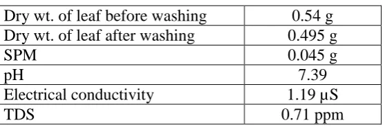
Dry weight of the leaf before washing was found to be 0.54 gm whereas that after washing was found to be 0.495gm, thus showing an SPM value of 0.045gm.The water with which the leaf was washed showed a pH of 7.39, electrical conductivity of1.19µS and a total dissolved solid (TDS) value of 0.71ppm. (Refer to Table 1).
Table 1: phyllosphere characteristics of the Adhatoda vasica leaf. Dry wt. of leaf before washing 0.54 g Dry wt. of leaf after washing 0.495 g
SPM 0.045 g
pH 7.39
Electrical conductivity 1.19 µS
TDS 0.71 ppm
Detection of phyllosphere contaminant
[image:5.595.161.440.496.588.2]Inductive coupled plasma spectroscopic analysis of the water with which the leaf was washed, reported the presence of 0.03mg/L of lead. (Refer to table 2).
Table 2: Inductive coupled plasma spectroscopy
Analysis Method Result Limit of Reporting Unit
Cadmium APHA 22nd EDN < 0.01 0.01 mg/L
In vitro slide bioassay
A marked increase in spore germination (22.12%) was seen when spore solution was supplemented with sugar solution, whereas a 31.97% decrease in the spore germination was observed in the presence of lead.
Among fungicides, Carbendazim was found to be more effective the percentage germination of spore (35.88%) as compared to Blitox (27.76%). (Refer to table 3 and Fig. 1).
Table 3: In vitro slide bioassay
Sample treatment Percentage germination
% increase or decrease
Germ tube length (µm)
Standard error Control
Fungal spore suspension + water
55.88% 14.35
Fungal spore suspension +
sugar solution 78% 22.12% increase 13.28 2.226
Test
Test 1 – Fungal spore suspension + fungicide 1
28.125% 27.76% decrease 14.14 3.30
Test 2 – Fungal spore
suspension + fungicide 2 20% 35.88% decrease 17.271 4.958 Test 3 – Fungal spore
suspension + sugar solution + fungicide 1
37.5% 18.38% decrease 17.35 2.226
Test 4 – Fungal spore suspension + sugar solution + fungicide 2
34.375% 21.51% decrease 18.18 2.226 Test 5 – Fungal spore
solution + metal (Pb) solution
[image:6.595.66.533.236.736.2]23.91% 31.97% decrease 17.35 2.226
Figure 1: percentage germination of spore
In vivo assay
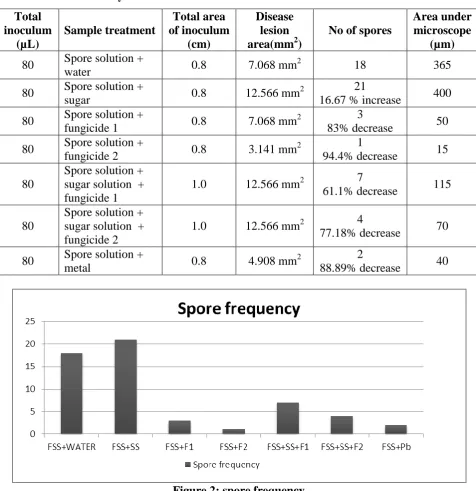
[image:7.595.60.537.222.713.2]In vivo analysis reported that Bavistin was most effective in reducing the spore frequency (94.4%). This was also confirmed by the fact that the treatment resulted in the decrease of the disease lesion area by 3.92mm2. Lead was found to be able to decrease the spore frequency substantially by 88.9%. Hence this assay substantiates the in vitro results, as reported in table 4 and Fig. 2.
Table 4: In vivo study based on Koch’s Postulates
Total inoculum (µL) Sample treatment Total area of inoculum (cm) Disease lesion area(mm2)
No of spores
Area under microscope
(µm) 80 Spore solution +
water 0.8 7.068 mm
2
18 365
80 Spore solution +
sugar 0.8 12.566 mm
2 21
16.67 % increase 400 80 Spore solution +
fungicide 1 0.8 7.068 mm
2 3
83% decrease 50 80 Spore solution +
fungicide 2 0.8 3.141 mm
2 1
94.4% decrease 15 80
Spore solution + sugar solution + fungicide 1
1.0 12.566 mm2 7
61.1% decrease 115
80
Spore solution + sugar solution + fungicide 2
1.0 12.566 mm2 4
77.18% decrease 70 80 Spore solution +
metal 0.8 4.908 mm
2 2
88.89% decrease 40
Figure 2: spore frequency
Fungal biomass reduction assay
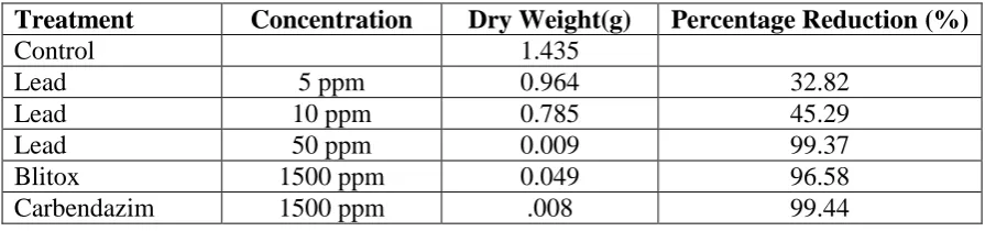
Estimation of the fungal biomass in the potato dextrose broth supplemented with various concentrations of lead, showed a decrease in the fungal biomass with increasing concentration of lead. (Refer to table 5) 10ppm of lead was found to reduce the biomass by 45.29%.
[image:8.595.74.522.264.369.2]The fungicide Bavistin was found to be more potent than fungicide Blitox. Carbendazim reduced the biomass by 99.4% when supplied at a concentration of 1500ppm.
Table 5: Fungal biomass reduction assay
Treatment Concentration Dry Weight(g) Percentage Reduction (%)
Control 1.435
Lead 5 ppm 0.964 32.82
Lead 10 ppm 0.785 45.29
Lead 50 ppm 0.009 99.37
Blitox 1500 ppm 0.049 96.58
Carbendazim 1500 ppm .008 99.44
Metal uptake assay
Metal uptake in the acidulated samples were checked according to the method as referred in APHA 22nd edition and the sample supplemented with 10 ppm of lead concentration showed an uptake of 111.50 mg/L. (as reported in table 6 ).
Table 6: Metal Uptake Assay
Analysis Method Result Limit of Reporting Unit Lead (Control) APHA 22nd EDN 24.68 0.01 mg/L Lead (10ppm) APHA 22nd EDN 111.50 0.01 mg/L
Detection of Vasicine content by HPLC and spectral analysis
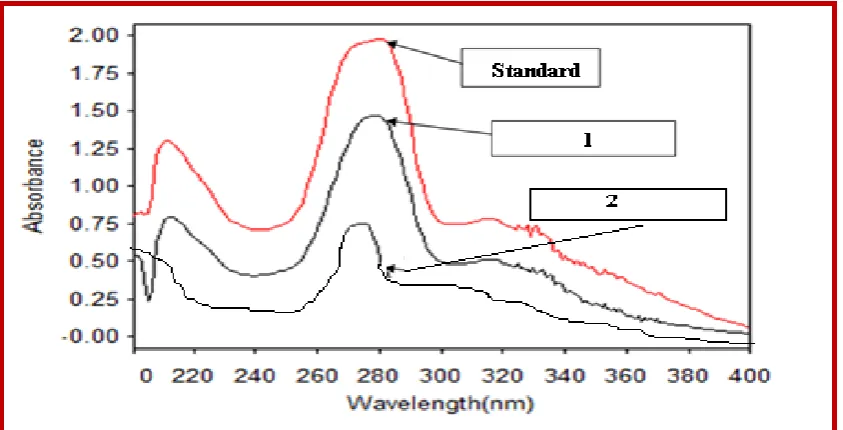
Figure 3: 1= Content in Non infected leaf. 2=Content in Infected Leaf
Rhizoctonia solani is known to be a potential pathogen causing foliar diseases in several plants which include medicinal plants (Janardhanan, 2002). A severe leaf spot disease caused by Colletotrichum gloeosporioides was reported on one of the most important medicinal plants, Adhatoda vasica, which was collected from Himachal Pradesh (Gautam, Avasthi, 2013). In 2006, Verma, Singh and Sharma discovered that R. solani caused leaf spot disease on A. vasica, commonly known as Malabar Nut.
The lesions on the Rhizoctonia spp infected Adhatoda vasica leaves were dark brown to black, round to irregular in shape and was distributed all over the leaf. Similar leaf spot symptoms were observed on Adhatoda vasica leaves collected from a high altitude location at Jammu.
Smiley, Wilkins and Klepper et al demonstrated the effects of fungicides on the growth of R.spp In 1978, Reddy and Bharathudu reported the susceptibility of R. spp to the fungicide Carbendazim. Substantial decrease in the fungal growth was observed in our work, when the isolate was treated with 1500 ppm concentration of Carbendazim.
Earlier HPTLC studies of Sharma, Pati et al (2008) showed reduction in alkaloid content in diseased leaf. Our study showed that the infection caused by R. spp on the A. vasica leaves drastically reduced the vasicine content by 60% which further emphasizes the capability of the isolate to reduce the medicinal values of the plant.
Present study revealed that the fungus which was isolated from a high altitude location was able to cause pathogenesis on leaves present at low altitude. The severe decrease in the amount of vasicine in the diseased leaf highlights the fact that the fungal isolate is a potential threat to this well known medicinal plant. However effective dose of the fungicide Carbendazim and lead salt can control the disease incidence.
CONCLUSION
Present study reveals that the fungus R. spp which was isolated from a high altitude location was able to cause pathogenesis on leaves present at low altitude. The leaf in general is contaminated with Lead and Cadmium of which Lead is the major contributor. This Lead can reduce spore germination by almost 32%.
Our study showed that the infection caused by R. spp on the A. vasica leaves drastically reduced the Vasicine content by almost 60% which further emphasizes the capability of the isolate to reduce the medicinal values of the plant.
The severe decrease in the amount of Vasicine in the diseased leaf highlights the fact that the fungus isolated is a potential threat to this well known medicinal plant. However effective dose of the fungicide Carbendazim and Lead salt can control the disease incidence.
ACKNOWLEDGMENT
REFERENCES
1. Verma, Singh and Sharma. First report of Rhizoctonia solani causing leaf spot of
Adhatoda vasica. Jaipur, India:New Disease Reports, 2006; 14: 39.
2. Reddy and Bharathudu. Effect of some fungicides on Rhizoctonia spp causing Damping Off of Groundnut. Andhra Pradesh, India: Proc. Indian natn Sci. Acad, 1978; B46: 405-409.
3. Janardhanan KK (2002). Diseases of Major Medicinal Plants. Delhi, India : Daya Publishing House.
4. Gautam and Avasthi. Colletotrichum gloeosporioides on Adhatoda vasica in India. Gwalior, India: Journal of Agricultural Technology, 2013; 9(3): 581-587.
5. Babich and Stotzky. Abiotic factors affecting the toxicity of lead to fungi. New York, New York: Applied and Environmental Microbiology, Sep 1979; 38(3): 506–513.
6. Smiley, Wilkins and Keppler. Impact of Fungicide Seed Treatments on Rhizoctonia Root Rot, Take-all, Eye Spot and Growth of Winter Wheat. Pendleton, Oregon: Plant Disease, 1990; 74: 782-787.
7. Pati PK, Sharma M, Sharma A, Salar RK, Gupta AP and Singh B. Studies on leaf spot disease of Withania somnifera and its impact on secondary metabolites. Indian Journal of Microbiology, 2008; 48(4): 432-437.