EFFECT OF SHOCK WAVE ON LOWER BACK MUSCLE SPASM
Abdulaziz Salem Alsorairi* and Dr. Junied
1
University of Hail.
2
College of Applied Medical Sciences, Physical Therapy Department.
ABSTRACT
Background: A muscle spasm is a sudden violent involuntary
contraction of a muscle or group of muscles. It is usually related to a
localized skeletal muscle injury from acute trauma and may also stem
from disorders such as hypocalcemia, hypokalemia or hyperkalemia,
chronic pain syndromes, or epilepsy. Pain and interference with
function attend muscle spasm, producing involuntary movement and
distortion. When a muscle goes into spasm, it freezes in contraction
and becomes a hard knotty mass, rather than normally contracting and
relaxing in quick succession. Objective: The aim of the present study
is to evaluate the role of shock wave on the treatment of patient with lower back muscle
spasm. Methodology: After a verbal explanation of the study protocol, the selected 20
participants were randomly divided into two groups; control group (G1, n = 8) (receive ways
to rest the back only) and treatment group (G2, n = 12). Each participant in G2 exposed to
shock wave once weekly for 4 weeks along the period of the study (1×1×4). Results: The
VAS readings indicated a significant difference at the 1% level (p = 0.003) between the two
groups, while the ODQ readings showed no significant difference (p = 0.123). Results
showed an improvement across two groups in pain and disability scores. In G1, the average
VAS was 6.02cm upon entry to the program and the average ODQ score was 43% before
treatment. Six weeks later, average scores were 3.87cm for the VAS and 36% for the ODQ.
The average VAS score for G2 was 6.19cm upon entry to the program, and ODQ results
showed that the average disability measure pre-intervention was 47%. Four weeks later,
average scores stood at 4.07cm and 35.4% for the VAS and ODQ respectively. Conclusion:
Exposure to shock wave once weekly has a better effect on improving pain and muscle
spasm.
KEYWORDS: muscle, spasm, chronic pain, shock wave, VAS.
Volume 6, Issue 6, 209-219. Research Article ISSN 2277–7105
Article Received on 12 April 2017,
Revised on 02 May 2017, Accepted on 23 May 2017
DOI: 10.20959/wjpr20176-8672
*Corresponding Author’
Abdulaziz Salem
Alsorairi
1. INTRODUCTION
The human body contains approximately 600 skeletal muscles. Skeletal muscle is voluntary,
meaning a person can contract it at will. Seen under a microscope, skeletal muscle fibers
show a pattern of cross-banding, which gives rise to its other name: striated muscle. The
striations are caused by the alignment of bands, the most prominent of which are the A bands,
I bands, and Z lines. The unit between two Z lines is called the sarcomere (Fig. 1).
Striated muscle is composed of two contractile proteins: actin and myosin. The thin filaments
are made of actin, which is attached to the Z lines and is found in both A bands and I bands.
The thick filaments, found in A bands, are made of myosin. In the process set forth in the
sliding filament theory, the sarcomere shortens and the Z lines move closer together when
muscle contracts. The filaments slide together because myosin attaches to, and pulls on, actin.
The myosin head attaches to the actin filament, forming a crossbridge. After formation of the
crossbridge, the myosin head bends, pulling on the actin filaments and causing them to slide.
The result is that the Z lines move closer together, the I band becomes shorter, and the A
band stays the same (Fig. 1). Muscle contraction is like climbing a rope. The crossbridge
cycle is one of grabbing, pulling, and releasing, repeated over and over.
Muscle contraction is triggered by a sudden inflow of calcium ion (Ca2+). In the resting state,
the protein tropomyosin winds around actin and covers the myosin-binding sites. The Ca2+
binds to a second protein, troponin; this action causes the tropomyosin to be pulled to the
side, exposing the myosin-binding sites. With the sites exposed, muscle contracts in the
presence of adenosine triphosphate (ATP). Muscle contraction stops when Ca2+ is removed
from the immediate environment of the myofilaments.
A muscle spasm is a sudden violent involuntary contraction of a muscle or group of muscles.
Spasm is usually related to a localized skeletal muscle injury from acute trauma. Spasms may
also stem from disorders such as hypocalcemia, hypokalemia or hyperkalemia, chronic pain
syndromes, or epilepsy. Pain and interference with function attend muscle spasm, producing
involuntary movement and distortion. When a muscle goes into spasm, it freezes in
contraction and becomes a hard knotty mass, rather than normally contracting and relaxing in
quick succession.
During spasm, the blood vessels that normally feed the muscles and supply oxygen constrict,
prolonged and strong muscular contraction, with relaxation occurring slowly. In the other
form of spasm, called clonic spasm, contractions of the affected muscles occur repeatedly,
forcibly and in quick succession, with equally sudden and frequent relaxations.
Shock waves, high-energy sound or pressure waves (like thunder following lightening or a
small, focused explosion), have little to do with electric shock. Shock waves can be generated
in many ways, but electrohydraulic devices have the greatest capacity to produce and project
high energy to a deep focal depth. ESWT can be highly focused and can achieve a focal point
beyond 10 cm into deeper tissues, depending on the treatment head used. ESWT differs from
radial pressure wave therapy, which does not deliver focused energy at the target; instead,
acoustic waves spread eccentrically from the applicator tip. ESWT is applied superficially,
with waves entering tissue and being absorbed or reflected. Energy is released when a wave
meets an area of high acoustic impedance (e.g., bone–tendon interface). Compressive and
tensile forces cause cavitation and mechanical microstress in cells and tissues, resulting in
modulation of inflammatory, angiogenic, and osteogenic proteins that assist the natural
healing process. Shockwave therapy applied to an area of chronic, non-healing tissue (Fig. 2)
may enable acute cytokines to be released and stimulate healing. The mechanism behind the
pain-relieving function may result from increased serotonin activity in the dorsal horn and
descending inhibition of pain signals.[3]
There are basically four different ways to produce the 'shock wave' (Fig. 2), which, without
getting technical about it are: spark discharge; piezoelectric; electromagnetic and pneumatic
(or electrohydrualic). The wave that is generated will vary in its energy content and also will
have different penetration characteristics in human tissue. In therapy the most commonly
employed generation method is based on the pneumatic system, and the key reason for this is
that a radial (dispersive) wave results. The focussed waves are essential for 'surgical'
interventions, but given their destructive nature, they are less appropriate for therapeutic uses.
Focussed waves are sometimes also referred to as 'hard' shockwaves, the radial or dispersive
Fig. 1: Showing the muscle structure. Fig.2: Showing the types of shock waves.
Since the late 1980s, focused shock waves therapy (SWT) has been widely and successfully
used in the treatment of pain in various musculoskeletal disorders. SWT devices use pressure
waves generated through electromagnetic, electrohydraulic and piezoelectric sources. These
waves have their point of higher pressure in the focus, which is placed within the treated
tissue; for this reason they are defined as focused shock waves.
In 1999, a new technology using a ballistic source to generate pressure waves was introduced.
This technology is called radial shock wave therapy (RSWT). The ballistic source consists of
a tube within which compressed air (1-4bar) is used to fire a bullet that strikes a metal
applicator placed on the patient’s skin. The applicator transforms the kinetic energy of the
bullet into radially expanding pressure waves with a low penetration power (less than 3 cm).
These unfocused shock waves have their point of highest pressure at the tip of the applicator,
outside the treated tissue.
It has been shown that both focused (FSWT) and unfocused (RSWT) shock waves produce
cavitation bubbles in the treated tissue. The cavitation is consequent to the negative phase of
the wave propagation. The rapid collapse of the cavitation bubbles leads to secondary
pressure waves. Cavitation-mediated mechanisms could have a central role in the action of
From a theoretical point of view, shock waves could be useful to treat dystonia and muscle
hypertonia in patients with UMNS. In accordance with the effects on tendon diseases, shock
waves could have a direct effect on muscle fibrosis and other non-reflex components of
muscle hypertonia, which are likely to be present also in some dystonic patients.
Furthermore, shock waves acting at the muscular level could modify the sensory inflow from
the treated muscle to the spinal cord, thus reducing spinal cord excitability and mitigating
spasticity.
The aim of the present study is to evaluate the role of shock wave on the treatment of patient
with lower back muscle spasm.
2. METHODS
Twenty Participants with a history of lower back muscle spasm prolonged more than 6
months ago, aged from 24 to 65 years, weight from 55 to 99 kg, and height from 159 to 188
Cm and body mass index (BMI) from 24.9 to 35.7, were selected from King Khalid Hospital,
after they agreed to participate in the study and then receiving explanations regarding the
purpose and procedures of the study, and signed an informed consent statement before
participation. At the time of the study the participants were not receiving any medical or
physical therapy.
After a verbal explanation of the study protocol, the selected 20 participants were randomly
divided into two groups; control group (G1, n = 8) (receive ways to rest the back only) and
treatment group (G2, n = 12). Each participant in G2 exposed to shock wave once weekly for
4 weeks along the period of the study (1×1×4).
Participants were excluded if they reported spinal, intra-abdominal or femoral surgery in the
past year, a history of trauma or accidents or had been diagnosed with rheumatoid arthritis,
ankylosing spondylitis, systemic lupus erythymatosus or osteoporosis. Those who had any
contraindications for physical tests, such as cardiovascular diseases or severe pulmonary
diseases, were also excluded.
The outcome measures used in this study were the Oswestry Disability Questionnaire (ODQ)
and the Visual Analogue Scale (VAS) both of which were scored by all participants who
Data analysis
Standard deviation and mean for the obtained data were calculated. Also, the data obtained
were analyzed by SPSS 13.0 software test to compare the data before and after treatment
within groups. The significant threshold set at p ˂ 0.05, or non-significant set at p ˃ 0.05.
3. RESULTS
The ODQ is used to score disability induced by lower back muscle spasm. It is a validated
tool that is designed to assess a patient’s level of function or disability, providing quantitative
data that are suitable for quality assurance and research purposes. The VAS scale is a valid
and reliable tool to rate pain intensities along a 10cm line. The patient is asked to put a mark
along this line to reflect the intensity of the pain.
A total of 20 participants were eligible to take part in this study. These were divided into 2
groups; control group G1 (n = 8) and treated group G2 (n = 12).
The VAS readings indicated a significant difference at the 1% level (p = 0.003) between the
two groups, while the ODQ readings showed no significant difference (p = 0.123). Results
showed an improvement across two groups in pain and disability scores, as illustrated in
Table 1 and Figures 3 & 4. In G1, the average VAS was 6.02cm upon entry to the programme
and the average ODQ score was 43% before treatment. Six weeks later, average scores were
3.87cm for the VAS and 36% for the ODQ. The average VAS score for G2 was 6.19cm upon
entry to the programme, and ODQ results showed that the average disability measure
pre-intervention was 47%. Four weeks later, average scores stood at 4.07cm and 35.4% for the
VAS and ODQ respectively (Table 1 and figures 3 & 4).
Although group G1 showed the best improvement in scores initially, group G2 scores
continued to improve over time, with patients doing equally as well as participants in Group
G1 after four weeks.
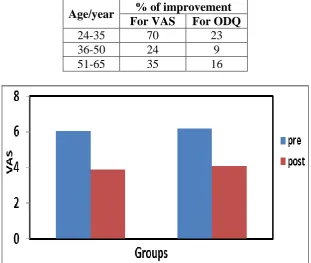
Age-related pre- and post-test differences were interesting. As shown in Figure 4, the
24-35-year-old age group improved by 70% on VAS scores and by 23% on ODQ scores. The
36-50-year-olds scored an average of 24% improvement on the VAS and 9% on the ODQ while
the 51-65-year-olds improved by 35% and 16% on the VAS and ODQ respectively (Table 2
Table 1: Showing the values of VAS and the percentage of ODQ before and after the treatment.
Groups VAS (cm) % of ODQ
Pre-treatment Post-treatment Pre-treatment Post-treatment
A 6.02 3.87 43 36
B 6.19 4.07 47 35.4
Table 2: Showing the percentage of improvement for VAS and ODQ related to age of participants.
Age/year % of improvement For VAS For ODQ
24-35 70 23
36-50 24 9
[image:7.595.82.512.112.171.2]51-65 35 16
Fig. 3: Showing the values of VAS pre and post-treatment.
[image:7.595.142.454.227.490.2] [image:7.595.140.457.532.718.2]Fig. 6: Showing the percentage of improvement for VAS and ODQ related to age of participants.
4. DISCUSSION
The aim of this study was to evaluate the effect of shock wave on participants with lower
back muscle spasm. A pre-test/post-test design was implemented over a period of four weeks.
Outcome measures consisted of ODQ and VAS scores.
Four weeks after programme, pain and disability scores improved in treated group (G2) that
received shock wave treatment once weekly for 4 weeks (1×1×4). At four weeks
post-intervention, Group G2 showed the most significant improvements in both ODQ and VAS
scores.
Participants in Group G2 who had exposed to shock wave had better VAS outcomes than
those in Group G1 (received ways for resting back). The opposite was true with the ODQ
results at 4 weeks. These findings are comparable to those of similar research studies in
which the effects of shock wave were investigated. The evidence is inconclusive as to which
skock wave is best and actually leans towards incorporating any general exercise programme
to improve functions of muscles.
It is noteworthy that the participants had been randomly assigned to two groups without
considering that age differences could affect outcomes. The distribution of ages between
groups appears to relate to the initial results and may have introduced a bias in favour of
Group G2 as age-related differences were striking. The 16-35-year-old participants showed
the greatest improvement, which finding could be due to several factors such as healing
[image:8.595.135.463.70.254.2]they assimilate exercises more easily. Also, they were more likely to be cases of first
incidence of lower back muscle spasm, which would be easier to treat than recurrent
episodes, or chronic muscle spasm.
The 36-50-year-old participants showed the least improvement. This may be because the
patients in this age group are likely to have had the greatest physical demands due to their
lifestyles at work and at home, and also the least time for their own well-being. The
51-65-year age group had a better outcome than their younger counterparts, which may reflect the
fact that they had more time for themselves and so were more likely to implement their home
exercises program (HEP). It would have been a good idea to record compliance to the HEP
with a diary. Another factor contributing to the results obtained for the older groups may have
been chronicity of pain.
5. CONCLUSION
The results of this study imply that exposure to shock wave once weekly have a better effect
on improving pain and muscle spasm. Interestingly, the control group G1 who had postural
re-education did as well as the exposed group. Age was not considered to be a factor when
allocating participants into groups. However, the younger age group showed marked
improvement with posture re-education and exposure. These results are clinically significant.
Further longitudinal studies in this area are called for, with a recommendation that
participants are followed up for at least one year post-intervention in order to find out which
approach has better long-term outcomes.
6. REFERENCES
1. J. W. Lance, “Symposium synopsis,” in Spasticity: Disordered Motor Control, R. G.
Feldman, R. R. Young, andW. P. Koella, Eds, 1980; 485–494.
2. T. D. Marcotte, T. J. Rosenthal, E. Roberts et al., “The contribution of cognition and
spasticity to driving performance in multiple sclerosis,” Archives of Physical Medicine
and Rehabilitation, 2008; 89(9): 1753–1758.
3. V. Dietz and W. Berger, “Normal and impaired regulation of muscle stiffness in gait: a
new hypothesis about muscle hypertonia,” Experimental Neurology, 1983; 79(3):
680–687.
4. J.-M. Gracies, “Pathophysiology of spastic paresis. I: paresis and soft tissue changes,”
5. F. Biering-Srensen, J. B. Nielsen, and K. Klinge, “Spasticity assessment: a review,”
Spinal Cord, 2006; 44(12): 708–722.
6. Hodges PW, Richardson CA: Inefficient muscular stabilization of the lumbar spine
associated with low back pain. A motor control evaluation of transversus abdominis.
Spine, 1996; 21: 2640-50.
7. Adams MA, Mannion AF, Dolan P: Personal risk factors for first-time low back pain.
Spine, 1999; 24: 2497-505.
8. Takala EP, Viikari-Juntura E: Do functional tests predict low back pain? Spine, 2000; 25:
2126-32.
9. Linton SJ, Halldén K: Can we screen for problematic back pain? A screening
questionnaire for predicting outcome in acute and sub-acute back pain. Clin J Pain, 1998;
14: 209-15.
10.P. Manganotti and E. Amelio, “Long-term effect of shock wave therapy on upper limb
hypertonia in patients affected by stroke,”Stroke, 2005; 36(9): 1967–1971.
11.S. W. Moon, J. H. Kim, M. J. Jung et al., “The effect of extracorporeal shock wave
therapy on lower limb spasticity in subacute stroke patients,” Annals of Rehabilitation
Medicine, 2013; 37(4): 461–470.
A. Santamato, M. F. Micello, F. Panza et al., “Extracorporeal shock wave therapy for the
treatment of post-stroke plantar flexormuscles spasticity: a prospective open-label study,”
Topics in Stroke Rehabilitation, 2014; 21(1): S17–S24.
A. Santamato, A. Notarnicola, F. Panza et al., “SBOTE study: extracorporeal shock wave
therapy versus electrical stimulation after botulinumtoxin type a injection for post-stroke
spasticity a prospective randomized trial,” Ultrasound in Medicine and Biology, 2013;
39(2): 283–291.
12.M. K. Sohn, K. H. Cho, Y. Kim, and S. L. Hwang, “Spasticity and electrophysiologic
changes after extracorporeal shock wave therapy on gastrocnemius,” Annals of Physical
and Rehabilitation Medicine, 2011; 35(5): 599–604.
13.F. Troncati, M. Paci, T. Myftari, and B. Lombardi, “Extracorporeal shock wave therapy
reduces upper limb spasticity and improves motricity in patients with chronic hemiplegia:
a case series,” NeuroRehabilitation, 2013; 33(3): 399–405.
14.M. I. Gonkova, E. M. Ilieva, G. Ferriero, and I. Chavdarov, “Effect of radial shockwave
therapy on muscle spasticity in children with cerebral palsy,” International Journal of
15.Y.W. Kim, J. C. Shin, J. Yoon, Y. Kim, and S. C. Lee, “Usefulness of radial
extracorporeal shock wave therapy for the spasticity of the subscapularis in patients with
stroke: a pilot study,” Chinese Medical Journal, 2013; 126(24): 4638–4643.
16.X. Vidal, A. Morral, L. Costa and M. Tur, “Radial extracorporeal shock wave therapy
(rESWT) in the treatment of spasticity in cerebral palsy: a randomized,