
organic papers
o800
Parvez, Hunt and Keay C21H20O3 DOI: 10.1107/S1600536801012508 Acta Cryst.(2001). E57, o800±o801 Acta Crystallographica Section EStructure Reports
Online
ISSN 1600-5368
2-(1-Naphthyl)cyclohexyl 3-furancarboxylate
Masood Parvez,* Ian R. Hunt and Brian A. Keay
Department of Chemistry, The University of Calgary, 2500 University Drive NW, Calgary, Alberta, Canada T2N 1N4
Correspondence e-mail: [email protected]
Key indicators Single-crystal X-ray study
T= 296 K
Mean(C±C) = 0.009 AÊ
Rfactor = 0.053
wRfactor = 0.242
Data-to-parameter ratio = 14.0
For details of how these key indicators were automatically derived from the article, see http://journals.iucr.org/e.
#2001 International Union of Crystallography Printed in Great Britain ± all rights reserved
The crystal structure of the title compound, C21H20O3,
contains molecules separated by normal van der Waals distances. The cyclohexyl ring adopts a classical chair conformation, while the furan and naphthyl rings are individually planar. The molecular dimensions are as expected.
Comment
3-Furoate trans-2-arylcyclohexanol esters were prepared as model compounds as part of an investigation to create a chiral auxiliary for use in an asymmetric intramolecular Diels±Alder reaction with a furandiene (IMDAF reaction) (Keay & Hunt, 1999). Molecular modeling had suggested that such systems might be able to block one face of the furandiene. The model compound, 2-(1-naphthyl)cyclohexyl 3-furancarboxylate, (I), was prepared and the structure determined in order to verify the molecular modeling results.
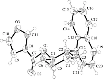
The crystal structure of (I) is composed of molecules (Fig. 1) which are separated by normal van der Waals distances. The molecular dimensions are normal and lie within expected values for corresponding bond distances and angles (Orpenet al., 1994). The C1±C6 cyclohexyl ring adopts a classical chair conformation, with puckering parameters (Cremer & Pople, 1975)Q= 0.563 (6) AÊ,= 177.7 (6)and!= 123 (21). The
naphthyl ring is essentially planar, with the maximum devia-tion of any atom from the mean plane being 0.027 (4) AÊ. The ®ve-membered furan ring is also planar.
Experimental
To a solution of 3-furoic acid (76 mg, 0.68 mmol) in dry methylene chloride (2.5 ml) and dry DMF (5ml), oxalyl chloride (120ml, 1.36 mmol) was added. The solution was re¯uxed overnight, then cooled and the solvent removed in vacuo. After redissolving the residue in dry methylene chloride (2.5 ml), a solution oftrans -2-(1-naphthyl)cyclohexanol (136 mg, 0.9 equivalents) in dry methylene chloride (2 ml) was added followed by dry triethylamine (189ml) and DMAP (9 mg). The reaction was stirred at room temperature for 36 h. After adding Et2O (25 ml), the solution was washed with 5%
HCl (10 ml) and water (20 ml), dried over MgSO4, then ®ltered and
evaporatedin vacuoto give the crude product as an oil. Puri®cation
viaa short column (silica gel, EtOAc) followed by radial chromato-graphy (EtOAc hexanes, 20:1 to 5:1), gave recovered trans -2-(1-naphthyl)cyclohexanol (41 mg) and the title product (I) (135 mg) as a white crystalline solid (m.p. 370 K).
Crystal data
C21H20O3
Mr= 320.37
Monoclinic,P21/c
a= 9.1991 (11) AÊ
b= 7.3840 (5) AÊ
c= 25.694 (3) AÊ = 100.030 (9)
V= 1718.6 (3) AÊ3
Z= 4
Dx= 1.238 Mg mÿ3
MoKradiation Cell parameters from 25
re¯ections = 10.0±15.0 = 0.08 mmÿ1
T= 296 (2) K Prism, colourless 0.400.300.24 mm
Data collection
Rigaku AFC-6Sdiffractometer !/2scans
3245 measured re¯ections 3043 independent re¯ections 917 re¯ections withI> 2(I)
Rint= 0.08 max= 25.0
h= 0!10
k= 0!8
l=ÿ30!30 3 standard re¯ections
every 200 re¯ections intensity decay: <0.1%
Re®nement
Re®nement onF2
R[F2> 2(F2)] = 0.053
wR(F2) = 0.242
S= 0.96 3043 re¯ections 217 parameters
H-atom parameters constrained
w= 1/[2(F
o2) + (0.1064P)2]
whereP= (Fo2+ 2Fc2)/3
(/)max< 0.001
max= 0.18 e AÊÿ3
min=ÿ0.24 e AÊÿ3
Most of the H atoms were located from difference maps. The H atoms were included at geometrically idealized positions with CÐH = 0.93±0.98 AÊ, in a riding mode with isotropic displacement parameters 1.2 times the displacement parameters of the atoms to which they were attached.
Data collection: MSC/AFC Diffractometer Control Software
(Molecular Structure Corporation, 1988); cell re®nement:MSC/AFC Diffractometer Control Software; data reduction: TEXSAN (Mole-cular Structure Corporation, 1994); program(s) used to solve struc-ture: SAPI91 (Fan, 1991); program(s) used to re®ne structure:
SHELXL97 (Sheldrick, 1997); molecular graphics: TEXSAN; soft-ware used to prepare material for publication: SHELXL97 (Shel-drick, 1997).
The authors thank the Natural Sciences and Engineering Research Council, Canada, for providing the diffractometer through an equipment grant to the University of Calgary.
References
Cremer, D. & Pople, J. A. (1975).J. Am. Chem. Soc.97, 1354±1358. Fan, H.-F. (1991).SAPI91. Rigaku Corporation, Tokyo, Japan.
Johnson, C. K. (1976).ORTEPII. Report ORNL-5138. Oak Ridge National Laboratory, Tennessee, USA.
Keay, B. A. & Hunt, I. R. (1999).Adv. Cycloaddit.6, 173±210.
Molecular Structure Corporation (1988).MSC/AFC Diffractometer Control Software. MSC, 3200 Research Forest Drive, The Woodlands, TX 77381, USA.
Molecular Structure Corporation (1994). TEXSAN. MSC, 3200 Research Forest Drive, The Woodlands, TX 77381, USA.
Orpen, A. G., Brammer, L., Allen, F. H., Kennard, O., Watson, D. G. & Taylor, R. (1994).Structure Correlation, Vol. 2, edited by H.-B. BuÈrgi & J. D. Dunitz, pp. 751±858. New York: VCH.
Sheldrick, G. M. (1997).SHELXL97. University of GoÈttingen, Germany. Figure 1
supporting information
sup-1
Acta Cryst. (2001). E57, o800–o801
supporting information
Acta Cryst. (2001). E57, o800–o801 [doi:10.1107/S1600536801012508]
2-(1-Naphthyl)cyclohexyl 3-furancarboxylate
Masood Parvez, Ian R. Hunt and Brian A. Keay
S1. Comment
3-Furoate trans-2-arylcyclohexanol esters were prepared as model compounds as part of an investigation to create a
chiral auxiliary for use in as asymmetric intramolecular Diels–Alder reaction with a furandiene (IMDAF reaction) (Keay
& Hunt, 1999). Molecular modeling had suggested that such systems might be able to block one face of the furandiene.
The model compound, 2-(1-naphthyl)cyclohexyl 3-furancarboxylate, (I), was prepared and the structure determined in
order to verify the molecular modeling results.
The asymmetric unit of (I) is composed of molecules (Fig. 1) which are separated by normal van der Waals distances.
The molecular dimensions are normal and lie within expected values for corresponding bond distances and angles (Orpen
et al., 1994). The C1–C6 cyclohexyl ring adopts a classical chair conformation, with puckering parameters (Cremer & Pople, 1975) Q = 0.563 (6) Å, θ = 177.7 (6)° and ω = 123 (21)°. The naphthyl ring is essentially planar, with the
maximum deviation of any atom from the mean plane being 0.027 (4) Å. The five-membered furan ring is also planar.
S2. Experimental
To a solution of 3-furoic acid (76 mg, 0.68 mmol) in dry methylene chloride (2.5 ml) and dry DMF (5 µl), oxalyl chloride
(120 µl, 1.36 mmol) was added. The solution was refluxed overnight, then cooled and the solvent removed in vacuo.
After redisolving the residue in dry methylene chloride (2.5 ml), a solution of trans-2-(1-naphthyl)cyclohexanol (136 mg,
0.9 equivalents) in dry methylene chloride (2 ml) was added followed by dry triethylamine (189 µl) and DMAP (9 mg).
The reaction was stirred at room temperature for 36 h. After adding Et2O (25 ml), the solution was washed with 5% HCl
(10 ml) and water (20 ml), dried over MgSO4, then filtered and evaporated in vacuo to give the crude product as an oil.
Purification via a short column (silica gel, EtOAc) followed by radial chromatography (EtOAc hexanes, 20:1 to 5:1),
gave recovered trans-2-(1-naphthyl)cyclohexanol (41 mg) and the title product (I) (135 mg) as a white crsytalline solid
(m.p. 370 K).
S3. Refinement
The space group, P21/c, was uniquely determined from the systematic absences. Most of the H atoms were located from
difference maps. The H atoms were included at geometrically idealized positions with C–H = 0.93–0.98 Å, in a riding
mode with isotropic displacement parameters 1.2 times the displacement parameters of the atoms to which they were
Figure 1
ORTEPII (Johnson, 1976) drawing of (I). Displacement ellipsoids have been plotted at the 30% probability level.
2-(1-Naphthyl)cyclohexyl 3-furancarboxylate
Crystal data C21H20O3 Mr = 320.37
Monoclinic, P21/c a = 9.1991 (11) Å b = 7.3840 (5) Å c = 25.694 (3) Å β = 100.030 (9)° V = 1718.6 (3) Å3 Z = 4
F(000) = 680 Dx = 1.238 Mg m−3
Mo Kα radiation, λ = 0.71069 Å Cell parameters from 25 reflections θ = 10.0–15.0°
µ = 0.08 mm−1 T = 296 K Prism, colourless 0.40 × 0.30 × 0.24 mm
Data collection Rigaku AFC-6S
diffractometer
Radiation source: fine-focus sealed tube Graphite monochromator
ω/2θ scans
3245 measured reflections 3043 independent reflections 917 reflections with I > 2σ(I)
Rint = 0.08
θmax = 25.0°, θmin = 2.5° h = 0→10
k = 0→8 l = −30→30
supporting information
sup-3
Acta Cryst. (2001). E57, o800–o801 Refinement
Refinement on F2 Least-squares matrix: full R[F2 > 2σ(F2)] = 0.053 wR(F2) = 0.242 S = 0.96 3043 reflections 217 parameters 0 restraints
Primary atom site location: structure-invariant direct methods
Secondary atom site location: difference Fourier map
Hydrogen site location: inferred from neighbouring sites
H-atom parameters constrained w = 1/[σ2(F
o2) + (0.1064P)2] where P = (Fo2 + 2Fc2)/3 (Δ/σ)max < 0.001
Δρmax = 0.18 e Å−3 Δρmin = −0.24 e Å−3
Fractional atomic coordinates and isotropic or equivalent isotropic displacement parameters (Å2)
x y z Uiso*/Ueq
H16 0.1443 0.2614 0.1440 0.094* C17 0.3437 (8) 0.3081 (8) 0.1837 (3) 0.0716 (19) H17 0.3149 0.4055 0.2025 0.086* C18 0.4943 (7) 0.2540 (7) 0.1937 (2) 0.0533 (15) C19 0.5977 (9) 0.3431 (8) 0.2311 (2) 0.0701 (18) H19 0.5690 0.4406 0.2498 0.084* C20 0.7396 (9) 0.2873 (9) 0.2400 (2) 0.078 (2) H20 0.8079 0.3452 0.2656 0.093* C21 0.7853 (7) 0.1430 (8) 0.2113 (2) 0.0611 (16) H21 0.8844 0.1101 0.2171 0.073*
Atomic displacement parameters (Å2)
U11 U22 U33 U12 U13 U23
O1 0.051 (2) 0.065 (3) 0.055 (2) 0.009 (2) 0.0110 (19) 0.009 (2) O2 0.055 (3) 0.095 (4) 0.097 (4) −0.006 (3) 0.020 (2) 0.026 (3) O3 0.084 (3) 0.062 (3) 0.061 (3) 0.010 (3) −0.003 (2) 0.005 (2) C1 0.046 (3) 0.059 (4) 0.045 (3) 0.006 (3) 0.010 (3) 0.005 (3) C2 0.046 (3) 0.052 (3) 0.046 (3) 0.003 (3) 0.008 (3) −0.005 (3) C3 0.077 (4) 0.059 (4) 0.074 (4) 0.017 (3) 0.032 (3) 0.013 (4) C4 0.080 (4) 0.067 (4) 0.091 (5) 0.027 (4) 0.026 (4) 0.018 (4) C5 0.071 (4) 0.069 (4) 0.100 (5) 0.020 (4) 0.039 (4) 0.011 (4) C6 0.065 (4) 0.070 (4) 0.059 (4) 0.015 (3) 0.029 (3) 0.005 (3) C7 0.064 (4) 0.055 (4) 0.050 (4) 0.000 (4) 0.024 (3) 0.000 (3) C8 0.060 (4) 0.050 (4) 0.045 (3) 0.010 (3) 0.013 (3) 0.003 (3) C9 0.074 (5) 0.074 (5) 0.064 (4) 0.009 (4) 0.027 (4) 0.010 (4) C10 0.108 (6) 0.067 (4) 0.059 (5) 0.015 (5) 0.031 (4) 0.015 (4) C11 0.075 (5) 0.049 (4) 0.047 (4) 0.006 (3) 0.005 (3) 0.006 (3) C12 0.056 (4) 0.045 (3) 0.043 (3) 0.000 (3) 0.016 (3) 0.006 (3) C13 0.052 (3) 0.043 (3) 0.041 (3) −0.003 (3) 0.014 (3) 0.000 (3) C14 0.055 (4) 0.061 (4) 0.059 (4) 0.005 (3) 0.012 (3) 0.001 (3) C15 0.061 (4) 0.079 (5) 0.091 (5) 0.000 (4) 0.020 (4) 0.000 (4) C16 0.059 (4) 0.080 (5) 0.101 (6) 0.017 (4) 0.031 (4) 0.010 (5) C17 0.090 (5) 0.050 (4) 0.085 (5) 0.017 (4) 0.047 (4) 0.008 (4) C18 0.072 (4) 0.043 (4) 0.053 (4) 0.007 (3) 0.031 (3) 0.006 (3) C19 0.112 (6) 0.049 (4) 0.053 (4) 0.005 (4) 0.027 (4) −0.007 (3) C20 0.107 (6) 0.068 (5) 0.056 (4) −0.009 (5) 0.006 (4) −0.019 (4) C21 0.066 (4) 0.063 (4) 0.052 (4) −0.005 (4) 0.003 (3) 0.005 (4)
Geometric parameters (Å, º)
supporting information
sup-5
Acta Cryst. (2001). E57, o800–o801
C1—H1 0.9800 C13—C14 1.390 (7) C2—C3 1.531 (7) C13—C18 1.428 (7) C2—C12 1.530 (7) C14—C15 1.371 (7) C2—H2 0.9800 C14—H14 0.9300 C3—C4 1.509 (8) C15—C16 1.401 (8) C3—H3A 0.9700 C15—H15 0.9300 C3—H3B 0.9700 C16—C17 1.328 (8) C4—C5 1.511 (7) C16—H16 0.9300 C4—H4A 0.9700 C17—C18 1.421 (8) C4—H4B 0.9700 C17—H17 0.9300 C5—C6 1.532 (7) C18—C19 1.394 (8) C5—H5A 0.9700 C19—C20 1.350 (8) C5—H5B 0.9700 C19—H19 0.9300 C6—H6A 0.9700 C20—C21 1.403 (8) C6—H6B 0.9700 C20—H20 0.9300 C7—C8 1.459 (8) C21—H21 0.9300 C8—C11 1.334 (7)
C6—C5—H5B 109.6 C19—C18—C13 120.7 (6) H5A—C5—H5B 108.1 C17—C18—C13 118.1 (6) C5—C6—C1 110.6 (4) C20—C19—C18 119.8 (6) C5—C6—H6A 109.5 C20—C19—H19 120.1 C1—C6—H6A 109.5 C18—C19—H19 120.1 C5—C6—H6B 109.5 C19—C20—C21 120.8 (6) C1—C6—H6B 109.5 C19—C20—H20 119.6 H6A—C6—H6B 108.1 C21—C20—H20 119.6 O2—C7—O1 123.2 (6) C12—C21—C20 121.6 (6) O2—C7—C8 125.9 (6) C12—C21—H21 119.2 O1—C7—C8 110.9 (5) C20—C21—H21 119.2 C11—C8—C9 107.0 (5)
![Crystal structure of 10 [(3 oxo 3H benzo[f]chromen 1 yl)methyl] 2 trifluoromethyl 9a,10 dihydrobenz[4,5]imidazo[1,2 a]pyrimidin 4(5aH) one](data:image/gif;base64,R0lGODlhAQABAIAAAP///wAAACH5BAEAAAAALAAAAAABAAEAAAICRAEAOw==)