organic papers
o872
S. Ramaswamyet al. C6H16N4O22+2NO3ÿ DOI: 10.1107/S1600536801013460 Acta Cryst.(2001). E57, o872±o874Acta Crystallographica Section E
Structure Reports
Online ISSN 1600-5368
L
-Argininium dinitrate
S. Ramaswamy,aB. Sridhar,bV.
Ramakrishnanaand R. K.
Rajaramb*
aLaser Laboratory, School of Physics, Madurai
Kamaraj University, Madurai 625 021, India, andbDepartment of Physics, Madurai Kamaraj
University, Madurai 625 021, India
Correspondence e-mail: [email protected]
Key indicators
Single-crystal X-ray study T= 293 K
Mean(C±C) = 0.010 AÊ Rfactor = 0.070 wRfactor = 0.218 Data-to-parameter ratio = 7.1
For details of how these key indicators were automatically derived from the article, see http://journals.iucr.org/e.
#2001 International Union of Crystallography Printed in Great Britain ± all rights reserved
In the title compound, C6H16N4O22+2NO3ÿ, the diprotonated
argininium molecule is linked by a strong OÐH O
[2.653 (7) AÊ] hydrogen bond to the nitrate anion. The single-bonded O atom of the carboxyl group exhibits a very unusual
cis conformation with respect to the -amino N atom.
Chelated three-centered hydrogen bonds are observed in the case of the Nand N"atoms with the nitrate anions. The
argininium molecules are connected by type A, B and D
interactions through nitrate anions.
Comment
l-Arginine is one of the essential amino acids widely
distrib-uted in biological substances. l-Arginine phosphate
mono-hydrate is found to exhibit interesting non-linear optical properties (Jiang et al., 1983). The strong basicity of the guanidyl group is responsible for the functions in living matter (Aokiet al., 1971). The crystal structures of l-arginine dihy-drate (Karle & Karle, 1964),l-arginine hydrochloride mono-hydrate (Dowet al., 1970),l-arginine phosphate monohydrate (Aokiet al., 1971),l-arginine perchlorate (Monacoet al., 1987;
Srinivasan & Rajaram, 1997) and l-arginine diarsenate
(Zalkinet al., 1989) have been reported. In the present study, the crystal structure of l-arginine dinitrate, (I), was under-taken to study conformational aspects.
The conformation of the arginine molecule may be char-acterized by three planar groups: (i) the carboxyl group, (ii) the side chain C atoms consisting of the-,-,- and-carbon (C12, C13, C14 and C15) and (iii) the guanidyl group including the-C atom (C15, N4, C16, N5 and N6) (Aokiet al., 1971). The conformation of the single-bonded carboxyl O atom iscis
with respect to the amino N atom [ÿ29.7 (10)] for the
diprotonated argininium molecule. In general, this confor-mation is found to betrans, whereas the present structure is different from earlier studies, viz. l-valine hydrochloride (Koetzleet al., 1974),dl-valine hydrochloride (Di Blasioet al., 1977),l-arginine diarsenate (Zalkin et al., 1989), bis(dl -me-thioninium) sulfate (Srinivasan et al., 2001), tri(l
nium) sulfate bisulfate (Sridharet al., 2001) and l-valine l -valinium perchlorate monohydrate (Pandiarajanet al., 2001).
The side-chain conformation angle1has agaucheII form
[ÿ62.5 (8)], while2and3are in thetransform [ÿ168.6 (6) and 178.0 (6)] for the argininium molecule (Fig. 1). The other conformation angle, which has the guanidyl group at the end of the residue,4[ÿ92.9 (10)], is as expected and51and52 are 6.6 (14) and ÿ172.6 (8), respectively. These conforma-tional angles are very similar to those onl-arginine diarsenate, except for4which is 148.8. The argininium molecule, in the present study, is in a slightly folded conformation.
The guanidyl group is protonated and exists as a guanidi-nium ion. The three CÐN bonds in this group are nearly equal in length with an average value of 1.313 (9) AÊ. The three NÐ CÐN angles are very close to 120, con®rming the planarity of the guanidyl group. Like other arginine molecules, the C15 atom is only slightly displaced [0.15 (2) AÊ] from the plane of the guanidyl group. The two crystallographically independent nitrate anions have similar geometries.
The carboxyl O atom of the diprotonated argininium cation
is linked through a strong OÐH O hydrogen bond
[2.653 (7) AÊ] with the nitrate anion (Table 2), while the amino±N and the N atoms of the guanidyl group are linked through normal hydrogen bonds with the two nitrate anions.
Chelated three-centered hydrogen bonds are observed in the
case of the N and N" atoms (N3ÐH3C O5ii and N3Ð
H3C O4ii, and N4ÐH4 O3iiiand N4ÐH4 O2iii; Jeffrey & Saenger, 1991). The O atom (O2) of the nitrate anion links the carboxyl O atom (O1B), N"and N1(N5) through a chain along thecaxis. The O atoms of the second nitrate anion (O5
and O4) link the -amino N atom and N2 (N6) through a
chain along the a axis. The intermolecular guanidyl±nitrate
interactions are observed as (i) typeDbetween N atoms",1
with O2; (ii) typeAbetween N atoms1,2 with O3 and O1
and (iii) typeBbetween N atoms",1 with O3 and O2 of the nitrate anions (Salunke & Vijayan, 1981). An intermolecular short contact of 2.903 (12) AÊ is observed between O4 and the carboxyl C11(1ÿx,1
2+y, 1ÿz) atom. Experimental
The title compound was crystallized in aqueous solution from a 1:2 stoichiometric ratio ofl-arginine and nitric acid. Colorless needle-shaped crystals were grown.
Crystal data C6H16N4O22+2NO3ÿ Mr= 300.25
Monoclinic,P21 a= 7.744 (5) AÊ
b= 7.284 (5) AÊ
c= 11.653 (5) AÊ
= 92.600 (5) V= 656.6 (7) AÊ3 Z= 2
Dx= 1.519 Mg mÿ3
Dm= 1.519 Mg mÿ3
Dmmeasured by ¯otation in a
mixture of carbon tetrachloride and xylene
CuKradiation Cell parameters from 25
re¯ections
= 14.9±26.9
= 1.22 mmÿ1 T= 293 (2) K Needle, colorless 0.030.020.01 mm Data collection
Enraf±Nonius sealed-tube diffractometer
!±2scans
Absorption correction: scan (Northet al., 1968)
Tmin= 0.967,Tmax= 0.984 1284 measured re¯ections 1284 independent re¯ections
1213 re¯ections withI> 2(I)
max= 68.0 h=ÿ9!9
k= 0!8
l= 0!14
3 standard re¯ections frequency: 60 min intensity decay: none Re®nement
Re®nement onF2 R[F2> 2(F2)] = 0.070 wR(F2) = 0.218 S= 1.08 1284 re¯ections 181 parameters
H-atom parameters constrained
w= 1/[2(F
o2) + (0.2P)2]
whereP= (Fo2+ 2Fc2)/3
(/)max< 0.001
max= 0.35 e AÊÿ3
min=ÿ0.44 e AÊÿ3
Table 1
Selected geometric parameters (AÊ,).
O1AÐC11 1.201 (9) O1BÐC11 1.320 (8) N4ÐC16 1.296 (9)
C16ÐN6 1.308 (9) C16ÐN5 1.336 (9)
N4ÐC16ÐN6 122.5 (6)
N4ÐC16ÐN5 119.4 (6) N6ÐC16ÐN5 118.0 (6) O1BÐC11ÐC12ÐN3 ÿ29.7 (10)
N3ÐC12ÐC13ÐC14 ÿ62.5 (8) C12ÐC13ÐC14ÐC15 ÿ168.6 (6) C13ÐC14ÐC15ÐN4 178.0 (6)
C14ÐC15ÐN4ÐC16 ÿ92.9 (10) C15ÐN4ÐC16ÐN6 6.6 (14) C15ÐN4ÐC16ÐN5 ÿ172.6 (8)
Acta Cryst.(2001). E57, o872±o874 S. Ramaswamyet al. C6H16N4O22+2NO3ÿ
o873
organic papers
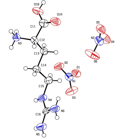
Figure 1
organic papers
o874
S. Ramaswamyet al. C6H16N4O22+2NO3ÿ Acta Cryst.(2001). E57, o872±o874Table 2
Hydrogen-bonding geometry (AÊ,).
DÐH A DÐH H A D A DÐH A
O1BÐH1B O2i 0.82 1.85 2.653 (7) 166
N3ÐH3A O1 0.89 2.08 2.950 (8) 165 N3ÐH3B O5 0.89 1.94 2.823 (9) 170 N3ÐH3C O5ii 0.89 2.00 2.855 (9) 161
N3ÐH3C O4ii 0.89 2.49 3.212 (9) 139
N4ÐH4 O3iii 0.86 2.22 3.010 (8) 153
N4ÐH4 O2iii 0.86 2.36 3.138 (8) 151
N5ÐH5A O2iii 0.86 2.34 3.086 (9) 146
N5ÐH5B O1iv 0.86 2.17 3.022 (8) 173
N6ÐH6A O4v 0.86 2.18 2.998 (9) 158
N6ÐH6B O3iv 0.86 2.10 2.957 (9) 177
Symmetry codes: (i) 1ÿx;1
2y;1ÿz; (ii) 1ÿx;yÿ12;1ÿz; (iii) 1ÿx;12y;2ÿz;
(iv)ÿx;1
2y;2ÿz; (v)xÿ1;y;z.
Data collection: CAD-4 Software (Enraf±Nonius, 1989); cell re®nement: CAD-4 Software; data reduction: CAD-4 Software; program(s) used to solve structure: SHELXS97 (Sheldrick, 1997); program(s) used to re®ne structure:SHELXL97 (Sheldrick, 1997); molecular graphics:PLATON(Spek, 1999); software used to prepare material for publication:SHELXL97.
The authors BS and RKR thank the Department of Science and Technology (DST), Government of India, for ®nancial support. One of the authors (SR) thanks the University Grants Commission, New Delhi, and the management of NMSSVN College, Madurai, for permitting him to pursue his doctoral research work under the Faculty Improvement Programme.
References
Aoki, K., Nagano, K. & Iitaka, Y. (1971).Acta Cryst.B27, 11±23.
Di Blasio, B., Napolitano, G. & Pedone, C. (1977).Acta Cryst.B33, 542±545. Dow, J., Jensen, L. H., Mazumdar, S. K., Srinivasan, R. & Ramachandran, G. N.
(1970).Acta Cryst.B26, 1662±1671.
Enraf±Nonius (1989).CAD-4Software. Version 5.0. Enraf±Nonius, Delft, The Netherlands.
Jeffrey, G. A. & Saenger, W. (1991). In Hydrogen Bonding in Biological Structures. Berlin, Heidelberg, New York: Springer-Verlag.
Jiang, M., Xu, D. & Tan, Z. (1983). Abstracts of the VIIth International Conference on Crystal Growth, Stuttgart, Germany, p. 2.67.
Johnson, C. K. (1976).ORTEPII. Oak Ridge National Laboratory, Tennessee, USA.
Karle, I. L. & Karle, J. (1964).Acta Cryst.17, 835±841.
Koetzle, T. F., Golic, L., Lehmann, M. S., Verbist, J. J. & Hamilton, W. C. (1974).J. Chem. Phys.60, 4690±4696.
Monaco, S. B., Davis, L. E., Velsko, S. P., Wang, F. T., Eimerl, D. & Zalkin, A. (1987).J. Cryst. Growth,85, 252±255.
North, A. C. T., Phillips, D. C. & Mathews, F. S. (1968).Acta Cryst.A24, 351± 359.
Pandiarajan, S., Sridhar, B. & Rajaram, R. K. (2001).Acta Cryst.E57, o466± o468.
Salunke, D. M. & Vijayan, M. (1981).Int. J. Pept. Protein Res.18, 348±351. Sheldrick, G. M. (1997). SHELXS97 and SHELXL97. University of
GoÈttingen, Germany.
Spek, A. L. (1999). PLATON for Windows. Utrecht University, The Netherlands.
Sridhar, B., Srinivasan, N. & Rajaram, R. K. (2001).Acta Cryst.E57, o558-± o560.
Srinivasan, N. & Rajaram, R. K. (1997).Z. Kristallogr.212, 311±312. Srinivasan, N., Sridhar, B. & Rajaram, R. K. (2001).Acta Cryst.E57, o746±
o748.
supporting information
sup-1
Acta Cryst. (2001). E57, o872–o874
supporting information
Acta Cryst. (2001). E57, o872–o874 [doi:10.1107/S1600536801013460]
L
-Argininium dinitrate
S. Ramaswamy, B. Sridhar, V. Ramakrishnan and R. K. Rajaram
S1. Comment
L-Arginine is one of the essential amino acids widely distributed in biological substances. L-Arginine phosphate
monohydrate is found to exhibit interesting non-linear optical properties (Jiang et al., 1983). The strong basicity of the
guanidyl group are responsible for the functions in living matter (Aoki et al., 1971). The crystal structures of L-arginine
dihydrate (Karle & Karle, 1964), L-arginine hydrochloride monohydrate (Dow et al., 1970), L-arginine phosphate
monohydrate (Aoki et al., 1971), L-arginine perchlorate (Monaco et al., 1987; Srinivasan & Rajaram, 1997) and L
-arginine diarsenate (Zalkin et al., 1989) have been reported. In the present study, the crystal structure of L-arginine
dinitrate, (I), was undertaken to study conformational aspects.
The conformation of the arginine molecule may be characterized by three planar groups: (i) the carboxyl group, (ii) the
side chain C atoms consisting of the α-, β-, γ- and δ-carbon (C12, C13, C14 and C15) and (iii) the guanidyl group
including the δ-carbon atom (C15, N4, C16, N5 and N6) (Aoki et al., 1971). The conformation of the single-bonded
carboxyl O atom is cis with respect to the amino N atom [-29.7 (10)°] for the diprotonated argininium molecule. In
general, this conformation is found to be trans, whereas the present structure is different from earlier studies, viz. L-valine
hydrochloride (Koetzle et al., 1974), DL-valine hydrochloride (Di Blasio et al., 1977), L-arginine diarsenate (Zalkin et
al., 1989), bis(DL-methioninium) sulfate (Srinivasan et al., 2001), tri(L-isoleucinium) sulfate bisulfate (Sridhar et al.,
2001) and L-valine L-valinium perchlorate monohydrate (Pandiarajan et al., 2001).
The side-chain conformation angle χ1 has a gauche II form [-62.5 (8)°], while χ2 and χ3 are in the trans form [-168.6 (6)
and 178.0 (6)°] for the argininium molecule (Fig. 1). The other conformation angle, which has guanidyl group at the end
of the residue, χ4 is [-92.9 (10)°] as expected and χ51 and χ52 are 6.6 (14) and -172.6 (8)°, respectively. These
conformational angles are very similar to L-arginine diarsenate, except for χ4 which is 148.8°. The argininium molecule,
in the present study, is in a slightly folded conformation.
The guanidyl group is protonated and exists as a guanidinium ion. The three C—N bonds in this group are nearly equal
in length with an average value of 1.313 (9) Å. The three N—C—N angles are very close to 120°, confirming the
planarity of the guanidyl group. Like other arginine molecules, the C15 atom is only slightly displaced [0.15 (2) Å] from
the plane of the guanidyl group. The two crystallographically independent nitrate anions have similar geometries.
The carboxyl O atom of the diprotonated argininium cation is linked through a strong O—H···O hydrogen bond
[2.653 (7) Å] with the nitrate anion (Fig. 2 and Table 2), while the amino-N and the N atoms of guanidyl group are linked
through normal hydrogen bonds with the two nitrate anions. Chelated three-centered hydrogen bonds are observed in the
case of the Nα and Nε atoms (N3—H3C···O5ii and N3—H3C···O4ii, and N4—H4···O3iii and N4—H4···O2iii; Jeffrey &
Saenger, 1991). The O atom (O2) of nitrate anion links the carboxyl O atom (O1B), Nε and Nη1 (N5) through a chain
along the z axis. The O atoms of the second nitrate anion (O5 and O4) links the α-amino N atom and Nη2 (N6) through a
chain along the x axis. The intermolecular guanidyl–nitrate interactions are observed as (i) type D between N atoms ε, η1
supporting information
sup-2
Acta Cryst. (2001). E57, o872–o874
the nitrate anions (Salunke & Vijayan, 1981). An intermolecular short contact of 2.903 (12) Å is observed between O4
and the carboxyl C11(1 - x, 1/2 + y, 1 - z) atom.
S2. Experimental
The title compound was crystallized in aqueous solution from 1:2 stoichiometric ratio of L-arginine and nitric acid.
[image:5.610.106.488.158.610.2]Colorless needle-shaped crystals were grown.
Figure 1
The molecular structure of the diprotonated argininium cation showing the atomic numbering scheme and 50%
supporting information
sup-3
Acta Cryst. (2001). E57, o872–o874 L-Argininium dinitrate
Crystal data
C6H16N4O22+·2NO3− Mr = 300.25
Monoclinic, P21 a = 7.744 (5) Å
b = 7.284 (5) Å
c = 11.653 (5) Å
β = 92.600 (5)°
V = 656.6 (7) Å3 Z = 2
F(000) = 316
Dx = 1.519 Mg m−3 Dm = 1.519 Mg m−3
Dm measured by flotation in a mixture of carbon
tetrachloride and xylene Cu Kα radiation, λ = 1.54180 Å Cell parameters from 25 reflections
θ = 14.9–26.9°
µ = 1.22 mm−1 T = 293 K Needles, colorless 0.03 × 0.02 × 0.01 mm
Data collection
Enraf-Nonius sealed tube diffractometer
Radiation source: fine-focus sealed tube Graphite monochromator
ω–2θ scans
Absorption correction: ψ scan (North et al., 1968)
Tmin = 0.967, Tmax = 0.984
1284 measured reflections
1284 independent reflections 1213 reflections with I > 2σ(I)
Rint = 0.000
θmax = 68.0°, θmin = 3.8°
h = −9→9
k = 0→8
l = 0→14
3 standard reflections every 60 min intensity decay: none
Refinement
Refinement on F2
Least-squares matrix: full
R[F2 > 2σ(F2)] = 0.070 wR(F2) = 0.218 S = 1.08 1284 reflections 181 parameters 1 restraint
Primary atom site location: structure-invariant direct methods
Secondary atom site location: difference Fourier map
Hydrogen site location: inferred from neighbouring sites
H-atom parameters constrained
w = 1/[σ2(F
o2) + (0.2P)2]
where P = (Fo2 + 2Fc2)/3
(Δ/σ)max < 0.001
Δρmax = 0.35 e Å−3
Δρmin = −0.44 e Å−3
Special details
Experimental. Intensity measurement was not carried out for the Friedel pairs.
Geometry. All e.s.d.'s (except the e.s.d. in the dihedral angle between two l.s. planes) are estimated using the full covariance matrix. The cell e.s.d.'s are taken into account individually in the estimation of e.s.d.'s in distances, angles and torsion angles; correlations between e.s.d.'s in cell parameters are only used when they are defined by crystal symmetry. An approximate (isotropic) treatment of cell e.s.d.'s is used for estimating e.s.d.'s involving l.s. planes.
Refinement. Refinement of F2 against ALL reflections. The weighted R-factor wR and goodness of fit S are based on F2,
conventional R-factors R are based on F, with F set to zero for negative F2. The threshold expression of F2 > σ(F2) is used
only for calculating R-factors(gt) etc. and is not relevant to the choice of reflections for refinement. R-factors based on F2
are statistically about twice as large as those based on F, and R- factors based on ALL data will be even larger.
Fractional atomic coordinates and isotropic or equivalent isotropic displacement parameters (Å2)
x y z Uiso*/Ueq
supporting information
sup-4
Acta Cryst. (2001). E57, o872–o874
O1 0.3517 (6) 0.1002 (8) 0.8013 (4) 0.0445 (13)
O2 0.6214 (6) 0.0334 (10) 0.8405 (4) 0.0531 (16)
O3 0.4728 (7) 0.1721 (12) 0.9646 (5) 0.065 (2)
N2 0.7063 (8) 0.4811 (11) 0.6596 (6) 0.0480 (16)
O4 0.8176 (8) 0.6055 (11) 0.6715 (5) 0.0661 (19)
O5 0.6552 (8) 0.4366 (9) 0.5605 (5) 0.0555 (16)
O6 0.6464 (11) 0.4076 (14) 0.7431 (6) 0.090 (3)
O1A 0.0856 (7) 0.5379 (11) 0.3654 (4) 0.0589 (17)
O1B 0.3654 (6) 0.4622 (11) 0.3822 (4) 0.0531 (17)
H1B 0.3669 0.5026 0.3166 0.080*
C11 0.2076 (9) 0.4760 (13) 0.4196 (6) 0.0420 (18)
C12 0.1966 (10) 0.4121 (11) 0.5432 (6) 0.0393 (16)
H12 0.0814 0.3603 0.5531 0.047*
N3 0.3284 (8) 0.2670 (9) 0.5704 (5) 0.0398 (14)
H3A 0.3205 0.2312 0.6430 0.060*
H3B 0.4336 0.3119 0.5605 0.060*
H3C 0.3096 0.1715 0.5239 0.060*
C13 0.2213 (9) 0.5750 (11) 0.6234 (6) 0.0386 (16)
H13A 0.1391 0.6701 0.6005 0.046*
H13B 0.3366 0.6244 0.6160 0.046*
C14 0.1973 (10) 0.5257 (11) 0.7482 (6) 0.0382 (16)
H14A 0.0919 0.4551 0.7543 0.046*
H14B 0.2934 0.4505 0.7766 0.046*
C15 0.1877 (10) 0.6979 (11) 0.8201 (6) 0.0415 (17)
H15A 0.2947 0.7658 0.8145 0.050*
H15B 0.0947 0.7745 0.7886 0.050*
N4 0.1587 (7) 0.6630 (11) 0.9416 (5) 0.0410 (15)
H4 0.2482 0.6442 0.9864 0.049*
C16 0.0089 (9) 0.6584 (11) 0.9869 (6) 0.0339 (15)
N5 0.0006 (8) 0.6455 (12) 1.1008 (5) 0.0504 (18)
H5A 0.0941 0.6403 1.1435 0.061*
H5B −0.0983 0.6423 1.1316 0.061*
N6 −0.1363 (8) 0.6680 (12) 0.9253 (5) 0.0496 (17)
H6A −0.1347 0.6778 0.8518 0.060*
H6B −0.2333 0.6645 0.9583 0.060*
Atomic displacement parameters (Å2)
U11 U22 U33 U12 U13 U23
N1 0.041 (3) 0.042 (3) 0.036 (3) −0.001 (3) −0.001 (2) 0.004 (3)
O1 0.038 (3) 0.054 (3) 0.042 (3) −0.007 (3) 0.004 (2) 0.006 (3)
O2 0.038 (3) 0.080 (4) 0.041 (3) 0.019 (3) 0.005 (2) −0.006 (3)
O3 0.043 (3) 0.101 (6) 0.052 (3) 0.009 (4) 0.003 (2) −0.032 (4)
N2 0.046 (4) 0.052 (4) 0.046 (4) −0.001 (3) −0.004 (3) 0.010 (3)
O4 0.065 (4) 0.076 (5) 0.056 (4) −0.026 (4) −0.010 (3) 0.015 (4)
O5 0.068 (4) 0.057 (4) 0.041 (3) −0.005 (3) −0.007 (3) 0.005 (3)
O6 0.111 (6) 0.113 (7) 0.047 (3) −0.054 (5) 0.006 (3) 0.024 (4)
supporting information
sup-5
Acta Cryst. (2001). E57, o872–o874
O1B 0.036 (3) 0.089 (5) 0.034 (2) −0.006 (3) 0.003 (2) 0.015 (3)
C11 0.033 (3) 0.058 (5) 0.034 (3) −0.005 (4) −0.006 (3) −0.001 (4)
C12 0.046 (4) 0.037 (4) 0.034 (3) 0.002 (3) 0.005 (3) 0.007 (3)
N3 0.052 (3) 0.035 (3) 0.033 (3) 0.000 (3) 0.005 (2) −0.001 (3)
C13 0.037 (3) 0.041 (4) 0.038 (4) 0.001 (3) 0.009 (3) 0.008 (3)
C14 0.049 (4) 0.038 (4) 0.028 (3) −0.001 (3) 0.008 (3) 0.003 (3)
C15 0.047 (4) 0.038 (4) 0.040 (4) −0.005 (3) 0.012 (3) −0.005 (3)
N4 0.030 (3) 0.060 (4) 0.034 (3) 0.010 (3) −0.001 (2) −0.008 (3)
C16 0.025 (3) 0.043 (4) 0.035 (3) 0.011 (3) 0.004 (2) −0.006 (3)
N5 0.040 (3) 0.070 (5) 0.042 (3) 0.005 (4) 0.007 (2) −0.006 (4)
N6 0.039 (3) 0.069 (5) 0.041 (3) 0.011 (4) 0.009 (2) −0.001 (4)
Geometric parameters (Å, º)
N1—O3 1.222 (8) C13—H13A 0.97
N1—O2 1.246 (8) C13—H13B 0.97
N1—O1 1.262 (7) C14—C15 1.512 (11)
N2—O6 1.221 (9) C14—H14A 0.97
N2—O5 1.246 (8) C14—H14B 0.97
N2—O4 1.254 (10) C15—N4 1.465 (9)
O1A—C11 1.201 (9) C15—H15A 0.97
O1B—C11 1.320 (8) C15—H15B 0.97
O1B—H1B 0.8200 N4—C16 1.296 (9)
C11—C12 1.520 (10) N4—H4 0.86
C12—N3 1.493 (10) C16—N6 1.308 (9)
C12—C13 1.517 (11) C16—N5 1.336 (9)
C12—H12 0.98 N5—H5A 0.86
N3—H3A 0.89 N5—H5B 0.86
N3—H3B 0.89 N6—H6A 0.86
N3—H3C 0.89 N6—H6B 0.86
C13—C14 1.518 (9)
O3—N1—O2 120.4 (6) C14—C13—H13B 109.0
O3—N1—O1 119.6 (6) H13A—C13—H13B 107.8
O2—N1—O1 120.0 (6) C15—C14—C13 110.2 (6)
O6—N2—O5 120.5 (8) C15—C14—H14A 109.6
O6—N2—O4 120.8 (7) C13—C14—H14A 109.6
O5—N2—O4 118.6 (7) C15—C14—H14B 109.6
C11—O1B—H1B 109.5 C13—C14—H14B 109.6
O1A—C11—O1B 124.9 (7) H14A—C14—H14B 108.1
O1A—C11—C12 122.5 (6) N4—C15—C14 113.9 (7)
O1B—C11—C12 112.5 (6) N4—C15—H15A 108.8
N3—C12—C13 110.9 (6) C14—C15—H15A 108.8
N3—C12—C11 110.5 (6) N4—C15—H15B 108.8
C13—C12—C11 109.5 (7) C14—C15—H15B 108.8
N3—C12—H12 108.6 H15A—C15—H15B 107.7
C13—C12—H12 108.6 C16—N4—C15 125.3 (6)
supporting information
sup-6
Acta Cryst. (2001). E57, o872–o874
C12—N3—H3A 109.5 C15—N4—H4 117.4
C12—N3—H3B 109.5 N4—C16—N6 122.5 (6)
H3A—N3—H3B 109.5 N4—C16—N5 119.4 (6)
C12—N3—H3C 109.5 N6—C16—N5 118.0 (6)
H3A—N3—H3C 109.5 C16—N5—H5A 120.0
H3B—N3—H3C 109.5 C16—N5—H5B 120.0
C12—C13—C14 112.8 (6) H5A—N5—H5B 120.0
C12—C13—H13A 109.0 C16—N6—H6A 120.0
C14—C13—H13A 109.0 C16—N6—H6B 120.0
C12—C13—H13B 109.0 H6A—N6—H6B 120.0
O1A—C11—C12—N3 152.9 (9) C12—C13—C14—C15 −168.6 (6)
O1B—C11—C12—N3 −29.7 (10) C13—C14—C15—N4 178.0 (6)
O1A—C11—C12—C13 −84.6 (10) C14—C15—N4—C16 −92.9 (10)
O1B—C11—C12—C13 92.8 (9) C15—N4—C16—N6 6.6 (14)
N3—C12—C13—C14 −62.5 (8) C15—N4—C16—N5 −172.6 (8)
C11—C12—C13—C14 175.3 (6)
Hydrogen-bond geometry (Å, º)
D—H···A D—H H···A D···A D—H···A
O1B—H1B···O2i 0.82 1.85 2.653 (7) 166
N3—H3A···O1 0.89 2.08 2.950 (8) 165
N3—H3B···O5 0.89 1.94 2.823 (9) 170
N3—H3C···O5ii 0.89 2.00 2.855 (9) 161
N3—H3C···O4ii 0.89 2.49 3.212 (9) 139
N4—H4···O3iii 0.86 2.22 3.010 (8) 153
N4—H4···O2iii 0.86 2.36 3.138 (8) 151
N5—H5A···O2iii 0.86 2.34 3.086 (9) 146
N5—H5B···O1iv 0.86 2.17 3.022 (8) 173
N6—H6A···O4v 0.86 2.18 2.998 (9) 158
N6—H6B···O3iv 0.86 2.10 2.957 (9) 177