Effect of altered haemodynamics on the developing mitral valve in chick
embryonic heart
Kar Lai Pang, Matthew Parnall, Siobhan Loughna
⁎
School of Life Sciences, Medical School, University of Nottingham, Nottingham NG7 2UH, UK
a b s t r a c t
a r t i c l e i n f o
Article history:
Received 17 February 2017 Received in revised form 23 May 2017 Accepted 29 May 2017
Available online 30 May 2017
Intracardiac haemodynamics is crucial for normal cardiogenesis, with recent evidence showing valvulogenesis is haemodynamically dependent and inextricably linked with shear stress. Although valve anomalies have been as-sociated with genetic mutations, often the cause is unknown. However, altered haemodynamics have been sug-gested as a pathogenic contributor to bicuspid aortic valve disease. Conversely, how abnormal haemodynamics impacts mitral valve development is still poorly understood. In order to analyse altered bloodflow, the outflow tract of the chick heart was constricted using a ligature to increase cardiac pressure overload. Outflow tract-banding was performed at HH21, with harvesting at crucial valve development stages (HH26, HH29 and HH35). Although normal valve morphology was found in HH26 outflow tract banded hearts, smaller and dys-morphic mitral valve primordia were seen upon altered haemodynamics in histological and stereological analysis at HH29 and HH35. A decrease in apoptosis, and aberrant expression of a shear stress responsive gene and extra-cellular matrix markers in the endocardial cushions were seen in the chick HH29 outflow tract banded hearts. In addition, dysregulation of extracellular matrix (ECM) proteinsfibrillin-2, type III collagen and tenascin were fur-ther demonstrated in more mature primordial mitral valve leaflets at HH35, with a concomitant decrease of ECM cross-linking enzyme, transglutaminase-2. These data provide compelling evidence that normal haemodynamics are a prerequisite for normal mitral valve morphogenesis, and abnormal bloodflow could be a contributing factor in mitral valve defects, with differentiation as a possible underlying mechanism.
© 2017 The Authors. Published by Elsevier Ltd. This is an open access article under the CC BY license (http://creativecommons.org/licenses/by/4.0/). Keywords:
Haemodynamics Cardiac valve development Mitral valve
Valve defect Valvulogenesis Outflow tract banding
1. Introduction
A mature heart has four cardiac valves; pulmonary and aortic valves (semilunar valves) and tricuspid (TV) and mitral valves (MV) (right and left atrioventricular valves, respectively). These valves ensure unidirec-tional bloodflow to both pulmonary and systemic systems. Aberrant developmental mechanisms can occur and give rise to congenital valve defects[1]. Congenital valve malformations have a prevalence of 2% of live births and occur in a number of genetic syndromes such as Marfan syndrome and trisomy 21, and can happen in isolation or in con-junction with other heart defects including hypoplastic left heart syn-drome and Tetralogy of Fallot[1–3]. Congenital MV anomalies were detected in 0.5% of 13,400 subjects in an echocardiographic study and are susceptible to subtle developmental abnormalities[4]. Although MV anomalies such as myxomatous valve disease, MV atresia, stenosis and prolapse often have an unknown aetiology, they have also been as-sociated with mutations infibrillin-1,filamin A andNOTCH1[5–9]. It is
known that haemodynamics plays an important role in normal valve morphogenesis and is associated with shear stress[10–13]. In recent years, the alteration of haemodynamics has been proposed to be a path-ogenic contributor to valve defects[14,15]. Absent atrioventricular (AV) valves were seen upon occlusion of haemodynamics in zebrafish[10], whereas constriction of bloodflow by placing a ligature around the out-flow tract (OFT-banding) increased mitral-aortic valve separation and valve regurgitation as well as affected OFT valve development in chick [16–19]. With regards the tricuspid valve, immature or abnormal TV valves were found upon morphological analysis of banded hearts in the chick[20,21].
The heart is thefirst organ to form as a linear tube and it consists of outer myocardium and inner endocardium which sandwich a layer of extracellular matrix (ECM) called cardiac jelly[3]. During looping of the chick heart tube at HH14, valvulogenesis initiates with ECM swell-ings that expand at the atrioventricular canal (AVC) and OFT, forming the endocardial cushions (EC), which act as primitive valves[22,23]. ECs form when a subset of endocardial cells lose intercellular adhesion and migrate into AV cushion mesenchyme, by endocardial-mesenchy-mal transformation (EMT)[22]. Subsequent growth and septation take place as the superior and inferior EC fuse to produce the central ⁎ Corresponding author.
E-mail address:[email protected](S. Loughna).
http://dx.doi.org/10.1016/j.yjmcc.2017.05.012
0022-2828/© 2017 The Authors. Published by Elsevier Ltd. This is an open access article under the CC BY license (http://creativecommons.org/licenses/by/4.0/). Contents lists available atScienceDirect
Journal of Molecular and Cellular Cardiology
mesenchymal mass[24,25]. This mass fuses with the dorsal mesenchy-mal protrusion and mesenchymesenchy-mal cap at the inferior part of the atrial septum primum at HH24 and the upper portion of the ventricular sep-tum at HH29/30 to fully septate the primitive atria and ventricles into four chambers[25–29].
During post-EMT (from HH26), distal outgrowth and maturation of the ECs take place via migration, apoptosis and proliferation with the expression of ECM proteins[30]. In addition, valve-associatedfibrous structures such as chordae tendinae form and the cushion mesenchy-mal cells differentiate into valvular interstitialfibroblasts[31]. The remodelled AV valve primordia eventually develop into mature struc-tures with three stratified layers containing different ECM components; atrialis (fibrillin-1, fibrillin-2, fibronectin), spongiosa (perlecan, aggrecan) andfibrosa (collagen type I and III, tenascin)[32,33]. Each layer has a specified role in withstanding the mechanical load in the heart[32–35]. With regards the valve leaflets, the septal leaflets of the TV form by delamination from the muscular ventricular septum, where-as the MV septal leaflets form by elongation and protrusion of the cush-ion mesenchyme into the ventricular lumen[36,37]. The lateral leaflets form due to proliferation of the AV mesenchyme from the lateral myo-cardial wall[38].
Herein, our aim was to characterize the effect of altered haemodynamics on the developing MV at the morphological and mo-lecular level. In the present study, we describe dysmorphic mitral valve primordia upon the alteration of haemodynamics using OFT-banding in the chick. In addition, the deformed HH29 primordial valves aberrantly expressed shear stress responsive genes alongside genes encoding ECM proteins, and a decrease in apoptosis was seen, factors possibly linked with failure of differentiation. Dysregulation of ECM pro-teins was further demonstrated in primordial valve leaflets at later stage of valve development, with a concomitant decrease of ECM cross-linking enzyme, transglutaminase-2. We provide evidence of novel roles for haemodynamics on the developing MV during post-EMT, and suggest abnormal bloodflow as a potential pathogenic contributor to valve defects.
2. Materials and methods
2.1. Outflow tract banding
White fertile chicken eggs (Gallus gallus; Dekalb white strain; Henry Stewart, UK) were incubated at 38 °C in a humidified atmosphere under constant rotation for 4 days until Hamburger and Hamilton stage 21 (HH21)[39]. After windowing, the inner shell membrane was removed to expose the heart. OFT-banding was performed as described using 10-0 nylon suture[19,21], using a double overhand knot snug around the OFT. Sham-operated embryos underwent the same procedure but had the suture removed immediately. Unoperated embryos were win-dowed, staged, and reincubated. All eggs were sealed and incubated for an additional 2–5 days until HH26-35. Animal work was performed in accordance with national (UK home office) and institutional regula-tions and ethical guidelines.
2.2. Embryo isolation
OFT-banded and control (sham and unoperated) embryos were iso-lated at HH26, HH29 and HH35 and external analysis was performed. Hearts were thenfixed in 4% PFA, washed twice in PBS, dehydrated in an ethanol series and wax-embedded in a transverse orientation. Unless otherwise specified, serial 8μm sections were taken (DSC1 microtome, Leica, Germany), dewaxed and rehydrated. For histological studies, sec-tions were stained with Alcian Blue (Sigma, UK) for 15 min at room temperature (RT) followed by Mayers haemalum (Raymond Lamb, UK). Images were acquired by a slide scanner (Nanozoomer 2.0-HT, Ha-mamatsu, Japan).
2.3. Stereology and morphometric measurement
Systematic random sampling[40]was used to assess tissue propor-tions throughout HH29 hearts (control [unoperated and sham] and OFT-banded; n = 12 controls and n = 7 OFT-banded). A 96-point grid was placed over everyfifth section on the AV valve region which com-prises the primordial AV septal and lateral leaflets in both right and left sides of the heart. Each point on the AV valve tissue was counted (223, 218 and 272 points for sham, unoperated and banded, respective-ly). Average tissue proportions were calculated and tested for statistical significance (see below). For HH29 valve region morphometric mea-surement, the endocardial cushion (μm2) and the raw length primordial MV septal leaflet (μm) were measured on every third section using nanozoomer viewing software (NDP.view2). Relative cushion area was determined by dividing the cushion area of OFT-banded hearts by that of control heart; valve length was determined using the same pro-cedure[41]. For quantification of embryo size, crown lump length, eye and eye lens diameter measurements were taken from HH29 embryos (n = 20) as previously described[42].
2.4. In situ hybridization
Riboprobes for shear stress responsive genes (Krüppel lung factor 2, KLF2; endothelin 1,EDN1) and ECM markers of endocardial cushion (versican,VCAN; T-box 20,TBX20) were designed in house. Aggrecan, periostin and positive controlGAPDHsequences were used as previous-ly reported[43–45]. Primers are listed inTable 1. All riboprobes were made with incorporation of Digoxigenin (DIG)-UTP (Roche) following manufacturer's instructions.KLF2,EDN1,TBX20and aggrecan antisense probes were synthesized with T7 polymerase from plasmids linearized withSpeI.VCANandGAPDHantisense probes were synthesized with SP6 polymerase from the plasmid linearized with Ncol, with periostin synthesized with SP6 polymerase from a plasmid linearized withPvuII. Paraffin-embedded neighbouring sections (8μm) from HH26 and HH29 hearts were mounted on Superfrost Plus microscope slides (Fish-er Scientific) and prepared for in situ hybridization as previously de-scribed[46]with some modifications. After deparaffinization, sections were treated with 20μg/mL proteinase K for 8 min at RT. After terminat-ing proteinase K reaction with glycine (2 mg/mL) and post-fixation with 4% PFA, sections were acetylated 10 min in 0.25% acetic anhydride in 0.1 M triethanolamine pH 8.0. Sections were pre-incubated with the hy-bridization mixture (50% formamide, 5 X SSC, 1 X Denhardt's solution, 10% Dextran sulfate, 0.1% Tween-20, 50μg/mL Heparin, 1 mg/mL tRNA) for 2 h at 67 °C, and reacted overnight at 67 °C with DIG-label RNA probes (4–7μL) added to the hybridization mix. After post hybrid-ization washes and blocking steps, the sections were incubated over-night at 4 °C with alkaline phosphatase-conjugated DIG antibody (1:2000, Roche). Sections were stained with BM purple at RT for colour development and mounted for imaging using Axioplan microscope (Zeiss).
2.5. Quantitative PCR analysis
triplicate within each PCR experiment. Each 20μL PCR reaction mixture consisted of 10μL of iTaq™universal SYBR® Green supermix (1×), 0.5 or 0.75μL of each forward and reverse primer (250 or 375 nM) forKLF2, periostin, aggrecan,EDN1,TBX20, transglutaminase 2 (TGM2),fibrillin 2 (FBN2), collagen type III alpha 1 chain (COL3A1), tenascin C (TNC) and 4 μL of diluted cDNA template dependant on individual genes (Table 2). Relative gene expression was quantified againstGAPDHandEEF1A1as reference genes. qPCR was performed on isolated AV canals forKLF2at HH29 andTGM2,FBN2,COL3A1,TNCat HH35. Otherwise, the qPCR was done in whole hearts for the remaining genes at HH35.
2.6. Apoptosis
ApopTag Peroxidase In Situ Apoptosis Detection Kit S7100 (Millipore, USA) was used to indicate apoptotic cells in accordance with manufacturer's instructions on 5μM serial sections. Imaging was performed using Zeiss Axio Scan Z1. Systematic random sampling[40] was utilised to count positive cells against total cell count in the endo-cardial cushions, primordial MV and TV septal and lateral leaflets to cal-culate proportions of apoptotic cells for statistical analysis (see below). A total of 224,749 cells were identified as either apoptosis‘positive’or ‘negative’in OFT-banded hearts at HH29 (n = 5 per group).
2.7. Fibrosis and immunohistochemical studies
HH35 OFT-banded and control hearts were serially sectioned (8μm), dewaxed and rehydrated in graded ethanol series and water, with neighbouring sections placed on adjacent slides to allow for the analysis of different stains. One set of the sections were mordanted in Bouin's fixative for 1 h at 56 °C and stained using Trichrome Stain (Masson) Kit (Sigma) based on manufacturer's instructions. Sections were differ-entiated in 1% acetic acid followed by dehydration. Images were taken using an Axioplan microscope (Zeiss).
[image:3.595.32.560.76.225.2]Forfluorescence immunohistochemistry, antigen retrieval was per-formed (microwaving for 10 min in 10 mM citric acid buffer pH 6.0) on three sets of neighbouring sections. They were blocked in 10% goat serum in 1% BSA/PBS for 2 h at RT with primary antibody incubation overnight at 4 °C. Primary antibodies used were JB3 (fibrillin-2; 1:50; DSHB, USA)[48], M1-B4 (tenascin; 1:50; DSHB) and 3B2 (type III colla-gen; 1:25; DSHB). Sections were incubated with secondary antibody (Alexa 488; 1:100, Molecular Probes; A21121) for 1 h at RT. Nuclei were counterstained with DAPI (1:1000; Sigma; D9542) and imaging was performed (Leica DMIRE2). Measurement of thefluorescent inten-sity (represented by grey values of all pixels within region of interest) was performed in the primordial MV septal leaflet using MetaExpress software (n = 5 per group). For the spatial-temporal expression profiles
Table 1
Primers pairs and annealing temperatures with amplicon size designed for riboprobe synthesis.
Primer GenBank accession number Primer sequence (5′to 3′) Product size (bp) Annealing temp (°C) Forward/reverse
KLF2 NM_001318423 ATGGTGAATGACTGCCACAC 515 62
ATGTGCCGCTTCATGTGC
EDN1 XM_418943.4 GAAGTGAACGCCGCATCG 528 62
GCTTTTCCAGATGCTTTGCC
TBX20 NM_204144.1 AGATATGCCTACCACCGCTC 783 62
ATGGTACCTTGGCATGTGGA
Aggrecan NM_204955.2 CTGCGTTCCCTGAGATTAC 963 62
TTGCCAGGTCGATCTCAC
Periostin NM_001030541 TAATGCTCTCCACCACCACA 1001 57.8
TCTGCTGGCTTGATGATTTG
VCAN NM_204787 GCAACAAACAATACAGCCCC 508 62
CTCTCTCAGCCGTATCCCAG
GAPDH NM_204305.1 GGGCTCATCTGAAGGGTGGTGCTA 810 62
GTGGGGGAGACAGAAGGGAACAGA
Table 2
Primers pairs used for qPCR.
Primer GenBank accession number Primer sequence (5′to 3′) Primer concentration (nM) Product size (bp) Forward/reverse
KLF2 NM_001318423 GCTTCTACCAGACAAACCCG 250 233
CAGGACTGGCCCATAACTGT
EDN1 XM_418943.4 GATGTGCCAGCCAGAGAGACAA 375 180
CAGCCTCCAGCCTCTTCATTTTC
TBX20 NM_204144.1 AGATATGCCTACCACCGCTCCT 375 178
TGATGTGGCCATGCTGATCCA
Aggrecan NM_204955.2 CGGCATCTGGACAAGAGACAGA 250 165
CTCCATTCAGACAGGGGCTTGA
Periostin NM_001030541 AGAACCTGATCTCATGGCAACCA 250 183
GGGTTGTAAAACGTCAGTGGAATCT
TGM2 NM_205448.1 CCAGCCCCACATGGAACAGA 375 128
CCACGCTGTCACCAGTCTCA
GAPDH NM_204305.1 AGACGGTGGATGGCCCCTCT 375 263
ACGGCAGGTCAGGTCAACAACA
EEF1A1 NM_001321516.1 GCTCTAACATGCCCTGGTTCAAG 375 188
TGGCTTCAGGACACCAGTTTC
FBN2 XM_004949379.2 GCCCATGTGAGCGGTGTGAA 250 194
CACTCGCCCTGTTCCATCCA
COL3A1 NM_205380 CTGGAAGGGCAGGGAACAAC 250 249
GGCATGGCTCTGGTTTCCAA
TNC NM_205456.4 GCTGAGGGTGGATGGCTACAG 250 168
[image:3.595.35.553.528.744.2]studies, unoperated control embryos (n = 2 per stage) at HH21, HH26, HH29 and HH35 were isolated, wax-embedded and sectioned as de-scribed above. Immunohistochemistry was performed using the anti-bodies described above.
2.8. Statistics
All data was analysed by parametric tests as the Shapiro-Wilk test values equalPN0.05. Levene's test was used to assess for equality of var-iances. IfPb0.05, two-tailed assuming equal variances was used to test for differences between group means on all samples (n≥3) in all exper-iments (SPSS V21 (SPSS, USA)); P b0.05 were considered to be significant.
3. Results
3.1. Alteration of haemodynamics results in valve morphogenetic abnormalities
To investigate the role of abnormal haemodynamics in the develop-ment of the primordial AV valve and its aligndevelop-ment with other septal components, OFT-banding of the chick embryonic heart was performed at HH21 (AVC EC is a localized protrusion of cardiac jelly; atrial and ven-tricular septation are ongoing). Harvesting was performed at different stages of post-EMT primordial valve development, at HH26, HH29 and HH35. Histological analysis revealed that the dysmorphic valve primor-dial leaflets were seen in 19 of the HH29 banded hearts (n = 31) and in most HH35 banded hearts (n = 6/7), compared to no abnormalities ob-served in controls at either HH29 (n = 37) or HH35 (n = 7) respective-ly. Morphologically, the valve leaflets appeared smaller (arrowhead in Fig. 1Af compared to d, e) or presented with a nodular thickening at the distal end of the valve leaflet (arrowhead inFig. 1Ai compared to g, h) in banded hearts. Also, the hearts which had dysmorphic AV valves also displayed a ventricular septal defect at HH29 (asterisk inFig. 1Af) and HH35 (data not shown). In contrast, the AV cushion was fully fused with the interventricular septum (IVS) in all controls (Fig. 1Ad, e for HH29) and HH35 (data not shown). The atrial septum and endocar-dial cushion at HH26 appeared morphologically normal in banded and control hearts (n = 5 per group; arrow inFig. 1Aa–c).
Stereological analysis (at HH29) showed that the primordial AV valve in controls represented 5.06 ± 0.39% (n = 12), whereas the valve region in the OFT-banded hearts accounted for 3.46 ± 0.11% (n = 7), a decrease of 31.62% (two-tailed Student's t-test; P = 0.002;Fig. 1Ba). This was supported by the morphometric measure-ment of the cushion area and raw primordial valve length at HH29; a smaller EC area was seen, which was 66% of the size of the controls (Pb0.001;Fig. 1Bb). Further, shorter primordial MV leaflets (Fig. 1Af), which were 82% of the length of controls (Pb0.001;Fig. 1Bc), were seen. However, the overall size of the embryo remained comparable between two groups (Fig. 1Ca, b), as shown by the similar crown rump length (3.74 ± 0.25 cm and 3.80 ± 0.27 cm in banded and sham, respectively;PN0.05). Further, the eye lens diameter presented 0.22 ± 0.01 cm in banded hearts and 0.23 ± 0.01 cm in sham (PN0.05), and the eye diameter measured 0.95 ± 0.06 cm in banded and 0.97 ± 0.06 cm in sham (PN0.05) (two-tailed Student'st-test). Upon Masson's Trichrome staining, deposition of collagen was not found in any of the primordial AV leaflets and EC in HH35 control or OFT-banded hearts (n = 5 per group; arrowhead inFig. 1Da, b; chick skin positive control Fig. 1Dc).
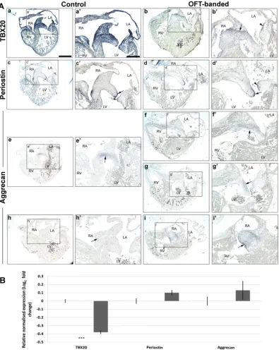
3.2. Aberrant expression of TBX20 and aggrecan in endocardial cushion and valve primordia of HH29 banded hearts
To further characterize the valve phenotype, theTBX20transcription factor and its downstream ECM valve markers (aggrecan and periostin) were examined on serial neighbouring sections throughout HH29
control (n = 9) and banded hearts (n = 7).GAPDHwas used as an ex-perimental control (all hearts expressed normal levels;Fig. 3Ae–f’).
TBX20was expressed intensely in the EC, all four primordial AV valve leaflets and in part of the atrial and ventricular myocardium in the controls (Fig. 2Aa, a’), as previously reported[49]. In contrast, a marked reduction ofTBX20expression was seen throughout the heart, including the cushion and all four primordial valve leaflets in banded hearts (n = 5/7;Fig. 2Ab, b’). However,TBX20expression was still distinct in the boundary of the EC subjacent to the atrial septum (arrow inFig. 2Ab’) and also delineated in the upper lining of the lateral leaflet of primordial MV (arrowhead inFig. 2Ab’). The qPCR quantified mRNA level ofTBX20 (n = 5 per group) demonstrated a 0.38-fold decrease (two-tailed Student'st-test;Pb0.001;Fig. 2B).
Consistent with the literature, punctuate periostin mRNA expression was detected throughout the endocardium of both atria and ventricles as well as the ventricular trabeculae in controls (data not shown)[31, 50]. Expression was further restricted to the endocardial lining of the atrialis side of the primordial MV septal (Fig. 2Ac, arrow inFig. 2Ac’) and lateral (arrowhead inFig. 2Ac’) leaflets. Additionally, abundant periostin mRNA was observed in the chordae tendinae of the left ven-tricular AV junction (data not shown). However, periostin mRNA was not found differentially expressed in the banded hearts by ISH or qPCR (Fig. 2Ad, d’, B).
At HH29, aggrecan was found localized in the cushion core, subja-cent to the atrial septum in the controls (Fig. 2Ae, e’), but not in the pos-terior part of the EC (Fig. 2Ah, h’) or to the valve primordia (Fig. 2Ae, e’, h, h’). Similarly, aggrecan was also expressed in the central cushion in the banded hearts (Fig. 2Af, f’). However, ectopic aggrecan expression was found infive banded hearts (n = 7) in the lateral part of the TV cushion (Fig. 2Ai, i’), and in the atrialis side of the primordial MV leaflet (Fig. 2Ag, g’). qPCR revealed no significant differences in aggrecan ex-pression (Fig. 2B).
3.3. Altered expression of shear stress responsive genes KLF2 and EDN1 in the OFT-banded heart at HH29
To investigate whether an abnormal primordial AV valve is associat-ed with alterassociat-ed shear stress expression, the expression of known shear-stress responsive genes (KLF2 andEDN1) [13] were analysed on neighbouring sections of banded and control hearts.KLF2in banded hearts showed a more intense and distinct expression at the endocardi-al lining of the atriendocardi-alis side of the primordiendocardi-al MV septendocardi-al and laterendocardi-al leaf-lets (n = 3/7; arrows inFig. 3Ab, b’) compared to controls (n = 7;Fig. 3Aa, a’). A significant 1.15-fold increase ofKLF2was shown in the band-ed hearts (n = 4 per group; two-tailband-ed Student'st-test;Pb0.01;Fig. 3B).
In control hearts, mRNA expression ofEDN1was detected in the lin-ing of the outer curvature of the right and left ventricular myocardium (n = 4;Fig. 3Ac, c’). However, the expression ofEDN1was asymmetrical in one of the banded hearts (n = 1/7), with expression increased in the RV compact myocardium (arrow inFig. 3Ad, d’) but was barely discern-ible in the LV ventricular myocardium (arrowhead inFig. 3Ad’); the re-maining banded hearts showed a general decrease ofEDN1expression in both ventricles (n = 4/7). This was further confirmed by significant downregulation of 0.65 fold ofEDN1mRNA (n = 5;Pb0.001;Fig. 3B).GAPDHwas used as a positive control; ubiquitous expression pat-terns were seen in both control (Fig. 3Ae, e’) and OFT-banded hearts (n = 7 per group;Fig. 3Af, f’).
3.4. Reduced apoptosis in cushion and primordial AV valves in HH29 OFT-banded hearts
4Ab’). In contrast, apoptotic cells occurred singly and scattered in the primordial MV (Fig. 4Aa”, b”) and TV leaflets (Fig. 4Aa”’, b”’) in both groups.
The most evident apoptotic region was the EC; 4.02 ± 0.56% apopto-tic cells were found in control hearts (Fig. 4Aa’), but only 1.69 ± 0.37% in the banded hearts (two-tailed Student'st-test;Pb0.01;Fig. 4Ab’, B). In primordial MV septal leaflets, 1.40 ± 0.22% of cells in banded hearts
[image:6.595.106.502.52.552.2](arrow inFig. 4Ab”) were undergoing apoptosis compared to 2.13 ± 0.16% in controls (Fig. 4Aa”, B;Pb0.05). Further, apoptotic cell numbers in the right lateral leaflet were significantly lower in the OFT-banded hearts (arrowhead inFig. 4Ab”’), with 1.22 ± 0.22% apoptotic cells com-pared to 3.12 ± 0.7% in controls (Fig. 4Aa”’, B;Pb0.05). However, the primordial TV septal (arrow inFig. 4Ab”’, B) and the primordial MV lat-eral leaflets (arrowhead in Fig. 4Ab”, B) showed a non-significant
reduction of apoptotic cells in the banded hearts compared to controls (arrow inFig. 4Aa”’, B and arrowhead inFig. 4Aa”, B, respectively;PN 0.05). The total apoptotic cells identified in the primordial valve region was significantly lower in banded hearts (1.53 ± 0.26%) compared to controls (2.89 ± 0.28%;Pb0.01;Fig. 4B).
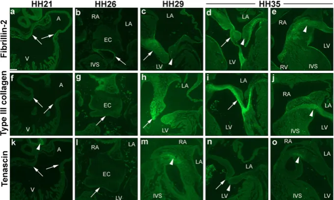
3.5. Spatial-temporal expression profiles of ECM valve proteins
With regards the expression of ECM proteins during early AV valvulogenesis,fibrillin-2 has partially been studied within the litera-ture in the chick[48], whereas the expression of both type III collagen and tenascin have only been studied in other species[51,52]. Therefore, immunohistochemical analyses were performed tofill these gaps in knowledge with a spatial-temporal expression analyses at crucial stages of early chick valvulogenesis (HH21, HH26, HH29 and HH35; n = 2 per stage). Fibrillin-2 was detected at all stages. At HH21, distinctfibrillin-2 immunoreactivity was seen at the endocardial lining of the AVC (Fig. 5a), and at HH26 was restricted to the border of the fused mesenchymalized EC (Fig. 5b). Positive immunostaining was not seen in any parts of the HH21 and HH26 developing heart with type III colla-gen (Fig. 5f, g) and tenascin (Fig. 5k, l). At HH29, stronger staining of fibrillin-2 was visible in the primordial MV leaflet (arrow inFig. 5c), with little to no immunoreactivity offibrillin-2 in the left lateral leaflet
(arrowhead inFig. 5c). At HH35, immunolocalisation offibrillin-2 was seen in a restricted pattern in both septal and lateral leaflets, and in the chordae tendinae (Fig. 5d). A fairly strong immunostaining of fibrillin-2 was seen in the central endocardial cushion at this stage (ar-rowhead inFig. 5e). Type III collagen was seen to a lesser degree in the HH29 primordial MV leaflet, being more centralized at the tip of the valve leaflet (Fig. 5h). However, strong staining of type III collagen was found extending from the septal leaflet to the chordae tendinae at HH35 (Fig. 5i), and in the core cushion (Fig. 5j). Positive staining of tenascin was detected in the core cushion at HH29 (Fig. 5m) and HH35 (Fig. 5o), but not in the primordial MV leaflets (Fig. 5n).
3.6. Decreased expression of ECM proteins in primordial AV valve in HH35 OFT-banded hearts
During post-EMT stages, the cushion mesenchyme normally differ-entiates into valve interstitial cells, the specialized valvularfibroblasts that expressfibrillary collagens, and chondroitin sulfate proteoglycans [3,34]. Therefore, in order to determine whether valve primordia were affected at later stage of valve development, type III collagen and tenascin immunohistochemistry were performed, as well as differentia-tion markerfibrillin-2[48].
Fibrillin-2 and type III collagen immunostaining were detected strongly throughout the leaflet in controls (n = 5;Fig. 6Aa, c respective-ly). A noticeable weaker and more diffuse staining offibrillin-2 (n = 3/5) and type III collagen (n = 4/5) was seen in the truncated primordi-al MV leaflets in the OFT-banded hearts (Fig. 6Ab, d). Likewise, a reduc-tion of type III collagen (n = 4/5) was seen in the chordae tendinae in the banded hearts (Fig. 6Af) when compared to controls (Fig. 6Ae). Sim-ilarly, a positive staining was seen in the central endocardial cushion area subjacent to the atrial septum with zonal restriction offibrillin-2 in controls (Fig. 6Ba), with decreased immunolocalisation in the core cushion of banded hearts (n = 3/5; arrow inFig. 6Bb). The positive staining of type III collagen was located in the central cushion in controls (arrow inFig. 6Bc). In contrast, little to no immunoreactivity of type III collagen was observed in the OFT-banded hearts (n = 4/5; arrowhead
[image:8.595.106.504.52.491.2]denotes equivalent region of the cushion inFig. 6Bc, d). Lastly, tenascin staining was in the centre of the cushion of controls (Fig. 6Be) but re-duced in all banded hearts (n = 5; arrow inFig. 6Bf). Thefluorescent in-tensity was further quantified in the primordial MV leaflets infibrillin-2, type III collagen and tenascin (Fig. 6C; n = 5). The average grey level of fibrillin-2 was 50.48 ± 1.71 in controls but 34.11 ± 8.02 in banded hearts, a 32.4% decrease offluorescent signal. However, this reduction was not significant (PN0.05). A non-significant 30.4% decrease offl uo-rescence intensity was seen for tenascin, where banded hearts and con-trol presented 16.1 ± 0.63 and 23.48 ± 3.14 respectively. The fluorescence intensity of type III collagen was 57.31 ± 6.39 in controls and 34.37 ± 3.92 in banded hearts, a significant reduction of 40% (two-tailed Student'st-test;Pb0.05). In order to support the decreased expression of these ECM proteins, mRNA level was quantitated by qPCR
at HH35 isolated valve region.FBN2andTNCboth showed a significant downregulation of 1.06 and 1.26 fold (n = 4 per group; two-tailed Student'st-test;Pb0.05;Fig. 6D). Further,COL3A1andTGM2 demon-strated a significant downregulation of 1.32 and 1.85 fold in banded hearts (n = 4 per group;Pb0.01;Fig. 6D).
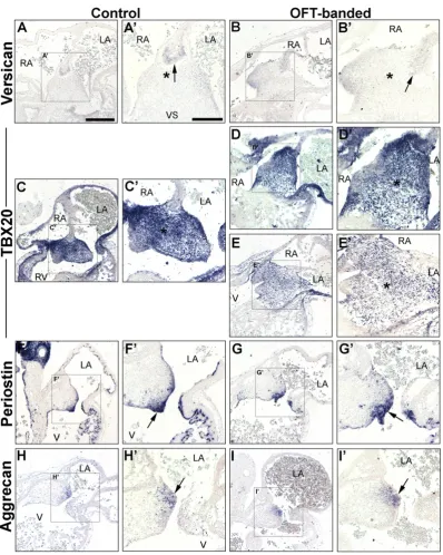
3.7. Morphologically normal endocardial cushions and atrial septation in HH26 OFT-banded hearts
Atrial septation was seen to be morphologically normal in banded hearts (n = 5), a process which completes around HH24[28]. However, the studies described above suggested that the defective valve primordia at HH29 and HH35 might form from a deformed EC. In order to examine this, ISH was performed on markers of the EC area on neighbouring sections at HH26 (post-EMT).
As expected, versican was expressed in the mesenchymal cap of the atrial septum that fused with the AV cushions in both controls (arrow in Fig. 7A, A’) and OFT-banded hearts (n = 5 per group;Fig. 7B, B’), confirming normal atrial septation had occurred [53]. TBX20was strongly expressed in the EC and part of the ventricular myocardium (Fig. 7C, C’) of controls. Less intenseTBX20expression was detected on serial sections in two of the banded hearts analysed (2/5;Fig. 7E, E’) compared to controls. Otherwise,TBX20expression was found normal (3/5;Fig. 7D, D’). At HH26, abundant periostin mRNA was detected in the endothelial lining and adjacent tissue of the AV cushions in controls (Fig. 7F, F’) and OFT-banded hearts (n = 5;Fig. 7G, G’). Aggrecan ex-pression was restricted to the lateral part of the left side of the endocar-dial cushion in both groups (arrow inFig. 7H, H’, I, I’).
4. Discussion
Valve defects constitute an important medical issue challenging our society. Though valve developmental defects may initially be asymp-tomatic, they are often progressive and contribute to valve disease later in life. Abnormal valves often display dysregulation of ECM pro-teins such as collagens and glycosaminoglycans, as seen in patients with myxomatous mitral valve and mitral valve prolapse [54].
Structural changes of ECM are associated with the aberrant re-expres-sion of early valve mesenchymal markers, implying the reactivation of the fetal gene program that is normally quiescent in adulthood[55,56]. Recently, valvulogenesis has been shown to be haemodynamically dependent, with occlusion of blood at either the inflow or outflow resulting in absent AV valve in zebrafish[10]. We delineated the role of abnormal haemodynamics in the developing primordial mitral AV valve and its alignment with other septal components at critical stages of chamber septation and valve development. A ligature was placed around the HH21 outflow region and harvested at HH26 (atrial septation complete, post-EMT with fusion of superior and inferior cush-ions), HH29 (fused cushions mature, primordial AV valve elongates and ventricular septation complete) and HH35 (remodelling). In this OFT-banding model, alteration of haemodynamics has previously been shown to result in increased ventricular pressure and wall shear stress, as well as higher peak bloodflow velocity in the OFT region[57–60]. Morphologically, the OFT-banded hearts exhibited right-shifted posi-tion of the OFT and appeared to be approximately 1.77 times larger than controls by HH29 (unpublished data;Fig. 1Cc, d), An enlarged heart is a characteristic of pressure-overloaded embryonic hearts[21, 58,61]. However, the crown rump length and eye diameter (as de-scribed here) together with cardiac output and heart rate[57,62], remained unchanged in this model.
The AV endocardial cushion was structurally normal at HH26 but dysmorphic and truncated primordial mitral valves were seen at HH29 and HH35, with a smaller primordial valve region. AV valves were generally immature in another altered haemodynamic model (vi-telline vein clip)[20], with the tricuspid valve morphologically abnor-mal upon banding[21].
[image:9.595.123.464.53.257.2]TBX20is important in promoting EC cell proliferation and migration, and ECM gene expression in vitro[43,44]. In addition,Tbx20plays a role in EC maturation and valve elongation[41]. In normal development, fu-sion of the superior and inferior AV cushions and their subsequent union with the dorsal mesenchymal cap of the primary atrial septum has occurred by HH26[26,63]. TBX20expression was analysed in order to elucidate if it was differentially expressed in the morphologi-cally normal OFT-banded HH26 early valve primordia. Interestingly,
despite no cushion structural abnormalities in HH26 banded hearts, de-creased expression ofTBX20was observed in the EC region in two of the five banded hearts (asterisk inFig. 7E, E’). Periostin and aggrecan were not differentially expressed in neighbouring sections. Periostin and aggrecan have been found downregulated in theTbx20knockdown [41]. The decrease in expression ofTBX20(−0.39 fold) in our study at HH29 suggests alteration of haemodynamics leads to reducedTBX20 and concurrent ectopic expression of its target aggrecan, which together engendered AV valve elongation defects. The data described here sup-ports the hypothesis thatTBX20acts upstream of aggrecan. Aggrecan is one of the main chondroitin sulfate proteoglycan constituents of the spongiosa layer in avian valves, and is required to withstand compres-sive forces against bloodflow[32,43], and reportedly has a role in valve cell differentiation[44,64].
Periostin has been previously reported for its multifaceted role in cell migration and differentiation; it promotes differentiation of prevalvular mesenchymal cells into collagen-producingfibroblastic cells termed‘valve interstitial cells’while repressing transformation
into myocyte lineages[31,44]. Periostin is also able to interact with other ECM components such as collagen, tenascin andfibronectin[65– 67]. Interestingly, although periostin was normally expressed, collagen III and tenascin were found to be differentially expressed. It can be spec-ulated that mRNA downregulation of one transcript variant might lead to a compensatory upregulation of another variant upon altered haemodynamics, as differential periostin isoform expression profiles were observed in periostin-null mice after myocardial infarction[68, 69].
[image:10.595.133.474.48.453.2]Remodelling of the AVC and OFT tissues occur through apoptosis [70]. Clustering of apoptotic cells is involved in cushion differentiation and outgrowth, as evidenced by a lack of apoptosis and overabundant cushions in the Nf-1 null mouse[71,72]. In contrast, our study showed a decrease in apoptosis in the primordial AV region and smaller cush-ions in banded hearts. This suggests normal levels of apoptosis are re-quired for EC differentiation[72]. In addition, the regulatory factors Bmp4,Bmp2andMsx2have been associated with cells undergoing apo-ptosis, and hence differentiation[73]. AsBmp2is considered to be a key
downstream targets ofTbx20in AVC development[74], it is a gene wor-thy of future investigation.
At a later stage of valvulogenesis, cushion mesenchyme is normally differentiated into valve interstitial cells, cells which express genes that encodefibrillary collagens, chondroitin sulfate proteoglycans and elastin, which are associated with stratified ECM of mature valves[3, 34,75]. Upon abnormal bloodflow, the differentiation process was pre-dicted to be affected in HH35 banded hearts, with weaker expression of COL3A,TNCandFBN2mRNA. Fibrillin-2 is known to be expressed in a subset of endothelial cells competent to transdifferentiate into cushion mesenchyme and connective tissuefibroblasts of the valve leaflets
[image:11.595.95.494.47.546.2][48]. Also,FBN2transcripts accumulate prior to tissue differentiation, decreasing rapidly thereafter during development[76]. We speculate that the decrease offibrillin-2 staining and its mRNA in our study sug-gests differentiation failed to occur. Similarly, in patients with mitral valve prolapse syndrome, a more diffuse and weaker staining pattern offibrillin and type III collagen were seen in the area of myxoid degen-eration of diseased MV leaflets[77]. Further, part of the EC is differenti-ated into valve leaflets and chordae tendinae during normal development, and the attachment of the papillary muscle to valve leaf-lets without chordae tendinae was seen in human fetal hearts at weeks 5–19 week, when differentiation failed to occur[78,79]. Additionally,
the downregulation in banded hearts of the gene transglutaminase-2 (TGM2), which encodes the TG2 ECM cross-linking protein, could lead to the decrease of the ECM proteinsfibrillin-2 and type III collagen. TG2 has been reported for its role in ECM condensation and organiza-tion by interacting withfilamin-A and serotonin[5]. TG2 has also been implicated in osteoblast differentiation, with its expression defining the border of differentiation; TG2 protein expression was absent in the cushion but present in the adjacent myocardium prior to cushion fu-sion[22,80]. It was also in almost all the interstitial cells following cush-ion fuscush-ion (HH27) and maturatcush-ion (HH35)[22]. Downregulation of TGM2in our study suggests aberrant differentiation of the cushion mes-enchyme, indicating that the proteolytically activate protein could be an area of future interest.
Normalfluidflow in the heart is important in shaping the EC into the developing cardiac valve leaflets[10,11]. Knockdown oftrpv4(encodes a mechanotransduction protein), which is upstream of the shear stress responsive geneklf2a, led to the development of dysmorphic valves in zebrafish[81]. In addition, theklf2anull mutant zebrafish displayed an array of valvular phenotypes, attributed to cell disorganization at the AVC and impairedfibronectin synthesis[12]. In addition, laminar shear stress is involved in endothelial differentiation[82,83]. In the OFT-banded heart, higher peak and end diastolic interventricular pres-sure was seen[18,57,58]. Further, increased peak bloodflow velocities near the banding site and regurgitation around the AV region were found at HH27[17,84]. Alteration of wall shear stresses, as a result of changes of bloodflow pattern, are known to alter shear stress respon-sive genesKLF2andEDN1[85], as did altering haemodynamics in the venous clip model [86]. Here, we support the notion of pressure overloading upon OFT-banding by showing the up- and down-regula-tion of theKLF2andEDN1shear stress genes respectively, in banded hearts. This might lead to aberrant differentiation of the EC cushion and decreased expression of the ECM proteins in the valve leaflet and hence a maturation arrest in valve development.
5. Concluding remarks
Fetal valvulogenesis consists of two main phases: EMT and post-EMT maturation, with the latter phase involving the remodelling of the cushion into mature valve leaflets. Defects arising in the post-EMT valve were associated with aberrant expression of early mesenchymal markers and dysregulation of ECM proteins such asfibrillin and collagen which were previously seen in patients with mitral valve prolapse syn-drome, with the underlying mechanism remaining poorly understood. In this study we show aberrant expression ofTBX20, aggrecan and shear stress responsive genes upon abnormal haemodynamics. The de-crease in apoptosis and the dysregulation of ECM proteins indicate the mechanism. Together, our data suggest that failure of differentiation of cushion mesenchyme into valvular interstitial cells caused by abnormal bloodflow, is a potential mechanism to give rise to valve defects post-EMT. Therefore, this study provides new insights into potential aetiol-ogies of human congenital valve malformations.
Funding
This work was supported by British Heart Foundation [grant number FS/12/44/29619 to S.L.].
Disclosures
No competing interests declared.
Acknowledgements
The authors would like to thank Professor David Sedmera, Professor David Brook, Dr. Catrin Rutland and Dr. Paul Scotting for technical ad-vice and helpful discussions, Nikki Asemota for some imaging, the SLIM
team University of Nottingham for imaging support and Joanne Marrison from University of York for her help in slide scanning using Zeiss Axio Scan Z1. We would also like to thank Dr. Catrin Rutland for reading the manuscript. The JB3 antibody, developed by Professor CD Little, the M1-B4 antibody, developed by Dr. DM Fambrough, and the 3B2 antibody developed by Dr. R Mayne, were obtained from the Devel-opmental Studies Hybridoma Bank, created by the NICHD of the NIH and maintained at The University of Iowa, Department of Biology, Iowa City, IA 52242.
References
[1] P.E. Seguela, L. Houyel, P. Acar, Congenital malformations of the mitral valve, Arch. Cardiovasc. Dis. 104 (8–9) (2011) 465–479,http://dx.doi.org/10.1016/j.acvd.2011. 06.004.
[2] J. Lincoln, K.E. Yutzey, Molecular and developmental mechanisms of congenital heart valve disease, Birth Defects Res. A Clin. Mol. Teratol. 91 (6) (2011) 526–534,
http://dx.doi.org/10.1002/bdra.20799.
[3] R.B. Hinton, K.E. Yutzey, Heart valve structure and function in development and dis-ease, Annu. Rev. Physiol. 73 (2011) 29–46, http://dx.doi.org/10.1146/annurev-physiol-012110-142145.
[4] A. Banerjee, T. Kohl, N.H. Silverman, Echocardiographic evaluation of congenital mi-tral valve anomalies in children, Am. J. Cardiol. 76 (17) (1995) 1284–1291.
[5] K. Sauls, et al., Developmental basis forfilamin-A-associated myxomatous mitral valve disease, Cardiovasc. Res. 96 (1) (2012) 109–119,http://dx.doi.org/10.1093/cvr/cvs238. [6] V. Garg, et al., Mutations in NOTCH1 cause aortic valve disease, Nature 437 (7056)
(2005) 270–274,http://dx.doi.org/10.1038/nature03940.
[7] F.N. Delling, R.S. Vasan, Epidemiology and pathophysiology of mitral valve prolapse: new insights into disease progression, genetics, and molecular basis, Circulation 129 (21) (2014) 2158–2170,http://dx.doi.org/10.1161/CIRCULATIONAHA.113.006702. [8] C. Dina, et al., Genetic association analyses highlight biological pathways underlying
mitral valve prolapse, Nat. Genet. 47 (10) (2015) 1206–1211,http://dx.doi.org/10. 1038/ng.3383.
[9] I.H. Melvinsdottir, A. Geirsson, Myxomatous leaflet biological aberrations and po-tential therapeutic targets, Ann. Cardiothorac. Surg. 4 (4) (2015) 355–357,http:// dx.doi.org/10.3978/j.issn.2225-319X.2014.09.24.
[10] J.R. Hove, et al., Intracardiacfluid forces are an essential epigenetic factor for embry-onic cardiogenesis, Nature 421 (6919) (2003) 172–177,http://dx.doi.org/10.1038/ nature01282.
[11] J. Vermot, et al., Reversing bloodflows act through klf2a to ensure normal valvulogenesis in the developing heart, PLoS Biol. 7 (11) (2009) e1000246,http:// dx.doi.org/10.1371/journal.pbio.1000246.
[12] E. Steed, et al., klf2a couples mechanotransduction and zebrafish valve morphogen-esis throughfibronectin synthesis, Nat. Commun. 7 (2016) 11646,http://dx.doi.org/ 10.1038/ncomms11646.
[13] B.C. Groenendijk, et al., Development-related changes in the expression of shear stress responsive genes KLF-2, ET-1, and NOS-3 in the developing cardiovascular system of chicken embryos, Dev. Dyn. 230 (1) (2004) 57–68,http://dx.doi.org/10. 1002/dvdy.20029.
[14] L. Sun, S. Chandra, P. Sucosky, Ex vivo evidence for the contribution of hemodynamic shear stress abnormalities to the early pathogenesis of calcific bicuspid aortic valve dis-ease, PLoS One 7 (10) (2012) e48843,http://dx.doi.org/10.1371/journal.pone.0048843. [15] S. Chandra, N.M. Rajamannan, P. Sucosky, Computational assessment of bicuspid aortic valve wall-shear stress: implications for calcific aortic valve disease, Biomech. Model. Mechanobiol. 11 (7) (2012) 1085–1096, http://dx.doi.org/10.1007/s10237-012-0375-x.
[16] V. Menon, et al., Altered hemodynamics in the embryonic heart affects outflow valve development, J. Cardiovasc. Dev. Dis. 2 (2) (2015) 108–124,http://dx.doi. org/10.3390/jcdd2020108.
[17] T.C. McQuinn, et al., High-frequency ultrasonographic imaging of avian cardiovascu-lar development, Dev. Dyn. 236 (12) (2007) 3503–3513,http://dx.doi.org/10.1002/ dvdy.21357.
[18] L. Shi, et al., Alterations in pulse wave propagation reflect the degree of outflow tract banding in HH18 chicken embryos, Am. J. Physiol. Heart Circ. Physiol. 305 (3) (2013) H386–H396,http://dx.doi.org/10.1152/ajpheart.00100.2013.
[19] E.B. Clark, N. Hu, G.C. Rosenquist, Effect of conotruncal constriction on aortic-mitral valve continuity in the stage 18, 21 and 24 chick embryo, Am. J. Cardiol. 53 (2) (1984) 324–327.
[20] B. Hogers, et al., Unilateral vitelline vein ligation alters intracardiac bloodflow pat-terns and morphogenesis in the chick embryo, Circ. Res. 80 (4) (1997) 473–481.
[21] D. Sedmera, et al., Remodeling of chick embryonic ventricular myoarchitecture under experimentally changed loading conditions, Anat. Rec. 254 (2) (1999) 238–252.
[22] A. de Vlaming, et al., Atrioventricular valve development: new perspectives on an old theme, Differentiation 84 (1) (2012) 103–116,http://dx.doi.org/10.1016/j.diff. 2012.04.001.
[23] T. Yamagishi, et al., Expression of tbx20 RNA during chick heart development, Dev. Dyn. 230 (3) (2004) 576–580,http://dx.doi.org/10.1002/dvdy.20076.
[24] S. Webb, N.A. Brown, R.H. Anderson, Formation of the atrioventricular septal struc-tures in the normal mouse, Circ. Res. 82 (6) (1998) 645–656.
[26] B.S. Snarr, et al., A spatiotemporal evaluation of the contribution of the dorsal mes-enchymal protrusion to cardiac development, Dev. Dyn. 236 (5) (2007) 1287–1294,
http://dx.doi.org/10.1002/dvdy.21074.
[27] G. Ben-Shachar, et al., Ventricular trabeculations in the chick embryo heart and their contribution to ventricular and muscular septal development, Circ. Res. 57 (5) (1985) 759–766.
[28]M.J. Hendrix, D.E. Morse, Atrial septation. I. Scanning electron microscopy in the chick, Dev. Biol. 57 (2) (1977) 345–363 (10.0012-1606(77)90220-2 [pii]).
[29] B.J. Martinsen, Reference guide to the stages of chick heart embryology, Dev. Dyn. 233 (4) (2005) 1217–1237,http://dx.doi.org/10.1002/dvdy.20468.
[30] K. Inai, et al., BMP-2 induces cell migration and periostin expression during atrio-ventricular valvulogenesis, Dev. Biol. 315 (2) (2008) 383–396,http://dx.doi.org/ 10.1016/j.ydbio.2007.12.028.
[31] R.A. Norris, et al., Periostin promotes afibroblastic lineage pathway in atrioventric-ular valve progenitor cells, Dev. Dyn. 238 (5) (2009) 1052–1063,http://dx.doi.org/ 10.1002/dvdy.21933.
[32] J. Lincoln, A.W. Lange, K.E. Yutzey, Hearts and bones: shared regulatory mechanisms in heart valve, cartilage, tendon, and bone development, Dev. Biol. 294 (2) (2006) 292–302,http://dx.doi.org/10.1016/j.ydbio.2006.03.027.
[33] M.D. Combs, K.E. Yutzey, Heart valve development: regulatory networks in develop-ment and disease, Circ. Res. 105 (5) (2009) 408–421,http://dx.doi.org/10.1161/ CIRCRESAHA.109.201566.
[34] R.B. Hinton Jr., et al., Extracellular matrix remodeling and organization in developing and diseased aortic valves, Circ. Res. 98 (11) (2006) 1431–1438,http://dx.doi.org/ 10.1161/01.RES.0000224114.65109.4e.
[35]M. Villegas Garcia, et al., Congenital diverticulum of the right ventricle associated with coarctation of aorta, atrial and ventricular septal defect and ductus, Eur. J. Echocardiogr. 2 (3) (2001) 205–206.
[36] F.J. de Lange, et al., Lineage and morphogenetic analysis of the cardiac valves, Circ. Res. 95 (6) (2004) 645–654,http://dx.doi.org/10.1161/01.RES.0000141429.13560.cb. [37] B.P. Kruithof, S.A. Krawitz, V. Gaussin, Atrioventricular valve development during
late embryonic and postnatal stages involves condensation and extracellular matrix remodeling, Dev. Biol. 302 (1) (2007) 208–217,http://dx.doi.org/10.1016/j.ydbio. 2006.09.024.
[38] J.T. Butcher, R.R. Markwald, Valvulogenesis: the moving target, Philos. Trans. R. Soc. Lond. Ser. B Biol. Sci. 362 (1484) (2007) 1489–1503,http://dx.doi.org/10.1098/rstb. 2007.2130.
[39]V. Hamburger, H.L. Hamilton, A series of normal stages in the development of the chick embryo, J. Exp. Morphol. 88 (1951) 49.
[40] T.M. Mayhew, The new stereological methods for interpreting functional morphol-ogy from slices of cells and organs, Exp. Physiol. 76 (5) (1991) 639–665.
[41] X. Cai, et al., Tbx20 acts upstream of Wnt signaling to regulate endocardial cushion formation and valve remodeling during mouse cardiogenesis, Development 140 (15) (2013) 3176–3187,http://dx.doi.org/10.1242/dev.092502.
[42] J. Mu, et al., In vivo quantification of embryonic and placental growth during gesta-tion in mice using micro-ultrasound, Reprod. Biol. Endocrinol. 6 (2008) 34,http:// dx.doi.org/10.1186/1477-7827-6-34.
[43] E.L. Shelton, K.E. Yutzey, Tbx20 regulation of endocardial cushion cell proliferation and extracellular matrix gene expression, Dev. Biol. 302 (2) (2007) 376–388,
http://dx.doi.org/10.1016/j.ydbio.2006.09.047.
[44] E.L. Shelton, K.E. Yutzey, Twist1 function in endocardial cushion cell proliferation, migration, and differentiation during heart valve development, Dev. Biol. 317 (1) (2008) 282–295,http://dx.doi.org/10.1016/j.ydbio.2008.02.037.
[45] J. Lin, C. Redies, Histological evidence: housekeeping genes beta-actin and GAPDH are of limited value for normalization of gene expression, Dev. Genes Evol. 222 (6) (2012) 369–376,http://dx.doi.org/10.1007/s00427-012-0420-x.
[46] S. Somi, et al., Dynamic patterns of expression of BMP isoforms 2, 4, 5, 6, and 7 dur-ing chicken heart development, Anat. Rec. A Discov. Mol. Cell. Evol. Biol. 279 (1) (2004) 636–651,http://dx.doi.org/10.1002/ar.a.20031.
[47] J. England, et al., Cardiac troponin T is necessary for normal development in the em-bryonic chick heart, J. Anat. (2016)http://dx.doi.org/10.1111/joa.12486.
[48]A.M. Wunsch, C.D. Little, R.R. Markwald, Cardiac endothelial heterogeneity defines valvular development as demonstrated by the diverse expression of JB3, an antigen of the endocardial cushion tissue, Dev. Biol. 165 (2) (1994) 585–601.
[49] T.F. Plageman Jr., K.E. Yutzey, Differential expression and function of Tbx5 and Tbx20 in cardiac development, J. Biol. Chem. 279 (18) (2004) 19026–19034.
[50] R.A. Norris, et al., Identification and detection of the periostin gene in cardiac devel-opment, Anat. Rec. A Discov. Mol. Cell. Evol. Biol. 281 (2) (2004) 1227–1233,http:// dx.doi.org/10.1002/ar.a.20135.
[51] K. Imanaka-Yoshida, et al., The dynamic expression of tenascin-C and tenascin-X dur-ing early heart development in the mouse, Differentiation 71 (4–5) (2003) 291–298.
[52] J.D. Peacock, et al., Temporal and spatial expression of collagens during murine atrioventricular heart valve development and maintenance, Dev. Dyn. 237 (10) (2008) 3051–3058,http://dx.doi.org/10.1002/dvdy.21719.
[53] D.J. Henderson, A.J. Copp, Versican expression is associated with chamber specifi ca-tion, septaca-tion, and valvulogenesis in the developing mouse heart, Circ. Res. 83 (5) (1998) 523–532.
[54] D.G. Greenhouse, et al., Mitral valve prolapse is associated with altered extracellular matrix gene expression patterns, Gene 586 (1) (2016) 56–61,http://dx.doi.org/10. 1016/j.gene.2016.04.004.
[55] D. MacGrogan, et al., How to make a heart valve: from embryonic development to bioengineering of living valve substitutes, Cold Spring Harb. Perspect. Med. 4 (11) (2014) a013912,http://dx.doi.org/10.1101/cshperspect.a013912.
[56] E.E. Wirrig, R.B. Hinton, K.E. Yutzey, Differential expression of cartilage and bone-re-lated proteins in pediatric and adult diseased aortic valves, J. Mol. Cell. Cardiol. 50 (3) (2011) 561–569,http://dx.doi.org/10.1016/j.yjmcc.2010.12.005.
[57] E.B. Clark, et al., Effect of increased pressure on ventricular growth in stage 21 chick embryos, Am. J. Phys. 257 (1 Pt 2) (1989) H55–H61.
[58] K. Tobita, et al., Regional passive ventricular stress-strain relations during develop-ment of altered loads in chick embryo, Am. J. Physiol. Heart Circ. Physiol. 282 (6) (2002) H2386–H2396,http://dx.doi.org/10.1152/ajpheart.00879.2001.
[59] V.K. Chivukula, et al., Effect of outflow tract banding on embryonic cardiac hemody-namics, J. Cardiovasc. Dev. Dis. 3 (1) (2016)http://dx.doi.org/10.3390/jcdd3010001. [60] M. Midgett, K. Thornburg, S. Rugonyi, Bloodflow patterns underlie developmental
heart defects, Am. J. Physiol. Heart Circ. Physiol. 312 (3) (2017) H632–H642,
http://dx.doi.org/10.1152/ajpheart.00641.2016.
[61] C.E. Hall, et al., Hemodynamic-dependent patterning of endothelin converting en-zyme 1 expression and differentiation of impulse-conducting Purkinjefibers in the embryonic heart, Development 131 (3) (2004) 581–592,http://dx.doi.org/10. 1242/dev.00947.
[62] B.B. Keller, M. Yoshigi, J.P. Tinney, Ventricular-vascular uncoupling by acute conotruncal occlusion in the stage 21 chick embryo, Am. J. Phys. 273 (6 Pt 2) (1997) H2861–H2866.
[63] S.E. Lindsey, J.T. Butcher, The cycle of form and function in cardiac valvulogenesis, Aswan Heart Centre Science & Practice Series 2011 (2) (2011) 10,http://dx.doi. org/10.5339/ahcsps.2011.10.
[64] M.K. Zanin, et al., Distinct spatial and temporal distributions of aggrecan and versican in the embryonic chick heart, Anat. Rec. 256 (4) (1999) 366–380.
[65] J.T. Butcher, et al., Periostin promotes atrioventricular mesenchyme matrix invasion and remodeling mediated by integrin signaling through Rho/PI 3-kinase, Dev. Biol. 302 (1) (2007) 256–266,http://dx.doi.org/10.1016/j.ydbio.2006.09.048. [66] R.A. Norris, et al., Periostin regulates collagenfibrillogenesis and the biomechanical
properties of connective tissues, J. Cell. Biochem. 101 (3) (2007) 695–711,http://dx. doi.org/10.1002/jcb.21224.
[67] I. Kii, et al., Incorporation of tenascin-C into the extracellular matrix by periostin un-derlies an extracellular meshwork architecture, J. Biol. Chem. 285 (3) (2010) 2028–2039,http://dx.doi.org/10.1074/jbc.M109.051961.
[68] M. Shimazaki, et al., Periostin is essential for cardiac healing after acute myocardial infarction, J. Cell Biol. 180 (2) (2008) i7,http://dx.doi.org/10.1083/jcb1802oia7. [69] S. Hoersch, M.A. Andrade-Navarro, Periostin shows increased evolutionary plasticity
in its alternatively spliced region, BMC Evol. Biol. 10 (1) (2010) 30,http://dx.doi.org/ 10.1186/1471-2148-10-30.
[70] T. Pexieder, The tissue dynamics of heart morphogenesis. I. The phenomena of cell death. B. Topography, Z Anat Entwicklungsgesch 138 (3) (1972) 241–253.
[71]M.M. Lakkis, J.A. Epstein, Neurofibromin modulation of ras activity is required for normal endocardial-mesenchymal transformation in the developing heart, Develop-ment 125 (22) (1998) 4359–4367.
[72] E. Abdelwahid, L.J. Pelliniemi, E. Jokinen, Cell death and differentiation in the devel-opment of the endocardial cushion of the embryonic heart, Microsc. Res. Tech. 58 (5) (2002) 395–403,http://dx.doi.org/10.1002/jemt.10159.
[73]E. Abdelwahid, et al., Overlapping and differential localization of Bmp-2, Bmp-4, Msx-2 and apoptosis in the endocardial cushion and adjacent tissues of the develop-ing mouse heart, Cell Tissue Res. 305 (1) (2001) 67–78.
[74] X. Cai, et al., Myocardial Tbx20 regulates early atrioventricular canal formation and endocardial epithelial-mesenchymal transition via Bmp2, Dev. Biol. 360 (2) (2011) 381–390,http://dx.doi.org/10.1016/j.ydbio.2011.09.023.
[75] A.H. Chester, P.M. Taylor, Molecular and functional characteristics of heart-valve in-terstitial cells, Philos. Trans. R. Soc. Lond. Ser. B Biol. Sci. 362 (1484) (2007) 1437–1443,http://dx.doi.org/10.1098/rstb.2007.2126.
[76]H. Zhang, W. Hu, F. Ramirez, Developmental expression offibrillin genes suggests heterogeneity of extracellular microfibrils, J. Cell Biol. 129 (4) (1995) 1165–1176.
[77] J.F. Nasuti, et al., Fibrillin and other matrix proteins in mitral valve prolapse syn-drome, Ann. Thorac. Surg. 77 (2) (2004) 532–536,http://dx.doi.org/10.1016/ S0003-4975(03)01584-4.
[78] P.W. Oosthoek, et al., Development of the papillary muscles of the mitral valve: mor-phogenetic background of parachute-like asymmetric mitral valves and other mitral valve anomalies, J. Thorac. Cardiovasc. Surg. 116 (1) (1998) 36–46,http://dx.doi. org/10.1016/S0022-5223(98)70240-5.
[79] I. Matsumaru, et al., Characteristics and treatment strategies of mitral regurgitation associated with undifferentiated papillary muscle, Gen. Thorac. Cardiovasc. Surg. 60 (7) (2012) 406–410,http://dx.doi.org/10.1007/s11748-012-0055-x.
[80]X. Yin, et al., Tissue transglutaminase (TG2) activity regulates osteoblast differentiation and mineralization in the SAOS-2 cell line, Braz. J. Med. Biol. Res. 45 (8) (2012) 693–700.
[81] E. Heckel, et al., Oscillatoryflow modulates mechanosensitive klf2a expression through trpv4 and trpp2 during heart valve development, Curr. Biol. 25 (10) (2015) 1354–1361,http://dx.doi.org/10.1016/j.cub.2015.03.038.
[82] H. Wang, et al., Shear stress induces endothelial differentiation from a murine em-bryonic mesenchymal progenitor cell line, Arterioscler. Thromb. Vasc. Biol. 25 (9) (2005) 1817–1823,http://dx.doi.org/10.1161/01.ATV.0000175840.90510.a8. [83] H. Wang, et al., Fluid shear stress regulates the expression of TGF-beta1 and its
sig-naling molecules in mouse embryo mesenchymal progenitor cells, J. Surg. Res. 150 (2) (2008) 266–270,http://dx.doi.org/10.1016/j.jss.2007.12.801.
[84] S. Rugonyi, et al., Changes in wall motion and bloodflow in the outflow tract of chick embryonic hearts observed with optical coherence tomography after outflow tract banding and vitelline-vein ligation, Phys. Med. Biol. 53 (18) (2008) 5077–5091,http://dx.doi.org/10.1088/0031-9155/53/18/015.
[85] B.C. Groenendijk, et al., The role of shear stress on ET-1, KLF2, and NOS-3 expression in the developing cardiovascular system of chicken embryos in a venous ligation model, Physiology (Bethesda) 22 (2007) 380–389,http://dx.doi.org/10.1152/ physiol.00023.2007.